Accupril

Accupril
Accupril dosages: 10 mg
Accupril packs: 30 pills, 60 pills, 90 pills, 120 pills

According to Steinhart and Kleinsasser treatment quadratus lumborum discount accupril 10 mg with amex, the sublingual glands must be routinely taken inside the resection to stop native recurrences treatment 911 accupril 10 mg discount with mastercard. However medications ritalin 10mg accupril generic, Clark and colleagues5 advocate that sublingual glands not be removed in early-stage T1/T2 lesions. They argue that removing offers little oncologic benefit whereas placing different anatomic buildings at risk, particularly the lingual nerve, the hypoglossal nerve, and the submandibular duct. Removal may improve the risk for fistula growth by weakening the tissue separating the oral cavity from the neck. Submandibular glands are generally eliminated as part of a selective neck dissection, although this seems to be extra of a holdover from the era of radical neck dissections. However, as a outcome of floor of mouth cancers are so close to the submandibular gland, it should be eliminated to ensure a complete 3D tumor clearance. The gland must also be excised in the case of direct tumor infiltration, although this was rare within the Steinhart and Kleinsasser series (1/253 glands). Fine needle aspiration can also be accomplished for small lesions; nonetheless, fine needle aspiration is mostly reserved for establishing analysis in scientific lymphadenopathy. Imaging for Floor of Mouth Cancers A good medical examination forms the spine of the decisionmaking process in an space where tumors are simply palpated and visualized. Smee and colleagues7 demonstrated the superiority of scientific examination in figuring out the staging and administration of sufferers with ground of mouth cancers. Imaging can help in staging, determine the extent of surgical resection, and make clear whether the mandible is to be thought of as part of the resection. Other normal imaging methods embody standard x-rays (mandible sequence, occlusal radiograph, and an orthopantomogram). Imaging of the neck for lymph node metastasis can also be one other important consideration for determining treatment and prognosis for sufferers. Staging Floor of Mouth Cancers Floor of mouth cancers are usually grouped beneath the staging system for cancers of the oral cavity. Depth of invasion is a extra subtle and reproducible methodology of measuring a tumor. These smaller stage cancers are typically resected via a transoral approach and infrequently contain a lip-split mandibulotomy approach. Lesions which are stuck to the mandible additionally involve elimination of half (marginal mandibulectomy) or all the mandible (composite resection). Conducting a marginal mandibulectomy when the tumor is adjacent to periosteum is now a extensively accepted follow, which was earlier advocated by Yco and Cruickshank. Local anesthesia (1% lidocaine with 1:a hundred,000 epinephrine) is injected across the tumor over the proposed boundary. Once the vasoconstrictor has taken impact, an incision is made with a scalpel blade through the mucosa and into the ground of mouth. The second part involves dissection over the proximal facet of the tumor over the ventral tongue and the lingual sulcus. The distal aspect of the duct is often included as a half of the surgical margin and due to this fact a sialodochoplasty is completed. Wong and colleagues9 found that 29% of patients who underwent a flooring of mouth or lateral tongue excision for carcinoma had symptoms related to their submandibular glands. These ranged from swelling to ache, and customarily have been self-limited in nature, though they offered significant misery for sufferers. Once the tumor is resected and margins are famous to be adverse, normally the location can be closed primarily or left to granulate. Alternatively, a cut up thickness skin graft or a full thickness pores and skin graft may be sutured over the defect with a bolster dressing. Surgery that entails a marginal mandibulectomy is began in an identical way with delineation of the tumor with a 1. The incision is made over the labial mucosa and brought down via the periosteum and onto bone. Typically the osteotomy, using a reciprocating saw, is made just below the roots and generally taken via the tooth roots if a height of 1 cm is to be preserved. Sharp line angles are avoided to forestall propagation of a fracture in the mandible. If the osteotomy is taken through the roots, then it is very essential to extract the remaining roots to prevent an infection or osteoradionecrosis of the jaw. For tumors deeper than 3 mm, tumors mounted to bone, or tumors that do have bone involvement a neck dissection is carried out as part of the surgical procedure. If the tumor crosses the midline due to the lymphatic drainage being on each side, a bilateral neck dissection is usually beneficial as a half of the oncologic surgical procedure. The neck dissection may be included as a continuous resection with the first specimen or it could be taken in a discontinuous trend. Primary closure for bigger lesions is tough and usually leads to wound breakdown with de-formation of the decrease lip providing a "sunken in" lip defect. To avoid such defects, a small pedicled flap corresponding to a nasolabial flap, facial artery myomucosal flap, or a submental island flap is used for closure. Management of the Neck in Floor of Mouth Carcinoma It is extensively accepted that the most typical failures of therapy in floor of mouth carcinoma are within the neck, adopted by recurrences on the website of the primary tumor. Given the central location of ground of mouth cancers, both ipsilateral and contralateral nodal websites must be thought-about. Several sequence have identified essentially the most frequent site of failure as an untreated contralateral neck. The threat of nodal disease can be elevated by the dimensions of the first tumor and by the depth of penetration. Most clinicians recommend elective treatment of the neck in oral cavity cancers when the risk of occult metastasis is greater than 20%. The debate over whether or not a neck dissection is important in those sufferers presenting with a ground of mouth most cancers and a N0 neck hinges around the risk of occult nodal disease. Reported incidences range from 4% to 46%, relying on the study, however usually seem to be larger than the 20% threshold for remedy of the neck electively. Citing a low share of occult metastases of their T1 patients (6%), the authors advised the "watchful ready" approach to the N0 neck in that patient group. This study supports more aggressive prophylactic remedy of the neck in earlystage lesions. Other authors have recommended similar rationales for treating the N0 neck, even in early disease, citing the low morbidity of neck dissection and the high rates of occult disease. Initial research included radical neck dissections whereas newer results have supported selective neck dissections preserving vital structures. The spinal accessory nerve, internal jugular vein, and sternocleidomastoid muscle are now not routinely sacrificed in N0 necks. Node-positive necks in ground of mouth cancers warrant neck dissections in all cases bilaterally due to the high fee of contralateral metastasis. Chemotherapy Systemic remedy for squamous cell cancer of the ground of mouth can be utilized in various different settings. In the adjuvant setting, it has a limited function within the remedy of lymph node�negative early-stage squamous cell cancers of the floor of mouth. For larger stage illness, the role of adjuvant chemoradiotherapy after major resection of sufferers with high-risk oral cavity, oropharynx, larynx, or hypopharynx squamous cell cancers has been evaluated in two giant randomized scientific trials, the results of which can be extrapolated to the patient with floor of mouth cancer. Combined analysis of those two trials identified the addition of cisplatin to radiotherapy to lead to improved total survival in patients with extracapsular nodal unfold or optimistic margins. For higher stage disease, concurrent chemoradiation provides advantage over radiation alone. The evaluation included ninety three randomized studies with greater than 16,000 patients with locally advanced head and neck squamous cell cancers; a small however vital enchancment in general survival (absolute advantage of four. The information could be extrapolated to patients with ground of mouth cancers in situations precluding the utilization of cisplatin, corresponding to efficiency status, age, and comorbid illness. Surgical resection remains the therapy of selection for patients with T1, T2, or T3 node-negative squamous cell cancers. At current, no data assist the use of adjuvant chemoradiotherapy in patients with resected node-negative T1, T2, or T3 cancers. For any T stage with node-positive disease, antagonistic threat options must be evaluated fastidiously upon pathologic examination. Adjuvant chemoradiotherapy is indicated for high-risk characteristics, corresponding to extracapsular nodal spread and positive margins, and may be given consideration for other options similar to a number of constructive nodes or perineural/perivascular/lymphatic invasion. The caveat is that these conclusions are limited by the fact that clinical trials evaluating chemotherapy embrace flooring of mouth squamous cell cancers as a subsite within the general category of oral cavity cancers and outcomes particular to sufferers with floor of mouth squamous cell cancers are generally unavailable. Systemic chemotherapy is helpful in sufferers with locally advanced and distant metastatic disease.
Durability of full responses in patients with metastatic most cancers treated with high-dose interleukin-2: identification of the antigens mediating response medications kosher for passover buy 10mg accupril overnight delivery. The National Cancer Data Base report on cutaneous and noncutaneous melanoma: a summary of 84 medications or drugs order accupril 10mg without a prescription,836 instances from the previous decade medications after stroke 10 mg accupril generic. The American College of Surgeons Commission on Cancer and the American Cancer Society. Primary mucosal melanoma of the pinnacle and neck: a proposal for microstaging localized, stage I (lymph node-negative) tumors. Malignant melanoma of nasal cavity and paranasal sinuses: report of 24 sufferers and literature review. Effect of surgical modality and hypofractionated split-course radiotherapy on local management and survival from sinonasal mucosal melanoma. Mucosal melanoma of the nose and paranasal sinuses, a contemporary expertise from the M. Prosthodontic therapy concerns for patients with oral sinonasal mucosal malignant melanoma: a clinical report. Evaluation of the prognostic impact of postoperative adjuvant radiotherapy on head and neck mucosal melanoma: a meta-analysis. Neck dissection and postoperative chemotherapy with dimethyl triazeno imidazole carboxamide and cisplatin protocol are helpful for oral mucosal melanoma. The parotid nodes are the commonest website of nodal involvement23; they drain the brow, temporal area, and anterior scalp and may also receive drainage from the face and ear. Evaluation of postoperative or post-treatment neck to assess recurrent and residual disease, especially at clinically occult sites. Goals of cross-sectional imaging in aggressive skin-based main cancers are as follows: 1. For optimum outcomes, dual-phase contrast-enhanced imaging of the neck is performed after intravenous injection of iodinated distinction. Computed tomography demonstrates ulcerated and infiltrative mass with deep extension to the left zygoma. T1-weighted axial (C) and T2 fat-saturated axial pictures (D) show deep infiltration in the masticator area with marked enhancement (E and F). It might show loss of cortical bone margins and widening of the neural foramina, which are usually seen in late stages of perineural unfold. Once squamous cell carcinoma of the skin has metastasized, the prognosis is poor, with fifty seven. The rationalization for T2 prolongation noted in muscle tissue present process denervation relates to the dimensions of the extracellular fluid space (extracellular water), which has a for much longer T2 prolongation than intracellular water. T1-weighted axial and T2 fat-saturated axial (A and B) images present deep tumor recurrence subdermal in location and involving the best hemimandible with erosion of each lingual and buccal cortex of the mandible (C). Tumor is hypercellular and aggressive, as seen by restricted diffusion on diffusion-weighted imaging sequence (E). Postoperative magnetic resonance image with marked artifact from steel hardware is suboptimal however exhibits no proof of residual tumor (F). Perineural spread of carcinoma may also be insidious, as a result of sufferers may be asymptomatic for years before the symptoms of perineural unfold manifest clinically. Perineural extension seen on imaging can be divided into three zones: zone 1, peripheral; zone 2, central and skull base; and zone three, cisternal (Table 44. Ultrasonography the neck is examined with a high-frequency linear array transducer ranging from 7. The lack of ionizing radiation; real-time image reconstruction; small measurement of the transducer; and suitability to be used with ancillary tools, corresponding to needle guides and cryogenic probes, make ultrasound a key modality for intraprocedural imaging and, particularly, image-guided procedures. In explicit, tumors with high glycolytic exercise will accumulate fludeoxyglucose F 18, as the preliminary glycolytic metabolite becomes trapped within the cell after preliminary phosphorylation. Scans carried out 12 or more weeks after the completion of definitive remedy have a moderately larger diagnostic accuracy. On postcontrast fat-saturated T1-weighted coronal photographs, the enhancement of the mass is seen past the skin attachment margins (C and D). Arrows level to perineural unfold alongside left V3 on the degree of foramen ovale (D). Note vital soft tissue infiltration on magnetic resonance imaging (D) with frank extracapsular spread well identified on T2-weighted fat-saturated axial and postcontrast T1-weighted fat-saturated coronal pictures (E and F). Optimal therapy will sometimes contain surgical resection of the tumor and invaded structures to obtain histologically clear margins, followed by reconstruction of the resultant defect. For small (<2 cm) well-defined major lesions, 3-mm margins will end in tumor clearance in 85% of circumstances. When surgical procedure is indicated, the purpose of surgical resection should be to acquire clear surgical margins whereas minimizing morbidity to the patient. In high-risk lesions, tumors with poorly defined margins, and people involving the central face, eyelids, nose, lips, and ears, Mohs micrographic surgical procedure should be thought-about when possible to facilitate satisfactory tumor clearance and preservation of uninvolved tissue. T1-weighted axial picture (A) confirms tumor recurrence at operative website with infiltration into deep lobe of parotid. Postcontrast T1-weighted axial pictures (C and D) present marked enhancement of left facial nerve (circle) in mastoid segment; additionally seen is enhancement alongside left V3 at foramen ovale (arrow, B) suggestive of perineural spread. T1-weighted fat-saturated postcontrast coronal image (A) demonstrates skin-based major with perineural unfold (B; arrow, C) along left V1. Note marked thickening and enhancement of left cavernous sinus and Meckel cave (D), explaining left sixth nerve palsy. Follow-up magnetic resonance image 1 12 months after radiotherapy exhibits marked enchancment. Denervation in left pterygoid muscle tissue is seen, but cavernous sinus illness is nearly resolved (E and F). The fundamental rules of removing of sufficient tissue margins or one anatomic boundary past the tumor holds true for most cutaneous malignancies. Advanced illness often necessitates the removing of intensive facial tissue which will include pores and skin, cartilage, muscle tissue involved in mastication and facial animation, the parotid gland, the facial nerve, and elements of the facial skeleton. Tumors that frankly invade the orbit or necessitate the sacrifice of intraocular muscles might warrant orbital exenteration to acquire enough margins or to keep away from the problems of everlasting untreatable ophthalmoplegia and diplopia. Cases requiring adjuvant radiotherapy close to the orbit and globe may benefit from evisceration or exenteration to keep away from a variety of the debilitating eye problems of radiotherapy similar to intractable pain. The trunk of the facial nerve could be successfully traced back via a lateral temporal bone dissection; the infraorbital nerve could be followed again to the inferior orbital fissure within the orbit. Cases of advanced malignancy involving the calvaria must be evaluated and handled along side neurosurgical colleagues. Preoperative imaging will assist assess for the involvement of dura, frank invasion of the brain parenchyma, and the involvement of the venous sinuses. The morbidity associated with resecting a few of these constructions may generally outweigh the likelihood of remedy. Postcontrast T1-weighted axial pictures illustrate marked tumor infiltration in right foramen oval and Meckel cave with perineural unfold to cisternal segment of proper trigeminal nerve up to pons (A and B). No different head and neck defects have such vital esthetic demands that have to be considered as extremely as practical ones. In addition, the involvement of important constructions concerned in the senses, facial function, and mastication provides another layer of complexity. These revolve around the orbital tissues, facial nerve, ears, nose, lips, and temporomandibular joint. The decision-making course of is as elementary to success as is the execution of the reconstructive methods. The surgeon have to be prepared to balance conflicting reconstructive requirements with techniques that each one have advantages and limitations. The reconstructive ladder reminds us to use the simplest approach attainable to obtain the specified targets. This is particularly true in cutaneous defects as a outcome of regionally out there tissues are often the closest match for those that need changing. Replacing tissue like for like will typically end in superior useful and esthetic outcomes. This may necessitate composite tissue techniques to exchange the bony and cartilaginous foundations, tissue quantity, and cutaneous or mucosal surfaces. Such procedures may be supplemented by facial nerve reconstruction and primary or secondary reanimation strategies to restore lost facial muscle movement. We will instead evaluate the overall principles of the administration of the commonest defects and provide some examples.
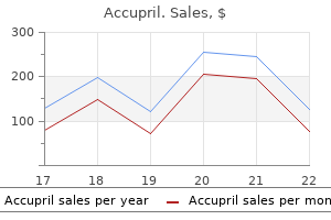
More pixels are available for viewing the vocal cord margins medications pain pills accupril 10 mg without a prescription, the first web site of sound production and the most probably location to determine the causes of hoarseness medications after stroke 10 mg accupril buy with visa. Left medicine and health purchase accupril 10 mg, Vocal cords are harder to see completely during the /a/ sound as the epiglottis and tongue base push posteriorly. They usually persuade themselves that irregular vocal sounds are the results of poor approach. In order to avoid audible impairments in their upper vocal range, they increase each their vocal twine tension and their subglottic pressure and consequently their volume. The increased subglottic strain overcomes small gaps, small elevations, and minor stiffness. By eliciting low-volume sound production from the affected person, smaller vocal impairments could be visually found through the examination. Eliciting larger volumes, especially at low pitch, can augment neurologic and muscular weakness visible findings by causing the weakened vocal twine to vibrate abnormally. A nontense thyroarytenoid muscle might break up into two or extra oscillatory segments, generating the looks of flutter on stroboscopy, and in addition could oscillate lateral to its axis, creating an infinite open part on stroboscopy. Tools for the Endoscopist Alter Pitch Alter Volume � High pitch � Highlights vocal margin � Augments stiffness � Low pitch � Highlights weakness � Typically removes compensation from superior laryngeal nerve (cricothyroid muscle) � Low quantity � Highlights gaps, stiffness, elevations � High quantity � Highlights weakness Quiet Observation Monitoring the vocal cords closely during quiet respiration can element delicate neurologic findings. Fasciculations are often noticed throughout quiet respiration when the larynx is relatively still. They are also visualized throughout the laryngeal ventricle on the superior surface of the vocal twine in a denervated thyroarytenoid muscle. During expiration, partial adduction usually occurs, whereas throughout inspiration, partial abduction happens. Left, At low pitch, vocal cords are quick and loose-marginal lesions disappear into the vocal twine mucosa. Right, At excessive pitch, vocal cords are long and tense-marginal lesions are pushed into the central glottic opening as a silhouette. Right, At low pitch, cricothyroid muscle compensation is removed, and the lack of tension and mass inside the right vocal twine becomes very obvious on stroboscopy, right here captured in the course of the open section. With acute denervation, the vary or diploma of motion is less on the injured side. Later in the healing process, as varying quantities of synkinetic reinnervation occur, the timing of motion can become asynchronous. Additionally, with synkinetic reinnervation, the resting position of the vocal course of might turn into extra medial. Height variations between the vocal processes can really only be appreciated nicely when the versatile endoscope is positioned between the arytenoids, almost parallel to the axis of the vocal cords. During deep inspiration, the ventricle on the facet of thyroarytenoid muscle atrophy appears to enlarge relative to the other facet. Left, At excessive pitch the cricothyroid muscle pulls the weak left vocal process towards the midline. Right, At low pitch, the right vocal course of pushes across the midline, whereas the left vocal process stays unrotated and very lateral with a big glottic hole. Left, During quiet respiration, the thyroarytenoid muscles are relaxed and the vocal cords and ventricles appear related in size. Right, During deep inspiration (sniffing), the proper vocal wire thins and the proper ventricle appears to enlarge, suggesting atrophy and paresis of the best thyroarytenoid muscle. Left, Typical endoscopic laryngeal view of a person with hoarseness; a common "redness" may be perceived. Right, Moving the endoscope close to the vocal cords, the dilated capillaries on the anterior vocal cord (which create the "redness" within the left photo) disappear beneath a white film as they pass posteriorly (arrow defines approximate transition point). Principles and follow of lasers in otorhinolaryngology and head and neck surgical procedure. The excessive practical and aesthetic calls for of this complicated anatomic region necessitate meticulous planning and exact execution by a multidisciplinary surgical staff. To improve the accuracy of preoperative modeling, scans are obtained with the affected person carrying fiducial markers on the face for reference. For resections with a dentoalveolar element that will have an effect on occlusion and orthognathic relationships, laser scanned occlusal molds or topographic occlusal imaging is obtained as properly. Soft tissue imaging and simulation software have been shown to be accurate enough for medical relevance,9 and could also be used as an help in remedy planning to anticipate the postoperative facial appearance ensuing from the planned skeletal adjustments. An on-line assembly is coordinated among the many engineers from the modeling company and the ablative, reconstructive, and prosthodontic teams. Using the digital mannequin, resection margins are determined and the engineer marks the specified slicing paths for the osteotomies. Following virtual resection of the diseased segment, the digital picture of the vascularized bone flap is superimposed on the ablative defect in the desired vascular and gentle tissue orientation. The engineers use the geometry of the resected section or mirror the contralateral disease-free skeleton to re-create the native bony construction and restore occlusal relationships. These strategies are also useful for secondary reconstructions wherein the resection or injury has already occurred and symmetry has been lost. The virtual surgery method allows for a larger number of segmental osteotomies to be deliberate and a number of osteotomy mixtures to be tried till the optimum permutation is identified to restore the craniofacial skeleton. Recent reviews recommend decreased operative times could also be achieved utilizing these applied sciences, regardless of increased reconstructive complexity. Modeling Phase Based on the surgical plan developed by the ablative, reconstructive, and prosthodontic groups in conjunction with the engineers from the vendor, stereolithographic manufacturing of the necessary elements is carried out. A mannequin of the native craniofacial skeleton is produced for intraoperative reference. These models and chopping guides facilitate the osteotomy process and allow for seamless integration between the ablative and reconstructive parts of the operation. To guarantee precise and stable intraoperative occlusal relationships, splints may also be designed and fabricated that allow alignment of the maxillary-mandibular section to the cranial base in all levels of freedom. This orthognathic positioning system eliminates the necessity for intraoperative placement of peripheral reference markers, ensures upkeep of condylar positioning in centric relation, bypasses the normal time-consuming strategy of mock surgery on mounted dental models, and obviates the need for fabrication of guiding intermediate splints. This process can enable for complicated and multipart jaw movements that combine orthognathic repositioning and ablative reconstruction with maximal accuracy. To facilitate skeletal plating after resection, a reconstruction plate template may be generated to permit for bending of the titanium plate preoperatively. Titanium plate contour and fixation factors are designed to optimize bony apposition, reduce periosteal stripping, avoid damage to nearby vulnerable structures. In addition to dental implant planning for delayed prosthodontic denture rehabilitation, certain cases may be designed with both the customized alloplastic implants and denture for instant placement, which allows for the creation of a "jaw in a day. If necessary, virtual modeling can also be linked with intraoperative navigation packages for even larger precision. B, Mandibular cutting information in place on the mannequin demonstrating proper splint orientation and path of the inferior alveolar nerve. B, the slicing information may be reinforced with a steel insert to forestall inadvertent damage to the plastic splint with the saw and subsequent deviation from the deliberate osteotomy orientation. It is important when planning the placement of these guides that they be sufficiently small not to encroach on gentle tissue margins. Once the slicing guides are secured, the resection is accomplished using a sagittal saw to precisely duplicate the angles of osteotomy planned on the computer mannequin. Resection ideally occurs concurrently with harvest of the deliberate free flap to be used for reconstruction using a twoteam approach. Flap harvest proceeds in standard style and incorporates the bony and gentle tissue components required for structural assist and protection of the oncologic defect. These virtually planned osteotomies not solely streamline the transition from resection to reconstruction, but additionally allow multiple reconstructive concerns to be taken under consideration. The chopping jigs are designed to optimize soft tissue location and orientation, maximize vascular pedicle length, go away sufficient bone at the donor web site, and provide best alignment for practical dental rehabilitation. Close communication have to be maintained between the ablative and reconstructive groups, particularly concerning any alterations to the preoperative plan. If the surgical plan is applied with out main intraoperative changes, the procedures can proceed independently to maximize operative effectivity and reduce anesthesia time. The ensuing bony segments are then secured to the customized reconstruction plate, creating the precise form required to fill the osseous defect. C, Complete fibula assemble assembled while still hooked up to its vascular pedicle on the leg. First the flapplate mixture is mounted in place, typically with minimal need for trimming to obtain flush bony apposition due to the cutting information precision. The microvascular anastomoses then happen with subsequent inset of the gentle tissue flap elements and closure.

Placement of a nasal endotracheal tube avoids inadvertent injury to the airway circuit and increases the working room for excision of an oral lesion medicine xyzal accupril 10 mg without prescription. Perioperative antibiotics are administered routinely as a end result of access is gained through a mix of transoral and transcervical approaches symptoms ebola generic accupril 10 mg online. A full-thickness incision through the decrease lip is performed and may be extended to the mandible relying on the placement of the surgical margins medications errors buy accupril 10mg low price. Closure of the wound is in a layered trend with care to guarantee approximation of the vermillion border. Management of the Primary Evaluation of the first tumor includes each the medical examination and review of imaging studies. The lesion is first outlined adopted by the delineation of the resection margin Approach Depending on the extent of the lesion, the method to surgical resection of squamous cell carcinoma of the buccal mucosa varies. Common instrumentation to increase this strategy is a chew block and mouth prop to preserve the mouth open during the procedure. For buccal lesions above the occlusal plane, a WeberFerguson approach may facilitate the elimination of a lesion that encroaches onto the maxillary gingiva or bony substructure. A lip cut up incision may be carried out for larger lesions that forestall enough transoral visualization of the tumor. Factors corresponding to trismus, microstomia, or posterior buccal location are indications for lip break up entry. In the normal epithelium, the intermediate and superficial layer cells comprise glycogen in their cytoplasm. Cancer and dysplastic cells include little or no glycogen due to the elevated glycolysis consequent to a dysfunctional mobile cycle. The margins surrounding the planned resection are sent for frozen margin evaluation. Care is taken to get hold of a frozen margin specimen of adequate size, roughly 3 mm in width along the remaining mucosal margin surrounding the resection specimen. The tissue margin is positioned on a non-adherent dressing and despatched for pathology evaluate with the mucosal floor dealing with upward for frozen section evaluation. To facilitate the elimination of the mucosal margins for frozen section, the posterior margin is obtained first, adopted by the anterior margins. When specimens are obtained from the anterior wound margins before the posterior margins, bleeding is usually a nuisance. The proximity of the tumor to adjacent bony structures, such because the dentoalveolar complicated of the mandible and/or maxilla, should be examined for the presence of erosions within the cortices, which would recommend bony involvement by tumor. Medullary involvement of the mandible or erosion into the sinus is an indication for a segmental resection and/or maxillectomy. Use of a marginal resection depends on the viability of residual basal bone, with mandibular bone lower than 10 mm being an indication for reinforcement with a reconstruction plate. These can embrace the inferior alveolar nerve, buccal nerve, and infraorbital nerve. For lesions with erosion into the maxillary sinus, the sinus lining ought to be sent as a specimen. The design of a marginal resection should incorporate smooth strains, with an effort to keep away from the placement of acute angles that could act as potential sites for stress/strain fractures to occur. The marginal resection could have an anterior boundary commonly requiring the extraction of enamel on the deliberate osteotomy website. A horizontal osteotomy is performed under the roots of the teeth with particular consideration to the amount of residual bone on the mandible. For the mandible, the posterior osteotomy generally travels by way of the sigmoid notch in a delicate curve. A maxillectomy could require separation of or osteotomy through the pterygoid plates to clear the posterior margin. Osteotomies may be created with a saw blade or fissure burr beneath copious irrigation. Following the marginal resection, sharp edges are smoothed with a burnishing burr or bone rasp to facilitate healing and closure. The duct can be spatulated and secured to the mucosa with a non-resorbable suture, corresponding to 6-0 nylon. The various is to clip the duct and to chemo-denervate the parotid gland, for instance, with Botox injections. It is believed that the buccinator is an anatomic barrier for the containment of cancerous cells, and the chance of native recurrence is elevated when the buccinator demonstrates signs of invasion by most cancers. Penetration of the buccinator probably locations most cancers cells into the buccal fats pad, enabling unfold through unpredictable patterns on this area that has no vital anatomic barriers. This aspect of buccal carcinoma is believed to contribute to the elevated risks of native and regional recurrence. The exterior skin must be examined for the possibility of tumor involvement, which might manifest as induration and lack of mobility of the subcutaneous and pores and skin layers. These options indicate the need for a full-thickness resection of the buccal mucosa and cheek. Lesions that encroach on 1 cm of the oral commissure risk improvement of microstomia as a end result of involvement of the lip ends in contracture and immobility of the lip and mouth actions. The buccal artery is usually encountered for lesions of the buccal mucosa and should be ligated to forestall postoperative bleeding. Management of the Neck Management of the neck relies on the chance of occult metastasis in lesions staged T1 and T2. Patients with a clinically unfavorable N0 neck are indicated for neck dissection if the primary buccal lesion is of T2 to T4 dimension. With T1 lesions which are less than 2 cm in greatest dimension, the most accessible predictor of occult metastasis is tumor thickness. Due to restricted information on cancers of the buccal mucosa, suggestions are generally extrapolated from studies of extra frequent subsites such as the tongue. Debate exists concerning the edge for an elective neck dissection: lesions less than 2 mm thick are generally noticed, and lesions thicker than 4 mm are generally indicated for elective neck dissection. These thresholds differ among institutions and range from greater than three mm to 5 mm. Reconstruction Reconstruction for T1 and T2 buccal carcinoma resection defects falls into three classes that embrace primary closure by way of local flap development, non-vascularized grafts, and microvascular free tissue switch. Local regional flaps may be raised or mucosal margins undermined to get hold of tension-free closure. Buccal fat pad advancement is often obtained because resection alone can draw out the buccal fat pad lobules. Gentle dissection and steerage can supply the fat pad to cover areas of the buccal mucosa for wound coverage. The buccal fat pad has five lobes with a wealthy vascular supply for a dependable source for wound protection. The use of the buccal fat pad as an oncologically viable reconstruction platform for buccal mucosal defects has not demonstrated a rise in local recurrence in contrast with different technique of reconstruction. Even with the loss of the buccinator muscle, the underlying buccal fat pad and/or subcutaneous tissues of the face are wealthy in vascular provide and collaterals and can help a graft. This graft would be applied immediately onto the wound bed and secured with chromic intestine suture to get rid of dead space. Microvascular free flap reconstruction can additionally be a viable option in areas of the buccal mucosa. For T2 lesions, resections involving solely gentle tissue are reconstructed with fasciocutaneous or myocutaneous flaps. In reconstruction of the anterior buccal mucosa by which the commissure of the mouth is compromised, the radial forearm flap can embody the harvest of the palmaris longus tendon to suspend the corner of the mouth for improved lip competence and symmetry. Alternative donor websites embody lateral arm, ulnar, and anterior lateral thigh free flaps. In a mandibular discontinuity defect, the fibula osteocutaneous flap would provide bony and soft tissue reconstruction. Vascular anastomosis is often accomplished between the facial artery and vein, with the vascular pedicle delivered into the neck on the medial or lateral facet of the mandible. Fracture of the residual mandible may end result after marginal resection if insufficient bone stays with out adequate support. A reconstruction plate can reinforce residual mandible bone if the bone is less than 10 mm in top. Care is taken to design a marginal resection with curved osteotomies to keep away from sharp angles within the native mandible as a outcome of these are areas of stress and tension, and may propagate a fracture extra readily than with curved line angles.
The tracheostomy tube could be eliminated when the airway is steady and the glottis is competent to prevent aspiration treatment receding gums buy 10mg accupril fast delivery. The adequacy of the airway may be ascertained via direct visualization with versatile endoscope medications for ptsd accupril 10 mg order with amex. It may be advisable to prepare patients with important laryngotracheal trauma for home tracheostomy care and to discharge them for subsequent follow-up and decannulation as an outpatient medicine pouch discount 10 mg accupril with amex. Complications � Accidental decannulation may be life threatening within the case of acute restore of a laryngotracheal injury. Prevention of stenosis via light dealing with of tissue, correct re-approximation of injured tissue, and judicious use of stents, keels, and other lumen-keepers seems to be the most dependable tactic. Eventual open exploration with adequate grafting could additionally be essential to create a everlasting lumen. One ought to notice that, although not universally accepted, surgical indications have evolved considerably. Conservative remedy is now not advocated for patients presenting a whole fracture of the thyroid cartilage (displaced or not) or sufferers with more than one fracture line via the cricoid. A vital number of these will respectively develop continual dysphonia or airway compromise as the fracture segments displace with the motion of the laryngeal and neck muscle tissue. Unusual presentation of blunt laryngeal injury with cricotracheal disruption by attempted hanging: a case report. Survival from accidental strangulation from a shawl resulting in laryngeal rupture and carotid artery stenosis: the "Isadora Duncan syndrome. Neck crepitance: evaluation and administration of suspected higher aerodigestive tract injury. The ratio is approximately three:1 (male to female) most likely as a end result of men participate in additional "at-risk" sports and actions. Editorial Comment this text presents a concise yet comprehensive evaluation of the analysis and administration of laryngeal fractures. As mentioned on this chapter, laryngeal fractures are rare occasions, albeit with potentially deadly problems, as a result of the loss of airway, and long-term sequelae that have an effect on all capabilities of the larynx. With the advent of laws mandating the use of safety belts, motor vehicle accidents have been replaced as the commonest reason for a laryngeal fracture by violent altercations and sports activities. Management of a patient suspected of having a laryngeal injury is geared to correct or forestall acute and future problems. By the method in which of illustration, a affected person with a historical past of a clothesline injury might undergo a partial or full laryngotracheal separation and recurrent laryngeal nerve avulsion. However, its range of harm and presentation varies extensively, with most of these sufferers dying before enough help may be offered, some surviving after establishing an emergency airway, and less generally the patient may appear secure but abruptly lose the airway upon extension of the neck. Laryngeal trauma within the pediatric inhabitants occurs less usually than in adults as a outcome of a. In youngsters, the larynotracheal complex has less calcification and thus is much less prone to fracture in response to bulk pressure. All of these statements are key elements of head and neck trauma analysis, besides a. Asking the patient to produce a high-pitch voice to consider for superior laryngeal nerve damage c. Endoscopic airway analysis of the nonintubated laryngeal trauma patient prior to sending the patient for radiologic analysis 3. All of these statements apply to open repair of median and paramedian laryngeal fractures, besides a. Efficacy of resorbable plates for discount and stabilization of laryngeal fractures. Patients usually have symptoms of airway obstruction, corresponding to biphasic stridor and dyspnea, with a traditional or practically regular voice. This is in distinction to patients with unilateral vocal fold immobility, who normally complain of a breathy voice and aspiration. It is usually associated with intubation, though extraesophageal reflux disease may be a cofactor. If that is noticed, immediate d�bridement, steroid injection, and antacid therapy may be associated with much less scar tissue formation and consequently much less airway stenosis. Laryngeal balloon dilation may also have a therapeutic position within the acute or subacute setting. The aim of therapy is to enhance the airway whereas minimizing adverse results on the voice. It permits for analysis of vocal fold movement during vocal fold adductory (saying /i/) and abductory (sniffing) duties. If the neurologic sample reveals a new injury with a chance of restoration, delaying harmful surgical procedure is prudent. The authors advocate that in an try and preserve the voice, surgery should contain the vocal fold with the worse neurologic standing. This might improve voice outcomes by not altering the vocal fold that has a better neurologic standing and thus possibly better muscle tone. Imaging research may be necessary in evaluating a patient with out obvious causes of vocal fold immobility. This helps obtain the correct "sniffing" position required for optimal laryngeal exposure in sufferers with a larger physique habitus. Proper surgeon ergonomic positioning can also be important to cut back surgeon musculoskeletal accidents. After laryngeal publicity is achieved, the right surgeon ergonomic place is achieved by transferring the bed angle (usually Trendelenburg, "head down") so the laryngoscope is forty degrees off the horizontal airplane. Relative contraindications include compromised pulmonary status, uncontrolled diabetes, and former radiation therapy. Patients must understand and settle for that to enhance their glottal airway, the quality of their voice may be adversely affected. Therefore patients ought to anticipate to have worse vocal quality at the expense of an improved airway. The patient ought to perceive that a quantity of procedures could additionally be essential to optimize the airway with the least potential impact on the voice. This allows the surgeon to enlarge the glottic airway in a staged, conservative fashion, which hopefully will reduce the unfavorable impression on both voice and swallowing. Rigid telescopes (0-, 30-, and 70-degree; 30 cm length, 5 to 10 mm diameter): these are used to better visualize the extent of lateral extension of the surgical area. Supraglottic jet ventilation by way of the laryngoscope is feasible however is hampered by subsequent laryngeal desiccation and motion from the air puffs. Intubation with intermittent extubation and subsequent apnea is much less generally used however is an effective choice for sufferers being treated with shorter period procedures, such as dilation. Tracheostomy provides probably the most stable airway and leaves the glottis devoid of accessory instrumentation. Possible placement of a tracheostomy at the time of glottic airway surgical procedure must be overtly discussed with the patient and listed on the surgical consent form. True vocal folds and arytenoids: Depending upon the place of the arytenoids and true vocal folds, the glottis, which is defined as the area between the vocal folds, may be too small for the affected person to breathe with out restriction. Scarring between the arytenoids can vary from mild to extreme, with full obliteration with scar of the respiratory glottis up to the vocal processes. Cricoid cartilage: the cricoid cartilage is a complete cartilaginous ring positioned under the true vocal folds. Microsuspension laryngoscopy Laser certification Balloon dilation Tracheotomy (see Chapter 19). Airway compromise: Inability to properly secure the airway prior to the start of surgical procedure requires the placement of an emergent surgical airway. A laser operator can be to be within the room always while the laser is in use. All working room workers must be skilled on what to do in case of airway fire-turn off oxygen while removing the endotracheal, Hunsaker, or tracheotomy tube; place saline in the airway; use bronchoscopy to evaluate for harm; and reintubate. In those cases, the patients must be prepared to accept long-term tracheostomy for airway management. When a bridge of scar tissue exists between the vocal processes with the presence of a posterior sinus tract, the bridge of scar tissue can be excised. Suspension laryngoscopy with exposure of the posterior glottis is carried out after the airway is secured. The mucosal integrity of the posterior glottis is assessed with the help of zero, 30, and 70-degree angled telescopes. The interarytenoid bridge of mucosa is excised utilizing laser or chilly knife method.
Syndromes

E and F medicine bg accupril 10mg buy low cost, Reconstruction is performed with bilateral Karapandzic flaps that keep the esthetics of the lower lip medicine xanax order accupril 10mg free shipping. E and F treatment water on the knee accupril 10 mg buy cheap line, the situation and measurement of the first most cancers of the lip led to planning with a block excision and reconstruction with a left reverse Karapandzic flap. Final histopathology recognized five of forty lymph nodes with metastatic illness, three of which exhibited extranodal extension of the metastatic cancer. Q, the patient underwent postoperative radiation remedy as a outcome of the extent of his disease. B, the margin required to take away this recurrent melanoma will sacrifice the left oral commissure. As such, a block excision is deliberate with instant reconstruction utilizing a right Karapandzic flap and a left Webster modification of the Bernard cheiloplasty. Management of the Cervical Lymph Nodes in Lip Cancer the incidence of cervical lymph node metastases related to lip most cancers is reported in 3-8% of sufferers. The tumor grade and the tumor thickness appear to be the most important predictive factors for lymph node involvement by lip cancer. Most early-stage lip cancers are identified as well-differentiated squamous cell carcinoma. These low-grade tumors not often metastasize to the cervical lymph nodes, and Szewczyk and colleagues20 determined that solely two of twenty-two neck dissections contained lymph node metastases from lowgrade tumors. In high-grade tumors, nonetheless, the incidence of nodal unfold was doubled with four out of 24 patients found to demonstrate lymph node metastases in neck dissection specimens. The depth of invasion by a lip most cancers or the tumor "thickness" additionally predicts the chance of unfold to regional nodes. In findings much like that for melanoma, the deeper a squamous cell carcinoma of the lip penetrates the conventional lip, the larger the risk of nodal illness. Several groups have found that a tumor thickness of greater than four mm places the affected person at a big danger for lymph node involvement. A giant German examine of lip cancer reviewed the influence of tumor grade and depth of invasion on the chance for node involvement. F and G, the flaps are developed and superior so as to create an anatomic closure. H, A 5-year postoperative look of the affected person reveals an esthetic reconstruction with minimal microstomia. Based on these findings the authors proposed a method for predicting lymph node involvement. Patients with well-differentiated grade 1 squamous cell carcinoma of the lip with a tumor thickness larger than 5 mm carry a big nodal threat and ought to be handled with neck dissection or irradiation of the regional nodes. Radiation Therapy Most squamous cell carcinomas of the lip current as surgically resectable tumors with effective management in 90�94% of sufferers. In these poor surgical candidates, definitive radiation therapy is an efficient therapy possibility with results identical to resection. Other sufferers have been treated with definitive radiation concentrating on solely the concerned lip. With a median follow-up of 55 months, wonderful native management was achieved in each groups, with local recurrences in only 3. The disease-free survival was not significantly totally different between patients who underwent surgery or obtained definitive radiation, and radiation therapy appeared to be more incessantly utilized in higher threat patients with larger tumors and high-grade lesions. A more recent Australian study19 examined ninety three patients with T1 or T2 lip cancer between 1980 and 2012. Patients were treated with definitive radiation therapy with native management reported at 95%, regional nodal failure reported in only 5%, and with a 10-year disease-free survival of 90%. In this study solely 3 of 93 patients treated by radiation therapy died because of lip cancer. Resection provides excellent native control in massive lip tumors however must be balanced against the useful and cosmetic outcomes. Tumors longer than 3 cm are difficult to resect with extensively adverse margins while nonetheless leaving a practical lip. Older sufferers may have extra tissue laxity that permits resection of barely larger tumors. However, anesthesia of the reconstructed lip limits full operate, and radiotherapy has been thought to supply a greater cosmetic and useful end result. Thanh Pham and Cross19 showed local recurrence in solely 5% of patients treated with definitive radiation and 40% of these sufferers presented with T2 lesions. As a outcome, current National Comprehensive Cancer Network treatment pointers recommend consideration of definitive radiation for tumors that occupy many of the lower lip. Radiation Technique Squamous cell carcinoma of the lip is a domestically invasive disease that hardly ever spreads to the regional lymph nodes. The seen main tumor is recognized as a gross tumor quantity, which is expanded by 1. An intraoral lead protect is usually used to defend the uninvolved gingiva and oral cavity from the radiation dose passing through the treated lip. Custom lead floor blocking can be used to defend the skin of the chin, face, and uninvolved lip. The normal radiation therapy is delivered at 2 Gy per fraction as daily remedy over 7 weeks to a complete dose of 70 Gy. As lengthy as 3 months could also be required following therapy for the handled tumor to fully resolve but native tumor management may be expected in up to 95% of patients, as discussed beforehand. Regional nodal failure is uncommon and disease-free survival at 10 years may be anticipated in 90% of patients. In older patients and those that stay a protracted distance from a radiation treatment center, a more hypofractionated radiation therapy protocol may be delivered. However, the larger dose per fraction can end result in severe dermatitis and mucositis requiring more than 1 month to resolve and leaving the patient at increased danger for epithelial atrophy and telangiectasia. Margin Status and Adjuvant Radiation Therapy the problem of margin standing and the necessity for adjuvant radiation therapy after resection is amongst the most complicated issues surrounding the administration of lip cancer. A repeat excision may be attempted; however, the surgical goal may be troublesome to define and the additional lack of lip tissue threatens cosmesis and function. No remedy failures have been seen in a latest examine by which lip most cancers was handled with surgical resection followed by adjuvant radiation to 66 Gy. No randomized trials have been conducted and no consensus exists as to the precise definition of a clear margin for lip cancer. Results from oral cavity cancer treatments assist a 5-mm surgical margin in order to be considered clear. B, At 5 years postoperatively, he showed no evidence of native or regional recurrent disease. Extrapolation of results from different head and neck sites is used to justify the addition of concurrent chemotherapy in locally advanced lip cancers with shut or optimistic margins, with extracapsular extension of nodes, or when a quantity of or bulky lymphadenopathy is present. Literature has been published by a quantity of groups24-26 supporting the efficacy of brachytherapy for the therapy of lip most cancers, with achievement of local control reported in 94�98% of instances. A dialogue of brachytherapy for lip cancer is past the scope of this chapter that will somewhat give consideration to external beam radiation as definitive remedy. However, a current Chinese examine discovered an increase in native recurrence and a decrease in overall survival in patients with close margins (less than or equal to 2 mm). An Australian report found that patients with lip most cancers with a surgical margin lower than 2 mm had increased recurrence charges. However, adjuvant radiation was able to control illness in these patients with shut margins, rising the recurrence-free survival to 92%. The surgical ablation of lip most cancers ought to be attempted with adverse margins whereas still preserving the optimal perform and cosmesis of the lip. If the margin is unfavorable by more than 5 mm, it appears clear that the patient can be safely noticed with low danger of failure. Patients fall into an equivocal zone when the resection margin is between 2 and 5 mm. In the absence of randomized trials, medical judgment and native experience are required. However, it could often be finest in these "close" margin sufferers to refer them for adjuvant radiation therapy with the knowledge that, with proper approach, irradiation can forestall recurrence and preserve lip perform in roughly 95% of sufferers. Chemotherapy Systemic remedy has a limited function in the remedy of lymph node-negative early-stage squamous cell cancers of the lip.
Additionally medicine for nausea buy cheap accupril 10 mg line, the retromandibular vein is one other different giant caliber recipient vein that can be utilized medicine 7 year program accupril 10 mg cheap amex. The vessel-depleted and irradiated neck poses additional reconstructive challenges symptoms intestinal blockage cheap 10mg accupril fast delivery, incessantly requiring the necessity for interpositional vein grafts to attain the closest available recipient vessels. Such circumstances are associated with increased risk for thrombosis and potential flap failure. Cephalic vein transposition also supplies enough recipient venous drainage when the jugular venous system is compromised. Its thin, pliable delicate tissue element allows it to conform well to the maxillary defect specifically permitting for each nasal and oral re-lining. Primary or delayed autologous bone grafts such as a "sandwich" approach enable for restoration of each exhausting and delicate tissue parts. This method should be used with warning in sufferers undergoing adjuvant radiation remedy as a result of the chance for bone graft loss and extrusion is larger. Cordeiro and colleagues24 reported the use of the osteocutaneous radial forearm flap for this objective whereby the vascularized radius was used to reconstruct the bony maxilla. However, the quantity of bone harvested is insufficient for prosthetic implantation, making dental rehabilitation unimaginable with out additional grafting. Depending on patient physique habitus, a thin pores and skin flap could additionally be harvested that can be utilized for intra-oral or nasal lining. In contrast, the myo-osseous flap harvested from the scapula tip bears nice resemblance in form to the maxilla and is well suited to palatomaxillary reconstruction, permitting for mucosalization of the muscle mattress. Prosthetic implantation is feasible but could require secondary bone graft augmentation. The iliac crest can be placed horizontally in low maxillectomy defects or vertically in high maxillectomy defects. Also, Brown40 confirmed that harvesting a pores and skin paddle was pointless in that the inner oblique used to reconstruct the palate epithelializes rapidly, retaining the qualities of oral mucosa. Futran and colleagues44 also reported glorious results by method of facial projection, speech, swallowing, and dental rehabilitation. It is thus evident from the abundance of literature pertaining to mid-face reconstruction that various microvascular flap options allow safe, reliable, and reproducible esthetic and functional maxillary reconstruction. Reconstructive Options for the Total or Subtotal Rhinectomy Defect Large ablative defects of the nasal complex�associated maxillectomy ought to be addressed after maxillary reconstruction. For ideal and steady nasal reconstruction, the underlying structural support offered by the maxilla must be restored first. The structural subunits of the nose are arguably essentially the most complicated throughout the face to reconstitute when absent, a fact that complicates nasal reconstruction. Complex 3D structure, multilayer construction, and functional significance spotlight the difficulties related to rebuilding nasal defects. Reconstruction of such through-and-through defects must employ an inside-out method, addressing the inner lining, bony and cartilaginous supportive framework, and at last pores and skin resurfacing of the exterior nostril. When available mucosal flaps are preferred so as to replace like with like inside the nasal cavity. Pedicled "turn-in" flaps when out there are easy and extremely dependable for this function. For the total nasal defect, the fasciocutaneous or osteocutaneous radial forearm free flap is right. This may be achieved with using autologous cut up calvarial bone and cartilage52 with the calvarial bone common into an L-strut or with the use of the osteocutaneous radial forearm free flap. Options here embody full- and split-thickness pores and skin grafts, each of which have poor skin shade match and inferior tissue quality. The gold normal and most well-described technique for pores and skin resurfacing of the nostril remains the paramedian forehead flap. Radiation Therapy Technique Radiotherapy is indicated in all curative cases of T4 carcinoma of the maxilla with invasion of the sinonasal area, either as main remedy or within the adjuvant setting. Adjuvant remedy must be initiated within 6 weeks of surgery, allowing for adequate therapeutic of surgical wounds. Oral stents, when tolerated, could be useful in displacing the tongue from the irradiated volume. In regionally superior or perineural illness that extends to the cranium base, magnetic resonance imaging can be helpful for therapy planning in both delineation of target volumes and intracranial normal tissue structures such because the mind, brainstem, visual pathways, temporal lobes, hippocampi, and hypothalamus-pituitary complex. Co-registering of presurgical imaging can additionally be helpful in delineating target volumes. Doses of 66 to 70 Gy are prescribed to the primary web site and high-risk regions of the neck (regions of extracapsular extension), doses of 60 Gy are prescribed to the optimistic region of the neck, and 45 to 50 Gy are prescribed to decrease threat areas of the neck over 6 to 7 weeks of radiotherapy delivered as quickly as daily using 2-Gy fractions. Hyperfractionation is also helpful when the target quantity lies close to the optic buildings, that are at decrease danger for toxicity when radiotherapy is delivered at a decrease dose per fraction. Many facilities have just lately adopted weekly cisplatin at 30 to 40 mg/m2 as an alternative. During therapy patients are monitored weekly for management of nutritional standing, hydration, mucositis, infection, skin reactions, and other acute effects of remedy. Intense supportive care is required, together with the utilization of feeding tubes in many patients, and therapy breaks must be minimized as a end result of rising the general treatment time has been associated with decreased locoregional illness control. Treatment outcomes in squamous cell carcinoma of the maxillary alveolus and palate: a population-based research. Maxillary squamous cell carcinoma: an 11-year retrospective study of 1 regional cancer heart. Clinicopathological characteristics and end result predictors in sufferers with squamous cell carcinoma of the hard palate. The determining threat factors for therapy outcomes in sufferers with squamous cell carcinoma of the onerous palate. Oral maxillary squamous cell carcinoma: administration of the clinically adverse neck. Cervical metastases of squamous cell carcinoma of the maxilla: a retrospective research of 9 years. High rates of regional failure in squamous cell carcinoma of the onerous palate and maxillary alveolus. Is neck dissection needed in squamous-cell carcinoma of the maxillary gingiva, alveolus and hard palate A 15-year review of midface reconstruction after whole and subtotal maxillectomy: part I. Vascularized iliac crest with inside indirect muscle for quick reconstruction after maxillectomy. The radial forearm osteocutaneous "sandwich" free flap for reconstruction of the bilateral subtotal maxillectomy defect. Outcome of simultaneous and staged microvascular free tissue switch linked to arteriovenous loops in areas missing recipient vessels. The osteocutaneous scapular free flap for mandibular and maxillary reconstruction. Maxillary reconstruction using the scapular tip free flap: a radiologic comparability of 3D morphology. The inside oblique-iliac crest osseomyocutaneous free flap in oromandibular reconstruction. Deep circumflex iliac artery free flap with internal indirect muscle as a model new method of immediate reconstruction of maxillectomy defect. The anatomical basis of the deep circumflex iliac artery perforator flap with iliac crest. Optimal use of microvascular free flaps, cartilage grafts, and a paramedian brow flap for aesthetic reconstruction of the nostril and adjacent facial units. Free radial forearm osteocutaneous perforator flap for reconstruction of complete nasal defects. Nasal reconstruction with the paramedian forehead flap using the aesthetic subunits principle. Maxillary sinus carcinomas: pure historical past and outcomes of postoperative radiotherapy. Does altered fractionation influence the chance of radiation-induced optic neuropathy and retinopathy
This is a preliminary examine medicine urinary tract infection cheap accupril 10 mg with amex, and additional research would be required to supply a definitive comparability between this feature and the gold normal of definitive surgical administration with or without adjuvant radiation therapy sewage treatment cheap accupril 10mg fast delivery. However medicine man movie buy accupril 10mg with amex, radiation therapy remains to be a remedy based mostly on the mechanical destruction of cancerous tissue. As a outcome, many patients have significant post-radiation scarring that can lead to clinically vital trismus and beauty defects, especially if the pores and skin is involved. Overall, the authors acknowledge the reality that not all patients have the option to bear surgical procedure with or without adjuvant radiation therapy, the current standard of care for T3/T4 buccal most cancers. The evidence offered here helps the utilization of definitive radiation or chemoradiation remedy for early-stage lesions, which have proven comparable outcomes to surgical procedure. Surveillance Buccal most cancers has been reported to have a number of the highest rates of recurrence by anatomic subsite. It is hypothesized that this is as a end result of of the shortage of anatomic barriers in this area once the most cancers penetrates the buccinator muscle and fascia. In the current literature, 5-year local management and overall management rates for buccal cancer have ranged between 57. The largest study within the United States, that by Diaz and colleagues, reported the median time to recur was 8 months, with the majority within 1 yr. As a end result, the authors suggest a follow-up go to each month for the primary year, every second month for the second 12 months, each third month for the third year, and each six months for the fourth and fifth years in accordance with the National Comprehensive Cancer Network guidelines. Often full visualization of the tumor resection web site is difficult due to bulky flaps or scarring and resultant trismus, particularly if adjunctive radiation remedy is indicated. Smit and associates reported on a cohort of postoperative oral squamous cell carcinoma patients and located that 70% of sufferers with recurrence reported pain as their first symptom. In patients who do experience local recurrence, salvage surgery presents the most effective probability at increased survival. Koo and colleagues reported an improved survival after recurrence for sufferers who obtained reoperation with or without adjuvant radiation therapy when in comparison with sufferers who obtained salvage chemoradiation therapy alone. However, the administration of regional recurrence in a beforehand dissected neck is more complicated as a result of the recurrence is usually in the surrounding gentle tissue and may be intimately concerned with the good vessels of the neck. In this case, imaging ought to be performed to consider the resectability of the lesion. If resection is feasible, the authors suggest surgical resection with adjuvant radiation therapy. Unfortunately, in research specific to buccal cancer, the reported charges of successful salvage are low, ranging between 9% and 22%. Regular follow-up and surveillance does more than simply verify for recurrence and supply an opportunity at salvage surgery. With common followup, we can proceed to present support and knowledge all through this course of. We must be reminded to always educate our patients on all therapy choices, including palliative care if it is applicable, so that we can best empower our sufferers in a joint decision-making approach. Changing tendencies in oral most cancers within the United States, 1935 to 1985: a Connecticut study. Squamous cell carcinoma of the oral gentle tissues: a statistical evaluation of 14,253 instances by age, intercourse, and race of sufferers. Treatment components related to survival in early-stage oral cavity most cancers: analysis of 6830 circumstances from the National Cancer Data Base. An epidemiological research of oral and pharyngeal most cancers in Central and South-East Asia. Squamous cell carcinoma of the buccal mucosa: outcomes of therapy in the fashionable period. Tobacco and alcohol related to the anatomical web site of oral squamous cell carcinoma. Malignant transformation and pure history of oral leukoplakia in fifty seven,518 industrial workers of Gujarat, India. The worth of follow-up in patients treated for squamous cell carcinoma of the pinnacle and neck. Good tumor management and survivals of squamous cell carcinoma of buccal mucosa handled with radical surgical procedure with or without neck dissection in Taiwan. Tumour thickness and relationship to locoregional failure in most cancers of the buccal mucosa. Development and validation of the neck dissection impairment index: a top quality of life measure. The significance of "positive" margins in surgically resected epidermoid carcinomas. Surgical margin determination in head and neck oncology: present scientific follow. Buccal mucosa carcinoma: surgical margin less than three mm, not 5 mm, predicts locoregional recurrence. Postoperative radiotherapy for persistent tumor on the surgical margin in head and neck cancers. Failure at the primary site following multimodality treatment in advanced head and neck cancer. Surgery versus surgical procedure and postoperative radiotherapy in squamous cell carcinoma of the buccal mucosa: a comparative examine. The influence of adjuvant radiotherapy on survival in T1-2N1 squamous cell carcinoma of the oral cavity. Five-year follow-up of concomitant accelerated hypofractionated radiation in advanced squamous cell carcinoma of the buccal mucosa: a retrospective cohort research. Role of high dose price interstitial brachytherapy in early and regionally superior squamous cell carcinoma of buccal mucosa. Clinical and Radiographic Features Maxillary malignancies typically stay asymptomatic or current with non-specific sinonasal signs till invasion into adjoining constructions occurs. Common signs embody facial or dental pain, unfastened enamel, nasal obstruction, and epistaxis. Less common presentations embody new onset facial asymmetry, displacement or protrusion of the globe with superior extension, and midface paresthesia with involvement of the trigeminal nerve. Plain dental radiographs, such as a panoramic radiograph, might present a "moth-eaten" destruction of lamina dura and surrounding bone, in addition to cloudy maxillary sinuses. The proximity of oral mucosa of the vestibule to the alveolar bone creates a challenge for radiologic evaluation, thus methods have been developed to differentiate between such constructions. Thus, Dillon and colleagues4 described putting a 2- � 2-cm gauze pad in the vestibule to present adequate displacement of oral mucosa. It is most commonly obtained for sufferers with T3 and T4 tumors or when metastasis is suspected. This entity has a higher propensity for distant metastases, specifically to the lung. Histopathologic evaluation reveals basaloidappearing nests with comedonecrosis and dysplastic floor mucosal epithelium. Intraorally it most commonly impacts the maxillary and mandibular vestibule, gingiva, buccal mucosa, tongue, and onerous palate. The site of chronic tobacco placement most often corresponds to the precise web site of lesion improvement. Adenosquamous Carcinoma Adenosquamous carcinoma was first identified by Gerughty and colleagues in 1968. Controversy existed over the definition of this entity, in that many people originally believed it was an Spindle Cell Carcinoma In the top and neck area, spindle cell carcinomas are most commonly found in the larynx. In the United States and Europe, 80% of paranasal sinus carcinomas involve the maxillary sinus. Evidence suggests a dose-response relationship to wood mud exposure and adenocarcinoma of the sinuses. This staff features a head and neck surgeon, reconstructive surgeon, dentist/prosthodontist, radiologist, medical oncologist, radiation oncologist, speech and swallow therapist, nutritionist, and social employee. Additional medical subspecialties may be needed relying on the structures affected by the malignancy. The experience of psychologists, habit companies, and palliative care providers must also be an integral a part of the top and neck cancer group. Separate N category approaches are given for patients handled with out cervical lymph node dissection (clinical N [cN]) and sufferers handled with cervical lymph node dissection (pathological N [pN]).
Safety and effectiveness of topical dry mouth products containing olive oil medicine kit for babies purchase 10mg accupril visa, betaine treatment 11mm kidney stone accupril 10mg buy fast delivery, and xylitol in reducing xerostomia for polypharmacy-induced dry mouth medicine evolution discount accupril 10 mg amex. Effectiveness of green tea mouthwash compared to chlorhexidine mouthwash in sufferers with acute pericoronitis: a randomized clinical trial. Antimicrobial properties of green tea extract in opposition to cariogenic microflora: an in vivo examine. The impact of thyme and tea tree oils on morphology and metabolism of Candida albicans. Taste disorders in cancer sufferers: pathogenesis, and method to assessment and administration. The impact of cancer remedy on the diets and food preferences of patients receiving outpatient therapy. The affiliation between malnutrition and psychological distress in sufferers with superior headand-neck cancer. Availability of outpatient scientific diet services for patients with cancer present process therapy at Comprehensive Cancer Centers. The changed meaning of meals: physical, social and emotional loss for patients having received radiation treatment for head and neck cancer. Effect of nutritional interventions on nutritional standing, high quality of life and mortality in patients with head and neck cancer receiving (chemo)radiotherapy: a systematic evaluate. A systematic evaluate of oral fungal infections in patients receiving cancer therapy. Successful therapy of invasive rhinopulmonary mucormycosis with an indolent presentation by mixed medical and surgical therapy. A systematic evaluate of viral infections related to oral involvement in cancer sufferers: a highlight on Herpesviridae. Regression of major recurrent aphthous ulcerations utilizing a combination of intralesional corticosteroids and levamisole: a case report. A systematic review of trismus induced by cancer therapies in head and neck most cancers patients. A cost-effectiveness evaluation of using TheraBite in a preventive exercise program for sufferers with advanced head and neck most cancers treated with concomitant chemoradiotherapy. The impact of exercise remedy in head and neck most cancers patients within the therapy of radiotherapy-induced trismus: a scientific review. Oral epithelial dysplasia and squamous cell carcinoma following allogeneic hematopoietic stem cell transplantation: clinical presentation and therapy outcomes. Incidence of second primary malignancies in sufferers with treated head and neck cancer: a complete evaluate of literature. Survival study and therapy technique for second primary malignancies in patients with head and neck squamous cell carcinoma and nasopharyngeal carcinoma. Prognosis of oral pre-malignant lesions: significance of clinical, histopathological, and molecular biological characteristics. Prevention of osteoradionecrosis: a randomized potential medical trial of hyperbaric oxygen versus penicillin. Osteoradionecrosis of the jaws as a side effect of radiotherapy of head and neck tumour patients-a report of a thirty 12 months retrospective evaluation. Osteoradionecrosis of the jaws-a present overview-part 1: physiopathology and threat and predisposing components. The radiation-induced fibroatrophic process: therapeutic perspective by way of the antioxidant pathway. Osteoradionecrosis in cancer patients: the evidence base for treatment-dependent frequency, current management strategies, and future research. Dental extractions in the irradiated head and neck patient: a retrospective evaluation of Memorial Sloan-Kettering Cancer Center protocols, standards, and finish outcomes. Lack of osteoradionecrosis of the mandible after intensity-modulated radiotherapy for head and neck cancer: doubtless contributions of each dental care and improved dose distributions. Dosimetric distribution to the tooth-bearing areas of the mandible following intensity-modulated radiation remedy for base of tongue cancer. Radiation-induced oral mucositis and periodontitis-proposal for an inter-relationship. Micromorphology of the dental pulp is very preserved in most cancers patients who underwent head and neck radiotherapy. Application of chlorhexidine, fluoride and synthetic saliva throughout radiotherapy: an in vitro study of microleakage in Class V restorations. Summary of Evidence-Based Oral Care Study Group, Multinational Association for Supportive Care in Cancer/ International Society of Oral Oncology medical follow guidelines for care of sufferers with different oral complications; 2010. In vitro effect of calcium-containing prescription-strength fluoride toothpastes on bovine enamel erosion under hyposalivation-simulating conditions. Effect of radiation dose on the prevalence of apical periodontitis-a dosimetric evaluation. Rehabilitation after mandibular reconstruction with fibula free-flap: medical outcome and quality of life assessment. Evaluation of the quality of life of patients with maxillofacial defects after prosthodontic therapy with obturator prostheses. Quality of life factors and survival after total or extended maxillectomy for sinonasal malignancies. Assessment of swallowing and masticatory efficiency in obturator wearers: a medical examine. Effect of prosthodontic rehabilitation of maxillary defects on hypernasality of speech. Understandability of speech predicts high quality of life among maxillectomy patients restored with obturator prosthesis. Cost analysis of dental companies needed earlier than hematopoietic cell transplantation. Treatment patterns and financial burden of metastatic and recurrent locally-advanced head and neck most cancers patients. The first crisis occurs when the person is given the preliminary analysis of cancer. At this time the catastrophic implications of the diagnosis itself and the menace of mortality eclipse all different considerations. The second crisis occurs throughout and after therapy as the person tries to take care of the instant unwanted effects of treatment. This could embody postoperative changes or, for the person present process radiation or chemoradiation, the gradual worsening of signs over time. The patient is faced with leaving the protective environment of the hospital to cope and modify at home. They should also understand how the rehabilitation course must be individualized to meet the precise needs, objectives, and needs of the individual. The presence of serious baseline dysphagia may inform selections concerning the need for short-term versus long-term enteral nutrition. This information is efficacious for consideration of therapy choices and planning for rehabilitation. Other important components include the availability of caregiver help throughout remedy and recovery. Oropharyngeal dysphagia following therapy for tumors of the higher aerodigestive tract is widespread. The test is performed in a radiology suite the place the individual is offered with a selection of barium consistencies to assess the flexibility to swallow foods and liquids. During the test a big selection of compensatory strategies can Voice and Speech Evaluation There are a number of protocols for the assessment of the voice, speech, and communication of the individual with head and neck cancer that could be used before, during, and after treatment. For the person with a voice disorder or dysphonia, a comprehensive voice evaluation will sometimes comprise endoscopic analysis of the larynx, acoustic and perceptual evaluation of the voice, and subjective self-assessment of vocal impairment. Videostroboscopy supplies an in depth evaluation of the vibratory properties of the vocal folds as nicely as the presence of hyperfunction throughout phonation. Typically acoustic vocal measurements are also collected as part of a complete voice evaluation. The particular person is audio-recorded whereas finishing a variety of vocal tasks, reading standardized passages, and during regular conversation, and these recordings are then analyzed utilizing acoustic software program.
Systemic treatment has been restricted to these sufferers with unresectable disease or with illness not amenable to other native interventions medicine 50 years ago accupril 10mg. Immune therapy trials have proven some promise and might be a subject of additional investigation going forward schedule 9 medications 10 mg accupril otc. Non-melanoma pores and skin cancer: an overindulged circumlocution articulating keratinocytic most cancers medicine 1900s spruce cough balsam fir buy accupril 10 mg line. How different wavelengths of the ultraviolet spectrum contribute to skin carcinogenesis: the function of cellular harm responses. Nonmelanoma cutaneous head and neck most cancers and Merkel cell carcinoma: current ideas, advances, and controversies. Trends of nonmelanoma pores and skin cancer from 1960 through 2000 in a Canadian population. The management of skin malignancy: to what extent should we depend on medical prognosis Update on metastatic basal cell carcinoma: a summary of printed circumstances from 1981 through 2011. Diagnosis and remedy of invasive squamous cell carcinoma of the skin: european consensusbased interdisciplinary guideline. Analysis of danger components figuring out prognosis of cutaneous squamous-cell carcinoma: a potential research. Pathologic occult neck disease in patients with metastatic cutaneous squamous cell carcinoma to the parotid. Incidence of cervical node involvement in metastatic cutaneous malignancy involving the parotid gland. Computed tomography of cervical lymph nodes: staging and administration of head and neck most cancers. Surgical resection for scientific perineural invasion from cutaneous squamous cell carcinoma of the head and neck. Multiprofessional tips for the administration of the affected person with primary cutaneous squamous cell carcinoma. Diameter of involved nerves predicts outcomes in cutaneous squamous cell carcinoma with perineural invasion: an investigator-blinded retrospective cohort study. Radiotherapy for perineural invasion in cutaneous head and neck carcinomas: toward a risk-adapted remedy strategy. Perineural infiltration of cutaneous squamous cell carcinoma and basal cell carcinoma without medical options. The position of postoperative radiotherapy for giant nerve perineural spread of cancer of the top and neck. Cutaneous head and neck malignancies with perineural spread to contralateral cranial nerves: an argument for extending postoperative radiotherapy volume. Incidence and survival for Merkel cell carcinoma in Queensland, Australia, 1993-2010. Adjuvant radiation remedy is related to improved survival in Merkel cell carcinoma of the pores and skin. Effect of radiotherapy dose and volume on relapse in Merkel cell cancer of the pores and skin. Merkel cell carcinoma: prognosis and therapy of sufferers from a single establishment. Adjuvant prophylactic regional radiotherapy versus remark in stage I Merkel cell carcinoma: a multicentric prospective randomized research. The initial expertise of electronic brachytherapy for the remedy of non-melanoma pores and skin cancer. Non-melanoma pores and skin most cancers handled with high-dose-rate brachytherapy and Valencia applicator in elderly patients: a retrospective case series. Effect of surgery on regular tissue toxicity in patients handled with accelerated radiotherapy. This variation in severity determines the therapeutic management of the condition. The first is a prefibrotic section by which changes in endothelial cells predominate with an acute inflammatory response. The second is a constitutive, organized phase by which abnormal fibroblastic activity predominates, and the extracellular matrix turns into disorganized. Finally, within the late fibroatrophic phase, tried tissue transforming types fragile healed tissues, which have a critical inherent danger for late reactivated inflammation within the event of local damage and which can result in necrosis in bone. The fibroatrophic principle supposes that the modifications in bone that trigger this process are very comparable to people who happen when bodily accidents have an result on different tissues within the body, such as the lungs and liver. Previous Classifications A number of classifications have been developed to aid management of the situation. Epstein and colleagues printed a classification in 1987,4 which can also be broadly used. Several publications describe its success alone or together with clodronate and vitamin E as an antioxidant to deal with fibrosis after radiotherapy and after chemical harm to a quantity of organs, though the exact mode of motion in ameliorating radiation fibrosis is unclear. Osteoradionecrosis-a evaluate of present ideas in defining the extent of the illness and a model new classification proposal. A New Classification of Osteoradionecrosis A classification was thus developed that takes into account the usage of pentoxifylline and vitamin E17 (Table 45. Easing sequestrated bone out in teams 1 to 3 with none additional treatment is often useful. Most of those with stage four illness required and consented to complex surgical administration. It is difficult to say whether or not the treatment stopped the situation progressing within the earlier levels. In the sequence reported by Epstein and colleagues,four 57% of instances that resolved with conservative treatment have been steady (15% complete decision and 42% stable). Other authors report comparable figures, although in some instances, conservative remedy concerned sequestrectomy and other minor operations. However, in the Epstein series, pathologic fractures developed in 23% of patients in the course of the study period and 19% of instances have been progressive. Delanian might well have achieved these spectacular figures by adding clodronate to the other two medication. Maxillofacial surgeons are averse to this medication in that it could possibly not often trigger osteonecrosis. A recent Cochrane evaluation talked about earlier has indicated that it could be of marginal benefit in these circumstances. Ideally, as a end result of in most instances just one aspect of the neck is affected severely by radiotherapy and sometimes surgical procedure, the other facet of the neck must be used for anastomosis of the pedicle. There is commonly a paucity of donor vessels within the neck where the radiotherapy harm is, and both the vein and artery could be extremely fragile and tear either throughout cautious exposure or suturing. The deep cervical vessels decrease within the neck are sometimes spared of radiotherapy damage and are a reasonable size. If venous drainage is a problem, the cephalic vein may be reduce just above the elbow and transposed superiorly into the neck. Use of a cephalic vein loop between the good vessels within the neck is a chance, however these are often too damaged to be viable. Lastly, the internal mammary vessels may also be harvested midway down the sternum and transposed into the neck. Although the artery is type of at all times sufficient in phrases of diameter and circulate, the venae comitantes are less constant. Second, it must be decided which bone may turn out to be osteoradionecrotic after surgery. This will make the reconstruction easier to execute, and better mandibular function could be anticipated if the pure condyle is used for articulation. If these enamel abut the area to be resected, then the issue can be resolved by extending the realm of bone to be resected. First, the ipsilateral neck must be opened even when the anastomosis is to be contralateral. Bearing in mind that radiotherapy damages all tissues, both onerous and gentle; ideally an ellipse of free vascularized pores and skin and subcutaneous tissue must be used to shut the neck. This acts as an island of latest tissue between the two sides of the neck incision, or any cervical fistula which could be present. Failure to do that may find yourself in a protracted course for the neck to heal in addition to an orocutaneous fistula.


