Microzide

Microzide
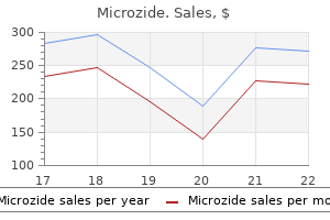
Microzide dosages: 25 mg
Microzide packs: 60 pills, 90 pills, 120 pills, 180 pills, 270 pills, 360 pills

Germ cell and trophoblastic tumors could be cured regardless of the presence of pulmonary metastasis arteria 3d medieval village purchase microzide 12.5mg visa. Lymphangitic lung metastases symbolize an emergent problem in analysis and management arteria carotis externa microzide 12.5 mg cheap without prescription. Hormonal manipulation is often ineffective or achieves a response too slowly to be useful blood pressure chart age nhs buy cheap microzide 12.5mg line. Symptoms from refractory lymphangitic lung metastases may be palliated by low-dose lung irradiation. Malignant tumors inflicting pleural effusions are as follows (in order of lowering frequency): lung cancer (especially adenocarcinoma), breast carcinoma, lymphoma, unknown major, gastric carcinoma, ovarian carcinoma, melanoma, and sarcoma. Pleural effusions are normally attributable to direct involvement of the pleura by tumor or by lymphatic or venous obstruction or both. Central effusions, significantly these caused by lymphoma or nerve tissue tumors, may be chylous and have high-triglyceride and low-cholesterol concentrations. Atelectasis, pneumonia, and extreme hypoalbuminemia that complicate malignancy may also cause pleural effusion. Patients with carcinomatous pleural effusions have a imply survival of 3 months from the time of diagnosis, however this varies with the responsiveness of the underlying tumor to systemic remedy. The differentiation of pleural fluid from pleural fibrosis or pulmonary consolidation is in all probability not potential by physical examination or chest radiographs. Ultrasonography is useful for figuring out and sampling small pockets of effusion. Dullness to percussion, decreased breath and voice sounds, decreased vocal fremitus, and egophony are the classic physical findings. Thoracentesis must be performed in any affected person with a suspected malignant, infectious, or empyemic pleural effusion. If the effusion appears chylous, triglyceride and cholesterol concentrations must be measured. Characteristics for exudates in numerous techniques are - Pleural fluid/serum ratio for protein >0. Repeated cytologic evaluation increases the yield if the first thoracentesis is negative. Leukocyte counts in malignant pleural fluid may be low or excessive; the predominant cells may be neutrophils, lymphocytes, or eosinophils. Pleural fluid lymphocytosis, particularly with lymphocyte counts representing about 95% of the total nucleated cells, suggests tuberculous pleurisy, lymphoma, sarcoidosis, chronic rheumatoid pleurisy, yellow nail syndrome, or chylothorax. Carcinomatous pleural effusions shall be lymphocyte-predominant in over one-half of instances. Pleural fluid eosinophilia (defined by pleural fluid eosinophils representing more than 10% of the total nucleated cells) often suggests a benign, self-limited disease. However, malignancy is as prevalent in eosinophilic as noneosinophilic pleural effusions. Eosinophilia can be related to drugs, an infection (fungal or parasitic), pneumothorax, hemothorax, pulmonary infarction, benign asbestos pleural effusion, and even prior thoracentesis. Pleural needle (Cope) biopsy is a blind procedure and is less sensitive than cytology. Among sufferers with a cytology-negative malignant effusion, the yield of this procedure is just about 7%. Malignant Pleural Effusions 705 be visualized, thus allowing direct biopsy of any pleural lesion. On event, it requires conversion to an open procedure if there are important adhesions or if there are undue dangers noted with the insertion of a thoracoscope. Respiratory insufficiency caused by malignant effusion could additionally be relieved by removing up to 1,500 mL of fluid by needle aspiration. Removal of excessive amounts of pleural fluid could be related to reactive pulmonary edema. In a small share of sufferers, no recurrence of the effusion develops after a single evacuation. In most instances, the effusion recurs, and extra definitive methods of remedy are required. Pleural effusion secondary to metastatic tumors which may be sensitive to chemotherapy (lymphoma; breast, ovarian, or testicular carcinoma) ought to be handled with acceptable mixtures of agents. The outcomes may be dramatic if the effusion presents early within the disease earlier than resistance to the chemotherapeutic medication develops. Pleural effusions that happen within the late or terminal stages are often immune to chemotherapy. A extra aggressive intervention could also be required if the malignant pleural effusion recurs rapidly. Placement of an indwelling pleural catheter with intermittent drainage by the affected person is the popular preliminary step for patients with recurrent malignant effusions in accordance with some authorities. This process is the least invasive possibility and requires little if any time in the hospital. Pleurodesis may develop spontaneously or may be tried later with a sclerosing agent if the indwelling catheter fails. Pleurodesis (visceroparietal pleural symphysis) is achieved with tube thoracostomy. The choice to endure chemical pleurodesis is commonly based mostly on a comparatively longer anticipated survival. Drainage procedure (1) the chest tube is inserted in the most dependent location, preferably on the anterior axillary line. The pleural fluid is first allowed to drain by way of a water seal gravity drainage system. The enlargement of the underlying lung is necessary to deliver the visceral and parietal pleural surfaces in close proximity in preparation for symphysis. Injecting sclerosing brokers without apposition of the pleural surfaces is ineffective and will end in loculation. Instilling sclerosing agents (1) the chest tube is first cross-clamped and is cleaned with antiseptic answer. The pleural fluid is then allowed to drain, ideally with adverse suction, till <100 mL drains in 24 hours. A nonfunctioning or blocked tube may produce complications (pain, atelectasis, and infection) and should be removed. Drugs and doses used for the remedy of malignant effusions are proven in Table 29. Asbestos-free, sterilized talc may be used as a powder at thoracotomy (poudrage) or thoracoscopy (insufflation) or as slurry via a chest tube. The last instance is related to efficacy charges of 90% to 100 percent in control of malignant pleural effusions. A meta-analysis of 10 randomized trials found that nonrecurrence of effusion was extra likely with talc than different sclerosants. The tetracycline by-product doxycycline is another sclerosant with reported success charges of about 80%. Bleomycin is the most commonly used of the antineoplastic agents for control of malignant pleural effusion. This agent is kind of 50% systemically absorbed but hardly ever causes systemic results. If the chest tube is obliterated (no fluid oscillation), insertion of a brand new chest tube is indicated. This symptom is self-limited and could also be advantageous because it additional clears atelectasis. Injection of radiopaque materials in to the pleural space adopted by upright and lateral decubitus chest radiographs may verify this problem. Empyema may be the result of both contamination or bronchopleural communication. The process carries high morbidity and mortality and is taken into account just for otherwise healthy sufferers in whom all the more conservative measures have failed. The pathogenesis of antineoplastic agent� induced lung damage is poorly understood. Many medication, in a growing list of offending brokers, have been related to pulmonary toxicity rarely (see legend to Table 29. Most alkylating brokers have been associated with the event of pulmonary fibrosis on rare events. Acute lung damage owing to hemolytic uremic syndrome may be seen with mitomycin C.
Patients with testicular torsion are more likely to have a young testicle arteria zarzad microzide 12.5 mg purchase without a prescription, an abnormal testicular lie hypertension 12080 generic microzide 25mg online, and/or an absent cremasteric reflex when compared with patients with epididymitis blood pressure medication and st john's wort microzide 12.5 mg generic free shipping. The presence of the cremasteric reflex is the most valuable scientific discovering in ruling out testicular torsion. Color Doppler ultrasonography is extremely useful in diagnosing the etiology of an acute scrotum, though, at occasions, diagnostic surgical exploration will be required for making a definitive prognosis. Centers for Disease Control and Prevention: Sexually transmitted illnesses remedy guidelines, 2010. Yang C Jr, Song B, Liu X, et al: Acute scrotum in children: an 18-year retrospective examine, Pediatr Emerg Care 27:270�274, 2011. Alternatively, he may be concerned about paresthesias and subtle genital lesions. He might want ache relief during a recurrence, or he may be struggling complications, such as superinfection or urinary retention. Often, with main infection, there are related systemic symptoms, such as fever, malaise, myalgias, and headache. Lesions may be tender and should be examined with gloves on, as a outcome of they shed infectious viral particles. The presence of multinucleate big cells with nuclear molding offers suggestive proof of herpes an infection. Send a serologic test for syphilis, and tradition any cervical or urethral discharge looking for other infections requiring different therapy. For the immunocompetent patient, prescribe acyclovir (Zovirax), 400 mg tid for 7 to 10 days. Alternative therapy regimens embody famciclovir (Famvir), 250 mg tid for 7 to 10 days, and valacyclovir (Valtrex), a thousand mg bid for 7 to 10 days. For recurrent infections, prescribe any of the next selections: acyclovir, four hundred mg tid for 5 days, or 800 mg bid for 5 days or 800 mg tid for 2 days; famciclovir, 125 mg bid or one thousand mg bid for 1 day or 500 mg as soon as, followed by 250 mg bid for two days; or valacyclovir, one thousand mg qd for five days or 500 mg bid for three days. The patient should be careful about touching lesions and washing hands, as a result of other pores and skin can be inoculated. Prescribe acyclovir, four hundred mg bid; famciclovir, 250 mg bid; or valacyclovir, 500 mg to 1 g qd. What Not To Do: Do not confuse these lesions with the painless, raised genital ulcer of syphilis or the erosive lesions of Stevens-Johnson syndrome, which will also contain at least one other mucous membrane, corresponding to oral mucosa, pharynx, larynx, lips, or conjunctiva. Discussion Infection is transmitted by direct contact with infected mucosa or secretions, and the incubation interval is 2 to 20 days. Lesions heal over a 3-week interval, though latent infection is established in dorsal root ganglia indefinitely and recurrent an infection is common. These infections are generally milder by means of length, extent of lesions, and pain and may be related to a prodrome of itching or burning ache. Diagnosis of herpes an infection could be made largely on clinical grounds with a typical historical past and bodily examination, although typical symptoms and indicators are absent in many contaminated sufferers. All the acyclic nucleoside analogue antiviral agents (acyclovir, valacyclovir, famciclovir) are equally effective in the remedy of an acute first episode of genital herpes an infection and within the episodic therapy of recurrent herpes. Acyclovir is the least expensive routine however is less convenient and should be taken extra usually than valacyclovir and famciclovir. Patients ought to be endorsed regarding the potential of transmission during periods of asymptomatic viral shedding and the necessity to abstain from sexual exercise with uninfected partners when lesions or prodromal symptoms are present. It is essential for the physician caring for these patients to present applicable psychological in addition to medical help. Suggested Readings Benedetti J, Corel L, Ashley R: Recurrence charges in genital herpes after symptomatic first-episode infection, Ann Intern Med 121:847�854, 1994. Warren T, Ebel C: Counseling the affected person who has genital herpes or genital human papillomavirus an infection, Infect Dis Clin North Am 19:459�476, 2005. Patients with phimosis could search acute medical care after they develop signs and symptoms of infection, such as pain and swelling of the foreskin and a purulent discharge. Pediatric patients with acute phimosis are both fussy or complain of penile ache over hours to days. Children also might develop hematuria or urinary retention because of obstruction or dysuria. The tight ring of preputial pores and skin (phimotic ring), which is caught behind the glans, creates a venous and lymphatic tourniquet that results in edematous swelling of the foreskin and glans. For paraphimosis, squeeze the glans firmly for a minimum of 10 minutes to reduce the edematous swelling. Wrap the shaft and swollen glans with a gauze pad adopted by a 2-inch elastic bandage to produce fixed, light compression. An alternative method for reducing the swelling is to apply an ice-filled surgical glove for five minutes. If that is unsuccessful, crush the dorsal foreskin with a straight hemostat alongside the midline and then make a linear incision through this crushed skin observe. The foreskin is then repositioned over the glans and, when possible, sutured in place. Topical antibiotics, corresponding to mupirocin (Bactroban), may be enough when poor hygiene leads to an infection in the pediatric affected person. Sexually transmitted illnesses must be suspected and treated appropriately in adolescents and adults (see Chapter 83). After the phimosis resolves, the foreskin ought to be retracted every day to prevent recurrence. What Not To Do: Do not confuse paraphimosis with, or overlook, a circumferential international body (such as a hair or rubber band). Diagnosis is made by history and bodily examination, though a radiograph could additionally be helpful if a constricting overseas body is suspected. Discussion Poor hygiene and chronic inflammation are the usual causes of stenosing fibrosis of the preputial opening. It can be normal for boys as much as 5 years of age not to be able to retract the foreskin completely. Repeated irritation, even via tension on a minimally restrictive aperture from regular erections, might exacerbate the fibrosis and lead to phimosis. When phimosis ends in acute urinary retention, the tip of a hemostat may be inserted in to the scarred end of the foreskin and gently opened, permitting the patient to void satisfactorily till urologic consultation may be obtained. In the case of uncared for paraphimosis, arterial occlusion might supervene, and ischemia, skin necrosis, and, finally, gangrene of the glans develop. One widespread iatrogenic explanation for paraphimosis is negligence in lowering the foreskin after retracting it to clear the glans and insert a Foley catheter. He also may have dysuria, urinary urgency and frequency, and indicators of obstruction to urinary move, starting from a weak stream to urinary retention. The infection might spread from or in to the contiguous urogenital tract (epididymis, bladder, urethra) or the bloodstream. For patients who appear to be poisonous with systemic signs, contemplate hospital admission for intravenous antimicrobials. An aminoglycoside and -lactam mixture or a fluoroquinolone may be administered, together with intravenous hydration. In severe cases, a suprapubic catheter may be preferable to a Foley catheter for bladder decompression and urinary drainage, as a outcome of it avoids trauma to the prostate with resulting ache and hematogenous unfold of infection. Rough treatment is unlikely to assist drain the an infection or produce the responsible organism within the urine but is likely to prolong or worsen a bacterial prostatitis or to precipitate bacteremia, urosepsis, or septic shock. Discussion Blood in the ejaculate may be a sign of irritation within the prostate and epididymis or, particularly in youthful males, may simply be a selflimiting sequela of vigorous sexual activity. For the therapy of bacterial prostatitis, solely trimethoprim and the fluoroquinolones possess each the appropriate bactericidal exercise and the power to diffuse in to the prostate. The prognosis of acute prostatitis largely relies on scientific indicators and symptoms and a limited number of laboratory findings. A copious, thick yellow-green discharge that stains underwear is characteristic of gonorrhea, whereas a thinner mucopurulent or white scant discharge with milder symptoms is attribute of Chlamydia. Urethritis in a lady may be asymptomatic or indistinguishable from cystitis or vaginitis. Female patients could not have the power to distinguish urethral discharge from vaginal discharge. In addition to elevated vaginal discharge, women who develop cervicitis might have intermenstrual bleeding, particularly postcoital spotting or dyspareunia and cervical friability. Determine the color, consistency, and quantity of any discharge in addition to any accompanying signs, similar to genital or belly discomfort and dysuria. Examine the complete genital area for lesions, and examine undergarments for discharge staining.
This is a systemic hyperinflammatory syndrome during which extreme stimulation of T cells and proliferation of these cells lead to blood pressure chart in pediatrics microzide 25 mg discount with visa disruption of immune regulation heart attack kush microzide 12.5mg order mastercard, cytokine storm blood pressure medication side effects safe microzide 25 mg, and systemic macrophage activation. Clinical findings embody fever, severe malaise, myalgias, and sometimes hepatosplenomegaly (which is less prevalent in adults than in children). Lymph node biopsy exhibits normal nodal architecture with hemophagocytic histiocytes. These cells can be seen on liver biopsies and may be evident in other effected organs. Blood research (1) Acute part reactants and proinflammatory cytokines are elevated. Alemtuzumab or allogeneic stem-cell transplantation could be considered for aggressive disease. Etoposide alone, antithymocyte globulin, and g-globulin have been used for much less severe manifestations. Sinus histiocytosis with huge lymphadenopathy (Rosai-Dorfman syndrome) is a polyclonal disorder manifested by large lymphadenopathy (particularly cervical) and is often self-limited. Lymphophagocytosis and erythrophagocytosis by histiocytes within the lymph node sinus are characteristic. Marked fibrosis within the capsular areas and distention and engorgement of medullary and subcapsular sinusoids by phagocytic histiocytes are usually diagnostic. The polyclonal histopathologic appearance of extranodal biopsies is very related to that of lymph nodes. Other issues of macrophages which are relevant to the differential prognosis of hemophagocytic histiocytosis embrace 1. Blood studies might reveal microcytosis and hypochromia (although this might be modified by the tendency towards macrocytosis induced by some chemotherapeutic agents). Important clues which will signify a recent hemorrhage are polychromasia (often outstanding 5 to 10 days after acute hemorrhage) or thrombocytosis (as a response to bleeding). Hypoferremia and hypertransferrinemia are often obfuscated in cancer sufferers by the presence of concomitant anemia of continual illness; serum ferritin levels are often more useful. Assays of soluble transferrin receptor (which is elevated in iron deficiency however not in anemia of continual diseases) could additionally be useful. Bone marrow examination demonstrating the absence of stainable iron is unreliable in patients with cancer. Anemia because of dietary deficiencies leads to megaloblastic anemia, macro-ovalocytosis, neutrophil hypersegmentation, and in extreme instances, pancytopenia. Folic acid deficiency is the commonest explanation for megaloblastic anemia in cancer patients. These cytokines can induce the production of hepcidin, which regulates the intestinal absorption and release from macrophages of iron. Parvovirus B19 is the etiologic agent of transient acute aplastic crises in patients with underlying hemolytic anemias. This complication can be seen in sufferers receiving chemotherapy, notably as treatment of leukemia. An acute an infection is manifested by worsening anemia, exanthem, and polyarthralgia. Bone marrow biopsy demonstrates markedly decreased-to-absent erythroid precursors and regular megakaryocytes and myeloid components. Patients with and with out thymoma have responded to remedy with cyclophosphamide, cyclosporine, or antithymocyte globulin. Autoimmune hemolysis because of IgG antibodies most commonly happens in patients with lymphoproliferative neoplasms. This complication has also been reported after remedy with varied cytostatic drugs. The IgG-coated erythrocytes are faraway from the circulation by the reticuloendothelial system, predominantly by the spleen (extravascular hemolysis). Patients with warm antibody autoimmune hemolysis usually have an insidious onset of severe anemia, delicate jaundice, and splenomegaly. Reticulocytes are sometimes elevated however could additionally be regular if some other explanation for anemia is also current. Patients with stable tumors related to immune hemolysis respond to prednisone occasionally. At 37�C, the IgM molecules dissociate from the cell, however the complement remains mounted. Overt hemolysis (often intravascular) is unusual besides in sufferers with very excessive titers (>1:10,000) of chilly agglutinins. Patients with excessive titers of chilly agglutinins might have acrocyanosis or Raynaud phenomenon. Chlorambucil or cyclophosphamide may be useful for sufferers with symptomatic continual cold agglutinin illness. Decreased manufacturing is by far the most typical explanation for thrombocytopenia in sufferers with most cancers. Splenic sequestration could cause thrombocytopenia, nearly at all times in association with anemia. Increased destruction of platelets is usually related to regular megakaryocytes in the bone marrow and decreased platelet life spans. Rituximab, single alkylating agents, or vinca alkaloids can efficiently obtain remission in some patients. Splenectomy could additionally be indicated in patients who fail these measures and have symptomatic thrombocytopenia or require comparatively excessive doses of prednisone chronically. Manifestations are often precipitated or exacerbated by transfusion of blood products. Therapy with cisplatin, bleomycin, cyclosporine, or gemcitabine has additionally been associated with this complication. Noncardiogenic pulmonary edema develops in 65% of patients with this syndrome, which is quickly lethal if not efficiently handled. Granulocytopenia in cancer sufferers is normally the end result of chemotherapy, radiotherapy, different medicine, severe an infection, or myelophthisis. An immune or cytokine foundation is involved in the granulocytopenia related to T-g-lymphoproliferative disease (syndrome of enormous granular T lymphocytes) and rare instances of thymoma. Experimental evidence additionally supports the existence of paraneoplastic suppression of granulopoiesis. Cultures should be obtained and broad spectrum antibiotics should be instituted at the first indicators of fever or infection; these can be modified later if a selected organism is recognized. Because fungal infections are also a threat with severe neutropenia, the addition of antifungal brokers must be thought of in sufferers who fail to reply to antibiotics if no different trigger is recognized. Monocytopenia is seen in all causes of aplastic anemia and is a constant discovering in bushy cell leukemia, for which it can represent an important diagnostic clue. When the hemoglobin stage that precipitated symptoms is decided, sufferers with chronic anemia are transfused prophylactically to exceed that degree. Febrile reactions occur in up to 80% of patients who receive a number of transfusions. The response normally starts shortly in to the transfusion, continues for two to 6 hours, and will persist for 12 hours. Some of those reactions are because of antibodies within the recipient directed towards immunoglobulin components and different proteins within the plasma of the donor. Major acute intravascular hemolytic transfusion reactions are most likely to occur as a outcome of human error throughout blood preparation or administration. Plasma is examined for confirmatory findings and in contrast with the pretransfusion specimen: increased free plasma hemoglobin (pink plasma) and methemalbumin (brown plasma). Detailed evaluation of antibodies evaluated in the cross-matching course of follows. Delayed hemolytic transfusion reactions occur 5 to 14 days after transfusion, notably in association with alloantibodies to antigens of the Kidd, Duffy, Kell, or Rh blood group systems. Hemolysis is extravascular and is manifested by jaundice and the absence of an enchancment of hemoglobin levels after transfusion.
This tumor is considered in the differential analysis of poorly differentiated blood pressure jadakiss lyrics microzide 12.5mg cheap mastercard, small blood pressure high bottom number 25 mg microzide discount with visa, blue round cell neoplasms arrhythmia medical definition 12.5mg microzide purchase with amex. Most patients present with Kadish stage B (within the paranasal cavity and paranasal sinuses) or C (extension past the paranasal cavity and paranasal sinuses). Presenting features are unilateral nasal obstruction, anosmia, epistaxis, rhinorrhea, sinus ache, headache, diplopia, or proptosis. Intracranial extension and orbital involvement are independent elements affecting consequence. The use of chemotherapy is anecdotal and is normally reserved for sufferers with high-grade tumors or advanced or relapsed illness. Nearly half originate in the head and neck area (particularly, at the carotid bifurcation and in the temporal bone), and the rest develop in the mediastinum, retroperitoneum, stomach, and pelvis. These unusual neoplasms are either familial (predominantly men) or nonfamilial (predominantly women). Familial paragangliomas are often related to mutations in succinate dehydrogenase complex subunit B, C, or D. They are multiple at a quantity of locations in 25% to 50% of the familial type and in 10% of the nonfamilial sort. Paragangliomas, that are normally thought of to be benign, are characterized by slow and inexorable development from the location of origin. About 5% of tumors are practical, manifest extreme secretion of neuropeptides and catecholamines, and produce a syndrome equivalent to pheochromocytoma. Paragangliomas must always be thought of as doubtlessly multiple, especially in sufferers with a household history of such tumors. Arteriography could also be helpful for tumor embolization done just earlier than surgical procedure or for evaluating contralateral crossover blood provide. These tumors have a rich blood supply; warning have to be exerted not to trigger hemorrhage during biopsy. Surgical extirpation is the treatment of alternative, particularly for small head and neck lesions, however technical expertise in vascular surgery is necessary. Metastatic malignant paraganglioma may reply to chemotherapy; the mix of cyclophosphamide, vincristine, and dacarbazine has been used most regularly. To do nothing is an acceptable possibility in some patients as a end result of these lesions are sometimes nicely tolerated for long intervals. Urachal Cancer 491 evolve slowly and are asymptomatic till late in the course of illness. Urachal adenocarcinoma should be differentiated from the uncommon adenocarcinoma of the bladder, which is a extra aggressive illness with worse prognosis. Presenting symptoms are painless hematuria, suprapubic mass, or passage of mucus within the urine. The presence of stippled calcification of a lower midline stomach wall mass is nearly pathognomonic for urachal carcinoma. Olfactory neuroblastoma: the 22-year experience at one comprehensive most cancers middle. Esthesioneuroblastoma: a population-based analysis of survival and prognostic factors. Treatment of malignant pheochromocytoma/paraganglioma with cyclophosphamide, vincristine, and dacarbazine: advice from a 22-year follow-up of 18 sufferers. Thymoma and immunodeficiency (Good syndrome): a report of 2 unusual instances and evaluation of the literature. Pure red cell aplasia related to thymoma: medical insights from a 50-year single-institution experience. Ameloblastic carcinosarcoma of the mandible arising in ameloblastic fibroma: a case report and evaluate of the literature. Metastatic adamantinoma responds to treatment with receptor tyrosine kinase inhibitor. New insights in to the hemangiopericytoma/solitary fibrous tumor spectrum of tumors. The site of origin could additionally be obscured by the extensiveness of metastases or by the atypical pattern of dissemination. About 35% of these patients have doubtlessly curable cancers of the higher aerodigestive tract. Biopsy of probably the most accessible web site must be performed before specialized blood or radiologic studies are accomplished; the histologic findings provide a useful information for a rational diagnostic workup. If a quantity of areas of tumor involvement are suggested by the findings from the screening evaluation, the preferred biopsy web site is that related to the least morbidity. The biopsy specimen ought to be positioned in a fixative that permits immunoperoxidase analysis. Morphologic clues could make sure anatomic sites extra doubtless and direct the sequence of investigation. Poorly differentiated tumors, together with adenocarcinomas, carcinomas, and small cell neoplasms, could additionally be indistinguishable by light microscopy. Squamous metaplasia overlying adenocarcinoma could additionally be misread as squamous cell most cancers. Extensive fibrosis, a common sequela of squamous cell carcinoma and breast adenocarcinoma, might masks the underlying tumor. Poorly differentiated, undifferentiated, or anaplastic carcinomas must be further evaluated with immunoperoxidase stains and, in particular circumstances, electron microscopy or molecular genetic analysis (if possible). Immunoperoxidase stains are useful for poorly differentiated neoplasms to verify the diagnosis of carcinoma, to determine patients with different neoplasms. The predominant tumors identified by particular antigens delineated by immunohistochemistry are shown in Appendix C2. A word of caution: these markers are stains connected to antibodies that have to be interpreted for positivity, negativity, and relevance; none of these results is perfect. Immunohistochemistry diagnostic algorithms (1) An immunohistochemistry diagnostic algorithm based on microscopic findings (spindle cell, epithelioid, small cell, or undifferentiated morphologies) is diagrammed in Appendix C3. More discriminatory immunostains can then be chosen primarily based on these phenotypes; examples of such are shown in Appendix C4. A few immunohistochemical markers have sufficient tissue-type specificity that allows for his or her use in attempting to establish a main tumor. The pure historical past, prognosis, and poor responsiveness to therapy are comparable for each of those histopathologies. The main website is decided antemortem in solely 15% of circumstances, even with exhaustive diagnostic efforts. When a major website is set, the websites of origin and relative frequencies are as follows: a. Lymphomas rarely are mistaken for adenocarcinomas, however the likelihood of confusion is elevated if the tissue obtained is small or of poor high quality. For instance, gastric lymphoma and anaplastic giant cell lymphoma are incessantly misdiagnosed as carcinoma. These sufferers, in particular, require special study with immunoperoxidase strategies. Other squamous cell cancer primary websites embrace the uterine cervix, penis, anus, rectum, esophagus, and, occasionally, urinary bladder. Squamous pores and skin cancer that arises in a chronic osteomyelitis fistula is in all probability not obvious till regional draining lymph nodes turn into concerned. It is important to distinguish melanoma from different histologies as a end result of metastases regularly contain lymph nodes alone, and these patients could additionally be cured with appropriate therapy. Polygonal cells with clear cytoplasm can represent artifactual changes, benign neoplasms, or malignancies. Seminomas, nonseminomatous germ cell carcinomas, lymphomas, and benign tumors can be clear cell tumors with a nearly equivalent clear cell appearance. Differentiation requires detailed analysis of scientific, histologic, immunohistochemical, and, often, electron microscopic options. Patients with metastases to lymph nodes alone have 5-year survival rates according to websites of involvement, which are as follows: a. Midline lymph node distribution with poorly differentiated adenocarcinoma, particularly in young men (30%) f. Except for peritoneal carcinomatosis in females, the median survival time for all sufferers ranges between <1 month and 5 months.
Patients who wear contact lenses should also be reevaluated in 24 hours and once more 3 to four days later blood pressure cuff microzide 12.5 mg for sale, even when they feel well arteria genus media microzide 25mg purchase without a prescription. Hard and gentle contact lenses can abrade the cornea and trigger a diffuse keratitis or corneal infiltrates and ulcers (see Chapter 15) blood pressure medication uk names microzide 12.5 mg sale. If the base of the abrasion turns into hazy, it might indicate the early growth of a corneal ulcer and demands instant ophthalmologic consultation. Le Sage N, Verreault R, Rochette L: Efficacy of eye patching for traumatic corneal abrasions: a managed clinical trial, Ann Emerg Med 28:129�134, 2001. The affected person will really feel a foreign-body sensation but will not be very correct in finding the foreign physique by sensation alone. On examination, usually occurring white papules inside the lids could be mistaken for foreign our bodies, and clear foreign our bodies may be invisible in the tear movie (until outlined by fluorescein dye). Most particles are easily identified as darkish specks, which are most often found underneath the upper eyelid. Instill topical anesthetic drops (proparacaine [Ophthaine, Alcaine, Ophthetic] or tetracaine [Pontocaine]). Perform a best-corrected visible acuity examination and funduscopy; study the cornea, anterior chamber, and tear movie with a shiny gentle (best carried out with a slit lamp), after which look at the conjunctival sacs. To examine the higher sac; maintain the proximal portion of the higher lid down with a cotton-tipped swab whereas pulling the lid out and up by its lashes, everting a lot of the lid, as the patient seems down. The stiff tarsal plate usually retains the higher lid everted after the swab is eliminated, as long as the patient continues wanting downward. Perform a fluorescein examination to disclose any corneal abrasions caused by the foreign physique. These vertical scratches happen when the lid closes over a coarse object and ought to be handled as described in Chapter sixteen. C, Push down on the applicator to reveal a foreign body hidden under the tarsal plate. Do not overlook an eyelash that has turned in and is rubbing on the floor of the eye. Discussion Good first assist (providing copious irrigation, pulling the higher lid down over the decrease lid, and avoiding rubbing of the eyes) will take care of most ocular international our bodies. The signs related to an intraocular overseas physique can be extremely subtle, causing only slight erythema and local discomfort. There could also be conjunctival chemosis, hyphema, localized cataract, or an iris damage with resultant pupil deformity. Techniques for conjunctival foreign physique elimination can be applied to locating a displaced contact lens (see Chapter 23), but remember that fluorescein dye absorbed by gentle contact lenses fades slowly. Simply apply an antibacterial ointment and, if more comfortable for the patient, patch the attention. Spontaneous opening will occur in 1 to 2 days, and a more thorough examination could be carried out at the moment if any discomfort persists. Other possibilities embrace particles from metal grinding, windblown grit, and wood or masonry from building websites. The affected person will complain of a foreign-body sensation and tearing and, presumably over time, will develop fixed pain, redness, and photophobia (posttraumatic iritis). Moderate- to high-velocity overseas our bodies (fragments chipped from a chisel when struck by a hammer or spray from a grinding wheel) could be superficially embedded on the corneal floor or lodged deep within the corneal stroma, the anterior chamber, and even the vitreous. Superficial overseas bodies may be visualized by easy sidelighting of the cornea or by slit-lamp examination. Deep overseas bodies could additionally be seen on funduscopy only as transferring shadows, with a slight or invisible puncture within the sclera. Perform a best-corrected visual acuity examination, funduscopy (looking for shadows), and bright-light anterior-chamber examination (slit lamp is best), and examine pupil symmetry and anterior chamber cell/flare (for iritis) and conjunctivae (for free international bodies). Under magnification, a superficial corneal foreign physique (usually metal or grit, but sometimes paint or plastic) shall be seen adherent to the corneal floor. With more serious punctures via the anterior corneal floor penetrating in to the anterior chamber, leakage of intraocular fluid from the puncture web site could be seen. Such a perforation requires immediate ophthalmologic intervention and software of a protective eye protect. The physician should request 3-mm sections by way of the orbit, except a international physique was seen on plain radiography, by which case 6-mm sections are acceptable. If an intraocular overseas physique is found, instant ophthalmologic consultation and intervention have to be obtained. Any intraocular overseas body can result in infection and endophthalmitis, a serious situation that may result in lack of the attention. Removal of the foreign physique leaves a defect that should be handled as a corneal abrasion (see Chapter 16). Use the needle to proceed scraping away this rust-impregnated corneal epithelium. This mechanical burr should be held in the same tangential method because the needle, as described earlier. If the extent of the corneal defect is unclear, perform a further fluorescein examination. Any giant corneal infiltrate or corneal ulcer or important anterior chamber response must be managed as a bacterial keratitis (see Chapter 15). Finish remedy with additional irrigation to loosen possible remaining fragments and with instillation of drops of a mydriatic (homatropine 5%) when photophobia and indicators of iritis are present (see Chapter 20). The first dose must be given before the patient is discharged from the medical facility. With deep central corneal international our bodies, the patient ought to be suggested of the potential for unavoidable scar formation and subsequent imaginative and prescient impairment. Make an appointment for ophthalmologic follow-up in 1 to 2 days to consider for complete therapeutic or any residual corneal staining. Do not overlook a overseas body lodged deep inside the globe; the delayed inflammatory response can lead to blindness and even lack of the eye. Do not go away an iron corneal international body in place without arranging for early ophthalmologic follow-up the following day. Discussion Superficial corneal overseas bodies are rather more frequent than deeply embedded corneal international bodies. Sometimes, the international physique may not be current on the time of the eye examination. It might have spontaneously dislodged, leaving only the resultant rust ring and/or punctate corneal abrasion with pain and attainable photophobia. Generally, superficial international bodies which are eliminated quickly after the damage go away no everlasting scarring or visual defect. The deeper the injury, the more the corneal stroma is concerned, and the longer the time interval between the harm and remedy, the higher the chance of problems. Suggested Reading Mueller J, McStay C: Ophthalmologic procedures within the emergency department, Emerg Med Clin North Am 26:1, 2008. In some circumstances, the criticism could additionally be that of generalized edema and erythema of the lid (cellulitis). What To Do: Examine the eye, together with assessment of best-corrected visual acuity and inversion of the lids (see Chapter 17 for technique). Tell her not to squeeze the stye, as a result of this will likely spread the infection in to surrounding tissues. Drainage must be achieved by making a small puncture on the level of most tissue thinning, where underlying pus is seen. What Not To Do: Do not overlook orbital or periorbital cellulitis, which is a severe infection and requires aggressive systemic antibiotic treatment. Discussion the terminology describing the two types of hordeolum has become confusing. Meibomian glands run vertically inside the tarsal plate, open at tiny puncta along the lid margin, and secrete oil to coat the tear movie. The glands of Zeiss and Moll are the sebaceous glands opening in to the follicles of the eyelashes. If the patient appears to have diffuse cellulitis of the lid, fever, and/or painful or restricted extraocular actions, posterior extension (creating an orbital cellulitis) should be suspected. He might have seen a pink-colored eye for a couple of days, suffered mild to reasonable trauma during the day past or two, or experienced no overt eye problems.
This adverse environment may cause melancholy of the myocardium hypertension teaching for patients discount 12.5mg microzide, worsening the scenario fetal arrhythmia 36 weeks microzide 25mg on-line. The delivery of oxygen from the blood to the lung declines because of increased useless space and increased ventilation/perfusion mismatch heart attack recovery 25mg microzide buy with visa. Gasping, deep (Kussmaul) respiration may be seen, because of the chemoreceptor response. If, for example, the heart is faced with a sudden 50% reduction in circulating haemoglobin it should double its output to maintain the established order, and in doing twice as a lot work would require twice as much oxygen. Since the blood now carries only half the oxygen it did, the coronary blood flow must improve four-fold. In becoming ischaemic, the bowel wall might turn into permeable to endotoxin, resulting in a superimposed septic shock. The therapy of hypovolaemia is by early infusion of intravenous fluid of the sort that stays within the circulatory house. Of these, colloids such as blood and plasma protein fraction will produce fast outcomes and will keep in the circulatory house for lots of hours. Severe fluid loss requires close monitoring of the circulation to guarantee speed and adequacy of analysis and therapy. Rather a sequence of measurements of multiple parameter over a time frame is required. In general the amount of monitoring required increases with the infirmity and the complexity of the scientific problem. From easy non-invasive measurements of pulse and blood strain we progress to extra invasive measurements such because the central venous pressure and pulmonary artery pressure. However, intermittent measurements could not always suffice, and steady monitoring could also be essential. A Electrocardiographic complexes from numerous positions in the horizontal plane and the sequence of activation from the atrium to ventricles B. It has quickly established itself as a useful aid to managing seriously ill patients. It depends on measurement of the different absorption of oxyhaemoglobin and deoxyhaemoglobin at different wavelengths. It then finds the points of most absorption (systole) and minimal absorption (diastole). The ratio of absorption on the two wavelengths is then in contrast Blood stress this ought to be measured at regular intervals. Further data may be deduced from steady remark of the stress trace: � � the speed of stress increase (up-slope) is proportional to myocardial contractility; and the world under the waveform is proportional to stroke quantity. Irregular pulse rhythms make prediction of maximum and minimal absorption tough. Other components that detrimentally have an result on efficiency are nail varnish, flickering lights, electrical interference. The precept depends on oxygenated haemoglobin absorbing more infrared light at 940 nm than at 660 nm. Low pressure, vasoconstriction, hypotension and venous pulsation can all intervene. Abnormal haemoglobins such as carboxy- and methaemoglobin and dyes such as methylene blue will all affect the pulsatile part and hence the accuracy of the algorithm utilized by the machine. Bilirubin provides falsely low readings, whereas carboxyhaemoglobin provides falsely high readings. Oxygen concentration in blood entering the lungs is CvO2, and blood leaving the lungs is CaO2. Once inserted, inflation of the balloon occludes the circulate from the best, allowing a fluid bridge to complete the connection to the left atrium. The pressure on the tip of the catheter at this stage would be the same as within the left atrium and thus will be intently associated to left ventricular filling strain. In practice the measurement is error susceptible, influenced by the catheter tip position relative to the left atrium, the section of respiration, presence or absence of positive finish expiratory stress and so on. The main benefit of the catheter is that the measurement of cardiac output turns into attainable. In apply a temperature dilution technique is used right now, with cold glucose solution injected proximally. Computerised integration of the temperature curve can present an instantaneous derivation of the cardiac output. Laboratory methods corresponding to these of Fick are impracticable but a brief description is given right here to aid an understanding of the physiology rules. Mixed venous blood is taken from a pulmonary artery catheter, and arterial blood from a peripheral artery. In recent years the risk of measuring cardiac output by the bedside without extremely invasive techniques has drawn closer. Simply put, the rate of blood circulate across the aorta is measured and cardiac output calculated from multiplying by the cross sectional area of the aorta (measured instantly or from a nomogram of top and weight). It is essential to notice that these methods are extremely error prone and must be interpreted with warning. This may be because of abnormal electrical conduction inside the heart (ventricular fibrillation, asystole) or to sudden loss of venous return (pulmonary embolus or shock). The loss of cerebral perfusion results in immediate lack of consciousness and the cessation of respiration. At normal temperatures the mind cells undergo irreversible harm inside three minutes. Causes of cardiac arrest the most common explanation for cardiac arrest in adults is ischaemic heart illness. A a lot smaller subgroup develops cardiac arrest as a outcome of particular circumstances such as drug overdose, trauma, or hypothermia. Ventricular fibrillation that is the commonest and most easily treatable form of arrest. It is normally attributable to myocardial infarction, but may also be caused by hyperkalaemia and electrical shock. The speedy uncoordinated contractions of the ventricle produce no output but luckily eat little oxygen. Pulseless electrical exercise this also carries a poor prognosis but may often point out a remedial cause that should be excluded. Forward blood circulate happens as a result of the veins at the thoracic inlet collapse throughout compression, while arteries stay patent. It has additionally been shown that the straightforward act of coughing can produce a life-sustaining circulation. Inflation of the lungs this may be achieved with expired air (mouth to mouth) however this accommodates only about 18% oxygen. It has been proven that cardiac outputs are potentially larger in the intubated topic, because the increased airway stress ends in extra blood being forced in to the left heart from the pulmonary circulation. Defibrillation this is the depolarisation of the myocardium by the passage of direct electric present and is required when the ventricles are fibrillating. This is the one most treatable explanation for cardiac arrest, and so delay must not be allowed (for every one minute delay, survival charges lower by 2�7%). If a adequate mass of myocardium is depolarised, then defibrillation will be successful. This depends on the current passed via the muscle (amperes) not the total vitality of the shock (joules). This will depend upon transthoracic impedance, skin resistance, physique size, electrode position and the energy of the shock. Electrode position is only essential inasmuch because it reflects the current handed by way of the guts. The best place is correct of the higher sternum and 5th left intercostal space in the midclavicular line. Other types of cardiac arrest carry a grave prognosis and depend upon the analysis of a treatable cause. Treatment of cardiac arrest the therapy of cardiac arrest involves two distinct ideas: restoration of the move of oxygenated blood to the brain as soon as attainable, and remedy of the underlying cause. Restoration of oxygenated blood circulate requires cardiac compression and decompression, whereas oxygenation of the blood requires inflation and deflation of the lungs.
Diabetes heart attack movie review 25mg microzide discount with mastercard, immune deficiency ailments blood pressure medication nausea 12.5mg microzide free shipping, and valvular coronary heart illness increase the danger for complications from bacteremia blood pressure below 60 microzide 12.5 mg cheap mastercard. Local extension of infection can lead to retropharyngeal abscess, Ludwig cellulitis, cavernous sinus thrombosis, osteomyelitis, mediastinitis, and pulmonary abscess, which are all critical problems requiring immediate consultation with an appropriate specialist. This situation is found in patients with continual periodontal illness and is the most typical dental abscess in adults. Treatment consists of native infiltrative anesthesia and drainage by subgingival curettage. Also, instruct the patient to rinse his mouth with heat salt water and consult a dentist for additional therapy. Severe periodontitis in a younger patient ought to elevate the potential for an underlying immune disorder. The ache may be gentle but is normally fairly intense and should radiate to the external neck, the throat, the ear, or the oral ground. The site appears pink and swollen, with a flap that may reveal a partial tooth eruption beneath it. Cervical lymphadenopathy, fever, and malaise may be current within the extra superior instances. Purulent material can be launched by inserting the catheter tip of the irrigating syringe beneath the tissue flap overlying the impacted molar. Use erythromycin, azithromycin, or clindamycin in penicillin-allergic people. Instruct the affected person concerning the importance of cleaning away any meals particles that collect beneath the gingival flap. This could be completed just by utilizing a gentle toothbrush or by utilizing water-jet irrigation. Have the patient rinse and swish with a hot, salty mouthwash after meals and a minimum of 4 instances per day. A follow-up visit with a dentist ought to be organized in order that the resolution of the acute infection can be noticed and in order that the patient could be evaluated to see if symptomatic remedy can suffice until eruption is complete or if surgical remedy to take away the gum flap or underlying tooth is necessary. This might spread a superficial infection in to the deep areas of the top and neck or observe a deep abscess posteriorly in to the carotid sheath. Discussion Pericoronitis is a particular sort of acute periodontal abscess that happens when gingival tissue (gum flap, or operculum) overlies a partially erupted or impacted tooth (usually a 3rd molar, also known as a wisdom tooth). Recurring acute signs are normally initiated by trauma inflicted by the opposing tooth or by impaction of food or debris underneath the flap of tissue that partially covers the erupting tooth. Inject local anesthetic, such as lidocaine (Xylocaine) 1% with epinephrine, immediately in to the overlying tissue, and then reduce it away utilizing the define of the tooth as a guide for the incision. Suggested Reading Sasaki I, Morihana T, Kaneko A, et al: Clinical evaluation of cefuroxime axetil in acute dental infections (in Japanese), Jpn J Antibiot 43:2035�2068, 1990. The extraction blood clot is absent from the tooth socket, the bony walls of that are denuded and exquisitely sensitive to even mild probing. What To Do: Treatment is optimized by first administering an anesthetic nerve block with a longacting native anesthetic, similar to bupivacaine (Marcaine) 0. An different to packing with iodoform gauze is to fill the socket with zinc oxide or, when obtainable, industrial dry socket dressing materials (Alvogyl). It has been reported that instant ache aid may be obtained by mixing this paste with one crushed aspirin and a pair of drops of eugenol. Placing a moistened, folded gauze pad over the location will forestall this material from coming out. The gauze packing should be removed and replaced each 24 hours till symptoms subside. Advise the patient that the dry socket paste will dissolve over the following few days and sure have to be replaced by the dentist at least another time in most cases. Scraping the socket can implant micro organism within the alveolar bone, setting the stage for osteomyelitis. Discussion Dry socket results from a pathologic course of combining lack of the therapeutic blood clot with a localized inflammation (alveolar osteitis). Fibrinolysis produced by bacterial activity may contribute to the production of the dry socket. It mostly occurs with troublesome extractions of the mandibular molars, especially third retained molars. This condition could additionally be promoted by smoking, spitting, or ingesting by way of a straw- activities that create negative pressure within the oral cavity. Thirty % of women taking oral contraceptives experience dry sockets after their surgical procedure. Suggested Readings Carvalho P, Mariano R, Okamo to T: Treatment of fibrinolytic alveolitis with rifamycin B diethylamide associated with Gelfoam: a histological research, Braz Dent J 8:3�8, 1997. Initially, the ache is decreased by warmth utility and increased by cold software, however as the condition progresses, warmth software worsens the pain, whereas application of ice dramatically relieves it. Severe pain might necessitate a nerve block with a long-acting local anesthetic, such as bupivacaine (Marcaine) zero. If a cavity is current, insert a small cotton pledget soaked in oil of cloves (eugenol). The cotton should fill the cavity loosely with out rising above the opening (where it would strike the opposing tooth). An different to eugenol is a pearl of benzonatate (Tessalon Perles), opened in order that the contents can soak the small cotton pledget. Alternatively, benzonatate pearls could be prescribed, and the affected person can chew them for repeated topical anesthesia. Refer the affected person to a dentist for definitive remedy (removal of caries, removing of pulp, or removing of the tooth) inside 12 hours. Reversible pulpitis is mild inflammation of the tooth pulp brought on by caries encroaching on the pulp. Pain is triggered by hot, cold, and sweet stimuli; lasts for a few seconds; and resolves spontaneously. Treatment includes removing of the carious tissue and alternative with a dental restoration or filling. Intractable pain often responds to nerve block techniques with injection of long-acting local anesthetics. If a patient refuses a nerve block or a nerve block fails to relieve the ache, consider the likelihood that the patient is seeking drugs. At the same time, do not neglect that some people have excessive phobias about dental injections. The only approach to definitively deal with the discomfort of irreversible pulpitis is root canal remedy (removal of the pulp and filling of the empty pulp chamber and canal) or extraction of the tooth. Patients must be warned to return in the event that they develop indicators of cellulitis, corresponding to facial swelling, fever, and malaise. What To Do: Assess the patient for any associated accidents, such as facial or mandibular fractures. Broken tooth fragments may be embedded in the gentle tissue, swallowed, or aspirated. Ultrasonography can be utilized to detect international our bodies that could be deeply imbedded and difficult to palpate. A chest radiograph examination can disclose tooth fragments that have been aspirated in to the bronchial tree. For uncomplicated Ellis class I fractures, the affected person can be instructed to file the rough edges with an emery board and then to see a dentist for reshaping and sharpening the damaged tooth or, for larger fractures, offering composite fillings to replace the broken portion. Provide ache medicines, instruct the patient to avoid hot and cold food or drink, and prepare for follow-up with a dentist the subsequent day. The tooth must be cleaned, then calcium hydroxide or moist cotton lined with foil be used as a temporary overlaying. Alternatively, medicalgrade cyanoacrylate (Dermabond) may be positioned on the uncovered pulp to lower the danger for infection and reduce the pain of an exposed nerve. Very free enamel must be pressed back in to their sockets (see Chapter 42) and wired or covered with a brief periodontal splint (Coe-Pak) for stability for up to 48 hours. The affected person should be scheduled for dental follow-up, definitive fixation, and a potential root canal.
A history of antituberculous therapy is the strongest predictor of the presence of resistance blood pressure chart generator microzide 12.5mg purchase amex. Radiographic features may be complicated in immunosuppressed sufferers hypertension 2013 guidelines cheap microzide 12.5mg, however arteria3d cartoon medieval pack microzide 12.5 mg order on line, in whom intrathoracic lymphadenopathy, pleural effusions, miliary infiltrates, or cavities could additionally be missing. Bacterial Infections 833 established by visualizing the organism in stained sputum smears or culturing M. Pleural fluid samples could yield the organism in as much as 30% of instances, and percutaneous needle pleural biopsies (three biopsies in three locations) provide up to a 75% yield. Culture of pericardial fluid could additionally be optimistic in as a lot as 50% of cases, and pericardial biopsy yields 80% optimistic results on both histology or tradition. Spinal fluid evaluation is variable, although mononuclear cell pleocytosis and low glucose concentrations are common. Most often, nevertheless, the prognosis is made on scientific grounds and therapy is empiric. After 2 months of remedy, the routine for sufferers with drug-sensitive organisms must be changed to isoniazid and rifampin administered every day for a further 4 months or until sputum cultures are adverse for three months. Alternative regimens are really helpful for patients who require immediately noticed remedy to guarantee compliance. The selection of brokers is decided by susceptibility testing, however until such results are available, the medicine most likely to be efficient embody pyrazinamide, streptomycin, ciprofloxacin, ofloxacin, cycloserine, and ethambutol. Such sufferers must be evaluated for the closeness of their contact with contaminated patients and their immune status. Treatment for dissemination should include clarithromycin (alternative is azithromycin) and ethambutol. When resistance to a two-drug regimen develops, one or two further medicine ought to be chosen from the following: rifabutin, a fluoroquinolone, or, in some cases, amikacin. Several types of cell-mediated immune defects have been described in association with nocardiosis. Nocardiosis could be asymptomatic, heal spontaneously, or produce a decrease lobe bronchopneumonia with cavities, abscesses, or empyema. Disseminated nocardiosis typically involves subcutaneous tissue, muscle, and brain. Sputum should also be examined with modified Ziehl-Neelsen stain, as a outcome of the organism is often weakly acid-fast. Sulfa medication have been the mainstay of remedy for Nocardia; in current times, the comfort, safety, and efficacy of the combination of sulfamethoxazole and trimethoprim (Bactrim or Septra) have led most consultants to think about this combination as the primary line of remedy. High initial daily doses (15 mg/kg of trimethoprim and 75 mg/kg of sulfamethoxazole per day) are used for severe infection, similar to disseminated infection or cerebral abscess. Poor response to therapy in some sufferers in the past could have been due to differing antimicrobial susceptibilities of these organisms. In addition, a variety of other conventional antibacterial brokers are lively towards many Nocardia isolates, and their use could also be helpful, depending on the character of the specific infection. Listeria monocytogenes is the most typical explanation for bacterial meningitis in patients with carcinoma and in sufferers receiving corticosteroids or different immunosuppressive therapy, particularly for lymphoma. Legionnaires disease can affect regular and immunosuppressed hosts, especially patients receiving glucocorticoids. The disease typically produces lobar pulmonic consolidation evolving from patchy infiltrates. Features that counsel Legionnaires disease embody nonproductive cough, pulmonary consolidation, diarrhea, hyponatremia, and confusion. Cultures on specialized media developed specifically for recovery of Legionella must be requested. This take a look at only detects Legionella pneumophila serogroup 1; nevertheless, serogroup 1 organisms cause the most extreme types of disease in people. Newer macrolides, corresponding to clarithromycin and azithromycin, and the socalled respiratory fluoroquinolones. Either azithromycin, 500 mg/d, or levofloxacin, 750 mg/d, are effective and welltolerated brokers. This toxin-mediated diarrheal disease of the colon is almost invariably associated with latest or concurrent use of antimicrobial therapy. The spread of this strain may be owing to resistance to newer fluoroquinolones, whereas the obvious enhance in virulence may end result from dysregulation of toxin manufacturing. If the diarrhea is trivial and the offending antimicrobial agent can be stopped, resolution of diarrhea occurs in approximately 90% of patients with out vital danger of relapse or recurrence. If the sickness is delicate to reasonable (high fever, marked leukocytosis, and abdominal pain or tenderness are absent), then remedy with metronidazole is suitable. Intravenously administered metronidazole penetrates the colonic lumen poorly and has not been established as reliably effective remedy for C. Fidaxomicin was not found to be superior to oral vancomycin however did have a considerably lower relapse price. Surgical consultation for consideration for colectomy is indicated with scientific proof of progressive toxicity, radiographic evidence of progressive colonic dilation, or concern relating to loss of bowel wall integrity. Some fungi trigger an infection in humans so rarely that identification of genus and species by a medical microbiology laboratory may not be potential, and if recognized by the laboratory, they will not be familiar, even to many clinically astute infectious diseases clinicians. In this section is a review of the extra common fungal pathogens, somewhat than an encyclopedic evaluate of the literature. Recovery of "nonpathogenic" fungi (or members of other classes of "nonpathogenic" microorganisms) from usually sterile body fluids, similar to spinal fluid or blood, or from biopsy samples ought to prompt the clinician to contemplate that "nonpathogenic" organism could be a pathogen. Unfortunately, the best downside faced by clinicians is the inability to detect fungal infection in neutropenic sufferers. The three main classes of antifungal brokers that are helpful within the patient with neutropenia and persistent fever are echinocandins, triazoles, and amphotericin B (AmB) preparations. Although the best clinical experience is with caspofungin, many consultants consider these three agents have similar antifungal actions and toxicities. The echinocandins have excellent in vitro exercise against most species of Candida (including fluconazole-resistant C. Triazoles are clinically helpful antifungal agents that have considerably differing spectra of exercise, opposed results, and potential for drug�drug interactions. In the previous, fluconazole had been thought of an appropriate different to AmB at institutions by which infections with sure Candida species (C. The primary value of fluconazole in neutropenic patients is for therapy of mucocutaneous Candida an infection and systemic infection documented to be caused by C. Its primary scientific utility is in opposition to the brokers of histoplasmosis and blastomycosis. It is essentially the most helpful of the triazoles for empiric and pathogen-specific treatment of fungal an infection in neutropenic patients. The potential for drug�drug interactions can be a drawback with the utilization of voriconazole. In chosen sufferers with appreciably impaired renal operate, a change to oral remedy with voriconazole may be possible. Additional research of this agent is needed to define its function in therapy of fungal infections in most cancers sufferers. Of importance, a variety of important drug�drug interactions involve voriconazole and the other triazoles. Most of those interactions involve cytochrome P450 isoforms and may lead to considerably elevated or decreased concentrations of voriconazole or of the interacting drug, resulting in either potential toxicity or lack of efficacy, respectively. In neutropenic sufferers, this concern is heightened by the frequent concomitant use of nephrotoxic chemotherapeutic brokers and antimicrobial agents. Their major drawbacks have been other potential unwanted side effects and their much higher cost. Reasonably clear pointers have been developed for antifungal remedy early in the middle of neutropenia, largely as a outcome of the best concern is for disseminated candidiasis. With extended broad-spectrum antibacterial therapy and empiric antifungal remedy, growing alternative and selective pressure are inclined to result in unusual infections. This makes microbiologic analysis of an infection crucial and will require invasive procedures to procure biopsy materials for culture and fungal staining. The technique for providing empiric antifungal therapy in most cancers patients with neutropenia relies on the following rules: 1. During the first 4 to 7 days of neutropenic fever, the fungal pathogens more than likely to be encountered are Candida spp.


