Mentax


Mentax
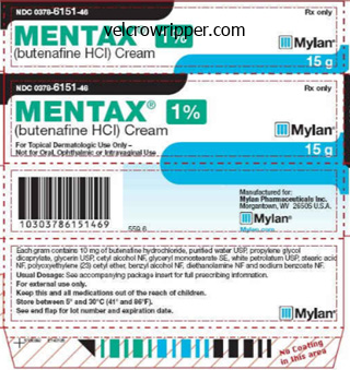
Mentax dosages: 15 mg
Mentax packs: 1 tubes, 2 tubes, 3 tubes, 4 tubes, 5 tubes, 6 tubes, 7 tubes, 8 tubes, 9 tubes, 10 tubes
Age of onset varies between 5 and 24 years and the clinical picture is generally pure fungus bob trusted 15 mg mentax. Features are quadriplegia fungus gnats in house mentax 15 mg with visa, aphasia will fungus gnats kill plants 15 mg mentax with mastercard, and impairment of imaginative and prescient and sphincter function. The medical image consisted of spastic paraplegia with distal motor neuropathy, mild cognitive dysfunction, and pes cavus. Onset of gait impairment diversified from eight to forty years, and hand amyotrophy was noted at 14�60 years. Other shows may be consistent with distal spinal muscular atrophy, pure spastic paraplegia, and peroneal muscular atrophy; the latter has been accompanied by pyramidal signs and signs [56]. In addition to spastic paraplegia, sufferers undergo from mental retardation, this form of spastic paraplegia was reported by Al-Yahyaee et al. Electron microscopic examination of leukocytes revealed electron-dense membranebound vacuoles. The medical picture consists of spastic paraplegia, pes cavus, and slowing of motor and sensory conduction within the lower extremities. Age at illness onset has ranged from 2 to 19, with difficulties in strolling and dysarthria. Drooling, pseudobulbar have an effect on, short stature, distal amyotrophy, and cerebellar indicators had been also noticed. Onset of disease for the extra pure forms is 25�45 years; advanced forms originate in childhood. Additional signs and signs have included skeletal abnormalities, cerebellar indicators, and polyneuropathy. Onset was in adulthood, with speech decline, personality changes, psychosis, and cerebellar dysfunction along with spastic paraplegia. In addition to spastic paraplegia, axonal neuropathy and cerebellar oculomotor dysfunction have been noticed. It was reported in 1944 [67] in a household with hypotonia at delivery, lack of ability to elevate the top, lowered motor milestones, generalized muscle atrophy, and hyperreflexia. Ataxia, dysarthria, athetoid actions, and spastic paraplegia had been also noticed. Onset is in infancy, and related indicators included sensorineural hearing deficits and persistent vomiting due to hiatal hernia. Onset of illness diversified between 12 and 21 years with distal sensory loss, delicate cerebellar signs, and saccadic ocular pursuits. Although originally described in a consanguineous Arab Israeli family in 1985, the locus was refined by Blumen et al. Onset is in infancy, with irregular pores and skin and hair pigmentation, skeletal deformities, and sensorimotor neuropathy. The illness starts in childhood, and associated options can embody spastic dysarthria and pseudobulbar signs. This identification stays questionable, and Rugarli and colleagues have reported that this mutation is a benign polymorphism [83]. The clinical picture consisted of spastic paraplegia, hyperreflexia, pes cavus, and atrophy of the decrease limb muscles. Additional signs and symptoms are clonus, dystonia, seizures, cognitive decline, and cerebellar signs. Other indicators included Babinski sign, involvement of upper extremities, and impairment of vibration sense. The medical picture consists of spastic paraplegia and peripheral sensorimotor neuropathy. This form of spastic paraplegia was reported in an Italian family by Orthmann-Murphy et al. The clinical picture consisted of spastic paraparesis, pes cavus, dysarthria, lack of delicate actions of fingers, and cerebellar signs. Spastic bladder, upper limb cerebellar dysmetria, congenital cataracts, and pes cavus were additionally observed. All patients developed cataracts, cerebellar ataxia, dementia, listening to loss, and axonal neuropathy. The clinical picture consists of spastic paraplegia, hyperreflexia, and delicate weakness within the upper extremities. Symptoms seen at delivery included microcephaly and hypotonia which ultimately evolved to hypertonia. Other signs include high palate, wide nasal bridge, short stature, hypermobility of joints, and genu recurvatum (a knee joint deformity, such that the knee bends backwards). Recently recognized instances have parkinsonism and retinal abnormalities as extra features [103]. The scientific image consists of dysmorphic features, developmental delay, brachycephaly, microcephaly, dental crowding, dysarthria, and dysmetria. The illness began at age 7 with impairment of vision, with walking difficulties a number of years later. Nerve biopsy revealed decreased numbers of large-diameter nerve fibers as properly as onion bulbs. The five affected siblings demonstrated indicators and signs of childish hypotonia, severe psychological retardation, quadriplegia, and strabismus. They also demonstrated signs of pseudobulbar palsy, compulsive laughter, and sphincter impairment. The medical picture consists of spastic paraplegia or quadriparesis, dystonic postures, dementia, and axonal neuropathy. In addition to spastic paraplegia, axonal demyelinating motor neuropathy and optic atrophy are present. The scientific picture consisted of quadriplegia, microcephaly, psychomotor retardation, seizures, and dysmorphic options. The illness began in infancy, and the medical image contains psychological retardation, contractures, pes equinovarus (club foot), microcephaly, dysmorphic features, inappropriate laughter, and brief stature. The medical image consists of quadriplegia, developmental delay, marked kyphosis, and pectus carinatum. Upper limb spasticity was also observed in addition to pes cavus, urinary problems, slight postural tremor, and impaired vibratory sensation. Patients also have loss of vibratory sensation at the ankles, and a couple of those affected have a gentle foot deformity. Age of onset was in infancy, and the scientific image in addition to spastic paraplegia consisted of pes equinovarus, amyotrophy, and a extreme sensorimotor polyneuropathy. Age of onset is early childhood, and related signs embrace optic atrophy, nystagmus, and mild neuropathy. Splice variants exist for each which lack a 32 amino acid stretch encoded by exon 4 [118]. The N-terminal half mediates interaction with proteins that recruit spastin to numerous mobile compartments. Disease onset was before the age of 1, and the clinical picture includes Achilles tendon contractures and amyotrophy. Age of onset diversified from infancy to eight years, and the clinical image is notable for slowly progressive lower extremity spasticity. The N-terminal half of spastin incorporates two distinct domains that may explain this isoform specificity. M87 spastin is required for completion of abscission on the terminal stage of cytokinesis, when the midbody is densely full of an anti-parallel array of microtubules. Thus, this exemplifies how microtubule regulation could be linked to membrane modeling occasions by way of spastin. Cells missing spastin have increased tubulation of endosomal tubular recycling compartments, with resulting defects in receptor sorting. In a zebrafish model, spastin was required for axon outgrowth during embryonic development [142], paying homage to effects of atlastin-1 depletion in rat cortical neurons [136]. Similar outcomes are obtained after depletion of atlastin, whereas overexpression of atlastin has the opposite effect. In animals, acetyl-CoA is crucial for maintaining a correct balance between carbohydrate and fat metabolism. Under normal circumstances, acetyl-CoA from fatty acid metabolism enters the citric acid cycle, contributing to the energy supply of the cell. Given the fundamental roles played by lipids and sterols in neuronal capabilities, it appears very likely that more genes might be identified within this category.
Syndromes
Associations with Turner syndrome antifungal scalp treatment generic 15 mg mentax mastercard, Noonan syndrome fungus documentary mentax 15 mg discount fast delivery, Down syndrome fungus diabetes order mentax 15 mg without a prescription, and fetal alcohol syndrome are nicely described. Inflammatory Lesions the commonest benign situation of the perivertebral area is spondylosis of the vertebral bodies. When a vertebral physique osteophyte is giant enough, it could simulate a submucosal pharyngeal or retropharyngeal mass and produce dysphagia. In cases of diffuse idiopathic skeletal hyperostosis and/or ossification of the posterior longitudinal ligament, there may be giant anterior osteophytes that coexist. These are unusual in sufferers with normal immune responses besides in those sufferers in whom surgical procedure on the cervical backbone has been carried out, in bacteremic patients, or in intravenous drug abusers. The radiographic findings of diskitis and osteomyelitis are discussed absolutely in Chapter 16. Rotatory subluxation of the atlantoaxial joint may coexist with retropharyngitis in the entity known as Grisel syndrome. Esophageal perforation could result in mediastinitis and infection of the perivertebral house. Acute calcific prevertebral tendinitis has findings of calcifications inside the tendons of the longus colli muscles and therefore represents a perivertebral course of. The affected person has neck ache and a fever and swollen prevertebral muscular tissues and improves with conservative management. It may current as a lateral neck mass and be misdiagnosed clinically as a lymph node. Most folks really feel this represents a low-grade soft-tissue tumor, not an inflammatory process. Imaging findings encompass (1) diffuse thickening and infiltration of the pores and skin and subcutaneous tissue (cellulitis); (2) diffuse enhancement and/or thickening of the superficial and deep cervical fasciae (fasciitis); (3) enhancement and thickening of the platysma, sternocleidomastoid muscle, or strap muscular tissues (myositis); and (4) fluid collections in a number of neck compartments. This disease may end in sloughing of tissue, gas containing abscesses, and pulmonary manifestations of adult respiratory misery syndrome, mediastinitis, and pneumonia. Aggressive fibromatosis (desmoid tumor) may infiltrate the muscles of the perivertebral space/posterior triangle. This lesion may be a precursor to or in the household of malignant fibrous histiocytoma. Fibrosing inflammatory pseudotumor can have an effect on the perivertebral house and spread to the skull base. Benign Neoplasms the benign neoplasms of the perivertebral area are relatively uncommon. They include lipomas, schwannomas from the cervical nerve branches, hemangiomas of the musculature, and benign bony tumors. This T2-weighted imaging scan demonstrates a high-intensity chordoma (C), arising on the degree of the atlas (C1), anteriorly displacing and stretching the longus coli muscle on the left (arrow) and infiltrating the muscle on the best with elevation of the nasopharyngeal delicate tissues. Posterior extension on the craniocervical junction leads to displacement and distortion of the brain stem (B). The differential diagnosis will embrace liposarcomas, which can or could not have nonfatty soft tissue related to them. Another bizarroma is Madelung disease, an entity with massive lipomatosis of the posterior neck. Excess fats could be seen predominantly in the posterior a part of the neck, beneath the trapezius and sternomastoid muscular tissues, in the supraclavicular fossa, between the paraspinal muscular tissues, and within the anterior a part of the neck (suprahyoid and infrahyoid). The patients could current with respiratory signs secondary to tracheal compression, neuropathies, weak spot, macrocytic anemia, and venous stasis. Chordoma Of the histologically benign bony neoplasms, chordomas preferentially have an result on the cervical perivertebral area. Chordomas, though histologically benign, must be thought-about malignant tumors as a outcome of they tend to invade aggressively into the skull base and metastasize in 7% to 20% of cases. Chordomas happen at the sacrum, the clivus, the C1-C2 region, in that order of frequency. It displaces the longus colli musculature anteriorly and may cross the boundaries of the C1-C2 area. The differential diagnosis includes chondrosarcoma, which has related features however hopefully is situated off of the midline. This one stumped us-a neonate with a palpable and calcified nodule in the left facet of the neck (arrow). Consider it to be paraspinal and you could get the right analysis: metastatic neuroblastoma. Malignant Neoplasms Metastasis Malignancies of the perivertebral area heart on the vertebral our bodies. Metastases from blood-borne sources are the commonest lesions in the vertebral body, usually from breast, lung, or kidney main tumors. Primary vertebral physique malignancies embrace osteosarcomas and Ewing sarcomas in addition to plasmacytomas. Invasion by Pancoast tumors extending from the lung apex can also current as a perivertebral or supraclavicular mass and will trigger a brachial plexopathy (see following discussion). Less commonly found soft-tissue neoplasms embody hemangiopericytomas and synovial sarcomas. A, Note the relationship of the brachial plexus (arrows) to the subclavian artery (a) on these coronal scans. C, Coronal and (D) axial T2-weighted photographs present left-sided pseudomeningoceles (arrows) because of avulsion of the left C7 and C8 nerve roots from delivery trauma. The expected sign of the C7 nerve root throughout the neural foramen is current on the proper (arrowhead) however absent on the left. Lymphadenopathy Lymphadenopathy is probably the most typical perivertebral area lesion. Lymphoma can also account for nodes within the posterior triangle (level V) of the neck or may present as a supraclavicular mass. Congenital Lesions Lymphatic and venous malformations are the commonest congenital lesions to affect the brachial plexus. The posterior neck is the most common website for cystic hygromas, followed by the axilla. Two kinds of perinatal injuries might have an effect on the brachial plexus: an Erb palsy and a Klumpke palsy. In the latter, the C7, C8, and even T1 roots are torn and subsequently the hand muscle tissue are affected. The Klumpke patient could have a coincident Horner syndrome secondary to involvement of the sympathetic nervous system buildings and/or stellate ganglion opposite the C7-T1 level. The brachial plexus is derived from the C5-T1 cervical nerve roots, which move inferolaterally to the axilla for supply to the upper extremity. The Trunks divide into Divisions (anterior and posterior), which then type Cords (lateral, posterior, and middle) on the Clavicle. At that time, the plexus runs with the axillary artery, posterior to the bigger axillary vein. Fat-suppressed T2-weighted imaging reveals left sided brachial plexus (arrow) inflammation that was viral in etiology. Nodular fasciitis is a nonspecific sclerosing inflammatory situation that may lead to lack of planes across the brachial plexus. A, There is an entire cervical rib on the best (arrows) and an incomplete one on the left. At surgery, a fibrous band across the left brachial plexus from the cervical rib stump to the manubrium was present. B, Left cervical rib attaching to C7 transverse course of with an exostosis-like process (arrowhead) compressing the brachial plexus (long arrow). Both are late to produce symptoms and are easily characterised by density (lipoma/fat) or shape (fusiform/schwannoma). Malignant Neoplasms Traumatic injuries in the adults are usually from "breaking a fall by a motocross racer" accidents. The incomplete cervical ribs usually have a band main from them to the clavicle that traps the plexus and due to this fact are extra typically symptomatic than the complete cervical ribs.
Type 2 is answerable for an infection within the neonatal period anti fungal die off 15 mg mentax effective, presumably acquired either transplacentally or throughout birth from moms with genital herpes fungus gnats in yard buy 15 mg mentax with mastercard. This pressure when contracted in utero might trigger a wide range of teratogenic problems together with intracranial calcifications antifungal shampoo for dogs discount mentax 15 mg otc, microcephaly, microphthalmia, and retinal dysplasia. Sequelae from neonatal herpes additionally embrace multicystic encephalomalacia, seizures, motor deficits, mental and motor retardation, and porencephaly. The features of intracranial neonatal herpes are completely different from these in adults and are summarized in Box 5-7. Calcification can seem between 17 and 21 days after illness onset and may be variable in location. Loss of gray-white contrast is an early abnormality that might be incorrectly interpreted as poor high quality images. The kind 1 virus is liable for the fulminant necrotizing encephalitis seen in children and adults. The scientific picture (just like "maintenance of certification examination encephalopathy") is certainly one of acute confusion and disorientation adopted rapidly by stupor and coma. Seizures, viral prodrome, fever (in more than 95% of cases), and headache are frequent presentations. Focal neurologic deficits such as cranial nerve palsies are found in lower than 30% of cases. Those patients with left temporal illness become symptomatic earlier because of their language impairment and thus could have more delicate imaging findings at the time of presentation. The virus asymmetrically attacks the temporal lobes, insula, orbitofrontal region, and cingulate gyrus. The areas of abnormality within the temporal lobe and insula abruptly end on the lateral putamen, which is characteristically spared. Gyriform enhancement may be visualized; nevertheless, enhancement varies with severity and stage of disease. Residual abnormalities include areas of low density and parenchymal loss at the web site of involvement. Herpes simplex encephalitis is a doubtlessly treatable encephalitis where outcome is determined by early prognosis. It is caused by reactivation of latent varicella zoster virus that has remained dormant in cranial nerve and dorsal root ganglia since primary varicella infection (chickenpox). The virus travels along the sensory nerve from the dorsal root ganglion to the skin producing a rash (shingles) with associated severe radicular pain and allodynia (sensitivity to touch). One medical presentation of intracranial involvement occurs in sufferers who initially have involvement of the ophthalmic division of the trigeminal nerve, herpes zoster ophthalmicus, and then develop contralateral hemiplegia a number of weeks later. Pathology reveals an occlusive granulomatous vasculitis involving the intracranial arteries and meningoencephalitis. Cerebral angiography can demonstrate the severe vasculitis attributable to herpes revealing multiple areas of segmental constriction, normally involving proximal segments Atrophy Encephalomalacia with cysts (late) Hydrocephalus Increased density in cortical grey matter Intracranial calcification from punctate to gyriform Microcephaly Microophthalmia Multiple cysts (usually in persons youthful than 18 years of age), whereas two thirds are the result of reactivation confirmed by the presence of preexisting antibodies. A potential clarification for the focality and latency of herpes simplex kind 1 might depend upon its recognized residence in the trigeminal ganglia. This latent virus underneath sure circumstances turns into reactivated and spreads alongside the trigeminal nerve fibers, which innervate the meninges of the anterior and center cranial fossae. A diffuse meningoencephalitis with a predominant lymphocytic infiltration is seen. The end in untreated circumstances, if the patient survives, is an atrophic cystic parenchyma. By the time the T2-weighted imaging scan is finished (D), each temporal lobe high sign is a fixture. The brightness pregadolinium suggests laminar necrosis (E), and every thing enhances on postgadolinium-bad prognosis (F). Case three: G, Even later one sees the low density on computed tomography, seen within the medial temporal and perisylvian regions bilaterally. Rarely, Zoster could produce myelitis secondary to neurotrophic spread of the virus. Patients may enhance (especially these that are immunocompetent) but are usually left with a residual neurologic deficit. This could additionally be because of reactivation of varicella zoster infection from the trigeminal ganglion resulting in the basal cisterns and from there the circle of Willis vessels. There is diffuse infiltration with lymphocytes, giant cells, and mononuclear cells, of small cerebral arteries and veins (<200 micrometers in diameter). Clinical manifestations include disorientation and impaired mental operate, with up to 25% of circumstances progressing to demise. These areas correspond to segmental abnormalities within the blood-brain barrier secondary to vasculitis. Associated medical signs embrace headache, nausea, vomiting, fever, nuchal rigidity, cerebellar ataxia, and parkinsonism. The differential diagnoses in such cases contain mind stem tumor, multiple sclerosis, Listeria an infection, and acute disseminated encephalomyelitis. Eastern Equine Encephalitis this mosquito-borne arboviral an infection presents with a short typical viral prodrome adopted by altered psychological status, seizures, and focal findings. Other less common areas of involvement are the periventricular and cortical areas. Japanese Encephalitis this illness occurs throughout Asia usually in the summer and early fall. Internal carotid angiogram with a number of areas of vasculitic narrowing (arrows) and vascular occlusion (small open arrow). Abnormal signal in the mind stem is seen on the fluid-attenuated inversion restoration. T2-weighted picture demonstrating increased depth in the deep gray, resulting in a patient with motor sway and stray. It can affect the mind stem, hippocampus, thalamus, basal ganglia, and white matter. Indeed, in some circumstances, despite neurologic disease, the imaging could also be somewhat unremarkable. Divide them into encephalitis, meningitis, pure white matter modifications, mass lesions (both infectious and neoplastic), and atrophy. Case 1: A, Brain stem irregular signal depth is well seen on the fluid-attenuated inversion recovery. E, After extremely lively antiretroviral therapy, notice that the gray matter disease fades. Meningitis is sometimes manifested by enhancement of the meninges, which can be associated with vasculitic modifications and ischemia. There is widespread thickening of the meninges and perivascular areas with a lymphocytic infiltration. Angiographic findings of syphilis are those of segmental constriction and occlusion of the supraclinoid carotid artery, the proximal anterior and middle cerebral segments, and involvement of the basilar artery (this giant and middle-sized vascular involvement is termed Heubner arteritis). On gadolinium (B) it even showed enhancement, which instructed demyelination development. It can additionally be seen in other kinds of immunosuppressed patients, particularly allograft recipients (heart, kidney). Most lesions enhance in a solid or ring style, though nonenhancing lymphoma has been reported. It is important to differentiate lymphoma from toxoplasmosis as a end result of the previous benefits from radiation remedy with greater than a fourfold improve in survival charges over the untreated. Both lesions might present with solid or multiple lesions, have lesions of various sizes (although toxoplasmosis abscesses are inclined to be more quite a few and smaller than lymphoma), and either ring-enhance or show solid enhancement. Improved specificity may be gained by performing a quantitative evaluation of the scans to exclude lesions with exercise not larger than that of scalp. Clinical findings embody the insidious onset of urinary incontinence, progressive spastic-ataxic paraparesis, and sensory loss. D, In a special affected person, a lot of edema seen associated with these lesions on T2 demonstrates postgadolinium ring enhancement (E) in a few. Strokes on imaging are seen in 17% of pediatric patients related to an 8% price of vascular stenosis. Adenopathy and adenoidal hypertrophy, as one would anticipate, are seen on the scans too. Transplacental transmission happens as a result of either recurrent or main maternal an infection.
The nasal septum is composed of three components: a cartilaginous anteroinferior portion; a bony posteroinferior portion often identified as the vomer; and a superoposterior bony portion antifungal treatment for thrush mentax 15 mg discount without prescription, the perpendicular plate of the ethmoid bone fungus around genital area order mentax 15 mg online. Nasal septal deviation facial fungus definition generic mentax 15 mg line, nonetheless, is frequent, and bony spurs typically develop on the apex of the deviation. Inflammation of agger nasi cells may be related to epiphora due to this close relationship. The duct subsequently runs within the anterior and inferior portions of the lateral nasal wall. It then passes lateral to the uncinate process in the infundibulum (fat black arrow) into the hiatus semilunaris (arrowheads) and then to the middle meatus. B, Note the anterior to posterior move of mucus from the frontal, ethmoid, and sphenoid sinuses on sagittal view schematic. Coronal computed tomographic picture reveals bilateral agger nasi air cells (labeled a on right). However, on the best aspect, the ostium is occluded (arrowhead) due to the presence of the larger Haller air cell. Rapid enlargement of the ethmoid air cells happens during ages zero to 4 years and once more with the adolescent development spurt from 8 to 12 years. This may also result in orbital cellulitis or subperiosteal orbital abscesses in children. The maxillary antrum can be current, though small at delivery, and development continues to age 14 years. B, More posteriorly in the same patient, the frontal recess is indicated (arrow), facilitating drainage from the frontal sinus (F) and ethmoid air cells (E) toward the middle meatus. The sphenoid sinus begins its pneumatization at roughly age 2 years and the expansion is slower and more delayed than the other sinuses. Similarly, the mucosa within the infant is considerably redundant and as a lot as 60% of asymptomatic infants can have full or close to full opacification of their sinuses. Some radiology departments additionally administer nasal spray decongestants or antihistamines to scale back reversible mucosal edema before the patients are positioned in the scanner. A width of less than eleven mm is indicative of congenital nasal piriform aperture stenosis. Abnormal dentition and a midline bony inferior palatal ridge are associated imaging findings. The most typical anomalies that result in infantile airway compromise embrace posterior choanal stenoses and atresias, dacryocystoceles, and stenosis of the piriform nasal aperture. Choanal Atresia Choanal atresia is often identified in infancy as a end result of neonates are obligate nose breathers as they suck on a bottle or breast (Box 12-1). A dacryocystocele or piriform aperture stenosis may mimic choanal atresia clinically. Congenital Sinonasal Masses There are several congenital lesions of the sinonasal cavity, together with congenital encephaloceles, dermoid/epidermoid cysts, sinus tracts, and nasal gliomas (Table 12-1). These lesions happen as an abnormality within the strategy of invagination of the neural plate. In embryogenesis, the dura of the brain contacts the dermis at the nasion area as the neural plate retracts. Dermoid cysts happen more commonly than tracts; however, tracts could cause more severe signs because of their intracranial connection in 25% of cases. It is unclear whether or not the maxillary sinus subsequent door is stuffed with tumor or secretions blocked inside the sinus due to obstruction at the ostiomeatal advanced. On the left aspect the nasal passageway narrows, the vomer is thicker on that aspect, and each a bony (posterior; white arrow) and soft tissue (anterior; black arrow) plug is seen. Hypoplastic Maxillary Antrum Congenital hypoplasia of the maxillary sinus occurs in 9% of sufferers. Bony changes that recommend the analysis of a hypoplastic antrum are listed in Box 12-2. In the differential prognosis of sinus hypoplasia is the "silent sinus syndrome" or maxillary sinus atelectasis. In this entity, ostial obstruction from continual sinusitis leads to persistent adverse strain, which outcomes in hypoventilation, which, over time, reduces the quantity of the sinus, hence "sinus atelectasis. The bacterial pathogens responsible for acute sinusitis embrace Streptococcus pneumoniae, Haemophilus influenzae, -hemolytic streptococcus, and Moraxella catarrhalis. In the chronic part Staphylococcus, Streptococcus, corynebacteria, Bacteroides, fusobacteria, and other anaerobes could also be responsible. The fungi that will infect the sinuses include Aspergillus, Mucor, Bipolaris, Drechslera, Curvularia, and Candida. The diagnosis of acute sinusitis is a clinical one, and imaging is usually not required. Imaging may be carried out to evaluate for recurrent sinus infections or atypical sinus infections, usually with the intention of surgical intervention. Is the uncinate process apposed to the medial orbital wall (an atelectatic infundibulum) Are there areas of dehiscence within the lamina papyracea, or do the orbital contents protrude into the ethmoid sinus (both of which can lead to unintentional orbital entry from the ethmoid sinus) Defects in the lamina papyracea have been reported in 5% to 10% of post-mortem specimens. The potential for intraorbital, intracranial, carotid, or optic nerve perforation at the time of surgery is decided by these anatomic variants, present in 4% to 15% of sufferers. Three percent of people have optic nerves that are in contact with the posterior ethmoid wall-most course alongside (90%) or through (6%) the sphenoid sinus. There have been limited stories of optic nerve transection throughout sphenoethmoidectomy from an intranasal method, and dehiscence of the sphenoid wall may be a predisposing factor. Overvigorous elimination of such an intersinus septum during surgery might end in carotid laceration, a real bloody mess. It is estimated that more than 31 million folks in the United States are affected by sinus inflammatory illness each year and that 16 million visits to primary care physicians yearly are for sinusitis and its issues. Overall, Americans spend more than $150 million per 12 months for over-the-counter cold and sinusitis medicines, $100 million of which is for antihistamine medications. Most circumstances of acute sinusitis are associated to an antecedent viral higher respiratory tract an infection. With mucosal congestion as a result of the viral infection, apposition of mucosal surfaces ends in obstruction of the normal flow of mucus, which ends up in retention of secretions, creating a favorable surroundings for bacterial superinfection. The ethmoid sinuses are mostly concerned in sinusitis, probably because of their place within the "line of fireplace" as inspired particles collide with and irritate the fragile ethmoid sinus lining. An intranasal meningoencephalocele is seen on coronal computed tomography in bone (A) and soft-tissue (B) home windows. There is a big deficiency on the cribriform plate (asterisk), allowing for herniation of brain tissue into the nasal cavity. Note the T2 hyperintensity within the herniated tissue, indicating dysplastic mind. Note the focal deficiency of the middle cranial fossa transmitting a small amount of mind tissue (arrow), making this a meningoencephalocele (M). F, For you nonbelievers on the market, slightly more superiorly in this same patient, axial T2 constructive interference in regular state imaging exhibits one other defect along the center cranial fossa, transmitting clearly dysplastic brain tissue (arrow) into the aerated however opacified sphenoid wing, once more cinching the analysis of meningoencephalocele (M). The constructive predictive worth of infundibular opacification for the presence of maxillary sinus inflammatory disease is roughly 80%. When the center meatus is opacified, the maxillary and ethmoid sinuses show inflammatory change in 84% and 82% of patients, respectively. B, On this coronal computed tomographic picture in a unique patient, the left maxillary sinus is completely opacified and smaller than the best. Note the slightly thickened partitions of the left maxillary sinus from continual inflammation. The orbital flooring of the left is depressed (arrow) in comparability with the conventional right facet. On this axial computed tomographic picture, the best septae in the sphenoid sinus connect to the medial wall of the best internal carotid artery (arrow). Overvigorous removing throughout sphenoid sinus surgery could cause a laceration in the carotid wall (ouch!
In the outpatient setting this means neurologists antifungal upholstery spray mentax 15 mg generic otc, psychiatrists fungus hydrangea leaves mentax 15 mg order mastercard, psychologists fungus rx mentax 15 mg buy fast delivery, genetic counsellors, and specialist nurses with assist group representation; complete recommendation and pointers have been revealed [119]. Family members and caregivers often endure from fatigue, loneliness, and stress-related sicknesses and they should be included within the support offered by the multidisciplinary team. This can then inform clinicians about how far to take medical interventions in advanced levels. Similarly, raising the risk of a trusted particular person to assume energy of attorney, to manage the funds and estate of the patient, only when she or he is no longer in a position, may be extremely useful and avoid appreciable angst, particularly if made while the patient still holds capability. Early involvement of a palliative care staff at this stage may be beneficial, Table 21. Domain Gait disturbance and chorea Examples of management measures Physiotherapy to optimize and strengthen gait and stability, and to assess for walking aids; occupational remedy assessment to modify residence surroundings to improve safety; weighted wrist bands to combat limb chorea Ensure daily has a structure to overcome apathy and issue in initiating actions (occupational therapy can advise on this); maintain routines to scale back want for flexibility Carers to assist at house; residential or nursing house care; day centers to maintain social interactions Speech and language therapy to optimize speech, and later in disease to assess for communication aids; guarantee affected person has time to comprehend and respond to speech, and that information is introduced merely Speech and language therapy to advise on most secure food consistencies at different stages of illness, and, in later illness, to advise on must consider enteral diet; dietician to optimize nutritional intake, especially sufficient calorie intake; reduce distractions to optimize swallowing safety Develop methods to cope with cognitive and/ or emotional challenges of disease using counseling or cognitive behavioral remedy Cognitive symptoms Social support Communication Nutrition Psychological issues Source: Novak & Tabrizi 2010 [114]. Insertion of a gastrostomy tube could be considered if vitamin is impaired; once more, if this is mentioned in advance, the opinion of the patient proves invaluable. Careful consideration to mouth care is necessary in any respect levels, provided that sufferers usually have xerostomia that may exacerbate dysphagia and dysarthria, and should neglect or be unable to attend to their own mouth care, significantly within the later levels of illness. Pain might arise from hyperkinetic movements and injury, or hypokinesis, dystonia, and spasticity, and these abnormalities of movement and muscle tone, as nicely as good ache management, should be addressed. Obviously, ache should also prompt examination for reversible causes such as occult fractures, ulcers, constipation, urinary retention, and an infection, amongst others. The predicted open studying frame yields a protein containing 3144 amino acid residues, with a predicted molecular mass of 348 kDa [123]. Most people with greater than 50 repeats develop the disease earlier than the age of 30. However this can be variable, and customarily such estimates are confined to the research setting when estimating years to onset in premanifest gene carriers. The sex of the transmitting parent was found to be important when it comes to repeat size expansion. Genetic counseling Genetic counseling is important every time genetic testing is being undertaken, regardless of whether a predictive or a diagnostic test is being contemplated. In broad phrases, an preliminary session would involve dedication of the extent of danger of the individual by way of evaluation of the household historical past, clinical history, and neurological examination. A psychological evaluation can be undertaken also to determine any patient with lively, untreated psychiatric disease corresponding to melancholy or anxiousness, to assess suicide danger in addition to set up the extent of emotional assist available. Each of the potential results and ensuing implications for the individual, family including children, employment, driving, and life Table 21. This is supported by the discovering that similar twins do show similar ages of illness onset but intriguingly can have completely different clinical phenotypes, while homozygosity largely eliminates any significant differences in age of onset [139�141]. A examine of the Venezuelan kindred revealed that both genetic and environmental factors modulate the age of onset of disease [142]. There was some interest in exploring the contribution of the scale of the normal allele in influencing the age of onset, nevertheless a linear regression based evaluation refuted an interplay between expanded and regular alleles, or certainly a second expanded allele [148]. Genetic analysis In clinical follow, genetic testing would be considered in three circumstances: (i) a confirmatory or diagnostic check, (ii) predictive testing of an asymptomatic individual recognized to be at risk, and (iii) prenatal testing. In addition, a optimistic predictive check places the extra burden of uncertainty over the timing of illness onset. Most clinicians are aware of the necessity for counseling of at-risk individuals for predictive testing, however the Source: Harper & Newcombe 1992 [150]. Strict confidentiality should be practised and permission should be obtained to discuss medical historical past with other family members, whose right to confidentiality and appropriate genetic counseling must equally be respected. A second consultation would then be arranged after no much less than a month to allow adequate time for the patient to adequately think about the knowledge given. Once the affected person has confirmed their understanding of the test and its scope, genetic testing could be undertaken following written, knowledgeable consent. The outcome could be given at a third consultation, after which a follow-up must also be carried out to assess how the patient is coping with the outcome. In those with a unfavorable gene check, improvement in psychological well-being is noted, though paradoxically around 10% have issue adjusting to an uncertain future free from illness rather than a clearer future afflicted with illness [152]. Reasons commonly cited for having predictive testing embrace wishing to relieve uncertainty, to inform choices about copy, and to plan for the future. Adequate genetic counseling and informed consent in these situations is equally important. Predictive genetic testing Prior to the availability of the genetic take a look at, surveys advised that 80% of these with a household historical past of the illness would take up predictive testing, however the demand has been decrease than anticipated. The reasons mostly given for needing a take a look at had been to relieve uncertainty, plan for the future, plan a household, and inform children [153]. This is to allow the person to think about all potential advantages and harms, for them personally and for others near them. What is totally clear from the extensive analysis that has been carried out on this space is the importance of providing correct data and pre- and post-test counseling and assist, and for mechanisms to be in place to ensure adequate safeguards in opposition to discrimination and breaches of confidentiality. An informed, competent adult should be free to make his or her personal choice, however in certain circumstances, similar to when the affected person is depressed, testing should be delayed. At the outset you will need to make explicitly clear that the process is embarked upon with the express intention of ending the being pregnant if the embryo is gene optimistic. Once the prognosis is revealed, common assessment ought to take place in accordance with occupational danger. It is prudent for them to inform their insurance coverage company as failure to disclose may invalidate their cowl. Chorionic villus sampling could be carried out at 8�10 weeks or alternatively amniocentesis could be undertaken at 15�18 weeks. Driving can current an increasing problem to sufferers because of a mix of impaired voluntary motor control and psychomotor processing, and diminished reaction instances. Generally, if concern is voiced by both the patient themselves or a associate or relative, or if medical examination signifies vital incoordination, bradykinesia, or perseveration, or judgment known as into query, the patient should be advised to stop driving and inform the related driver licensing authority. Often a fitness to drive test could be organized at the discretion of the licensing authority. It is predominantly a cytoplasmic protein, which is amenable to cleavage by proteases. In specific phrases, neuronal loss and atrophy happen particularly within the neostriatum � the caudate and putamen � though the previous is affected to a larger extent. As the illness progresses, generalized mind atrophy ensues such that at post mortem the brain is between 300 and 400 g lighter than the common grownup mind weight for affected person age [159]. Interneurons are generally spared; in contrast, the striatal medium spiny neurons, which comprise 90% of the striatal neuronal population, are selectively lost. These are predominantly the encephalin-containing projections to the exterior globus pallidum somewhat than the substance P neurons that connect with the interior globus pallidum. From a functional neuroanatomical perspective, the prevalence of chorea early in illness might mirror preferential injury to the oblique pathway of basal ganglia-thalamocortical circuitry [160]. In later disease the direct pathway is affected in addition to cortical neurons, which can contribute to the loss of motor control, abnormalities of eye movements, and neuropsychiatric symptomatology. At a microscopic degree, cortical neurons show decreased staining of nerve fibers, neurofilaments, tubulin, and microtubule-associated protein 2 and diminished complexin 2 concentrations, suggesting impairment of synaptic operate, cytoskeleton, and axonal transport [170, 171]. The most well known neuropathological classification is the Vonsattel grade. These repeats are composed of two antiparallel a-helices with a helical hairpin configuration [175], which assemble into a superhelical structure with a continuous hydrophobic core. The protein is generally cytoplasmic, with membrane attachment via palmitoylation at cysteine 214 [180]. A putative nuclear export signal is current close to the C-terminus but a classical nuclear localization sign has not been recognized. Homozygous topics appear to be regular developmentally but may have an earlier illness onset and extra severe phenotype, though at a microscopic level their early neuronal biology and circuitry remains unstudied. Pathogenic species can be monomeric or, extra doubtless (and as shown), type small oligomers. Toxic results in the cytoplasm embody inhibition of chaperones, proteasomes, and autophagy, which can trigger accumulation of abnormally folded proteins and other cellular constituents. Pathognomonic inclusion bodies are discovered within the nucleus (and small inclusions are additionally present in cytoplasmic regions). Increased stimulation of extrasynaptic glutamate receptors takes place, and reuptake of glutamate by glia is diminished, leading to excitotoxicity and enhanced susceptibility to metabolic toxic results. Cleavage is mediated by particular enzymes including caspases, that are primarily associated with apoptosis, and the place experimental paradigms in mice have demonstrated amelioration of phenotype if cleavage is pharmacologically or genetically prevented. Later work nevertheless has solid doubt on caspase activity producing fragment toxicity though it could have a task in clearance [189].
Veronica. Mentax.
Source: http://www.rxlist.com/script/main/art.asp?articlekey=96175
Large persistent hematomas produce nonspecific areas of hypodensity with focal quantity loss fungus japan train 15 mg mentax discount mastercard. Small hemorrhages within the posterior fossa fungus gnats miracle gro discount 15 mg mentax visa, the anterior inferior frontal and temporal lobes (the commonest locations for contusions) and adjacent to the calvarium are tough to detect because of artifact from adjoining bone and/or partial volume results antifungal weight loss mentax 15 mg proven. In circumstances where hemorrhage is suspected but not confirmed, coronal and sagittal reformations of the initial knowledge set can help to confirm or exclude hemorrhage in problematic areas. The use of intravenous iodinated contrast in patients with hemorrhage is unnecessary in most situations. If contrast is given, intraparenchymal hemorrhage is commonly associated with a peripheral rim of enhancement from approximately 6 days to 6 weeks after the preliminary event. This enhancement is the end result of breakdown of the blood-brain barrier on the margin of the hematoma due to toxic/inflammatory results of blood products. Detection of related and underlying lesions is facilitated and the intensity pattern on a quantity of sequences permits for accurate "aging" of the hematoma. B, Computed tomography angiography source image reveals "dot sign" (arrow) throughout the anterior portion of the hematoma, indicative of acute extravasation and active hemorrhage. The development of imaging hallmarks is nicely understood, though the precise time intervals for these changes are variable. Warning: this discussion would possibly bring again some painful recollections from your natural chemistry days. Structure of hemoglobin: Hemoglobin, the first oxygen service in the bloodstream, consists of four protein subunits. Each subunit contains one heme molecule consisting of a porphyrin ring and an iron atom (Fe2+), which sits close to the center of the porphyrin ring and binds to oxygen (O2). When a hemoglobin subunit loses its O2, it forms deoxyhemoglobin, which has four unpaired electrons and may usually be oxidized to methemoglobin in a reversible style. Susceptibility results: When placed in a magnetic field, certain substances generate an additional smaller magnetic area, which both provides to or subtracts from the externally utilized field. Paramagnetic supplies similar to deoxyhemoglobin and methemoglobin have unpaired electrons that generate a lot larger native fields surrounding the paramagnetic molecule that add to the externally applied field. As protons transfer (diffuse) through these locally various gradients, they accumulate a special quantity of part change depending on the time spent at different effective field strengths. Proton-electron dipole-dipole interplay:The paramagnetic iron atom in methemoglobin generates a neighborhood field roughly one thousand times higher than the local area generated by the proton nucleus. When water binds heme, each T1 and T2 are shortened by proton-electron dipoledipole interaction. In abstract, two key results are created by the hemoglobin molecule: (1) the paramagnetic impact secondary to the iron within the heme molecule, which can produce susceptibility effects/proton rest enhancement within the case of intracellular deoxyhemoglobin, methemoglobin and hemosiderin leading to T2 shortening, and (2) proton-electron dipole-dipole interactions with methemoglobin (both intracellular and extracellular) resulting in T1 shortening. Temporal Changes in Parenchymal Hemorrhage Temporal modifications in parenchymal attenuation and sign are summarized in Table 3-3. A, Computed tomography scan a number of hours after onset of new subject reduce reveals bilateral occipital hematomas. The right hematoma has a focus of hyperintensity in its lateral margin (arrow) indicative of acute to subacute hematoma. D, Gradient-echo scan reveals that left hematoma has a hypointense margin and a comparatively hyperintense middle. Hyperacute Hemorrhage (0 to four Hours) In the primary 3 to 6 hours after extravasation the intact red cells comprise mostly diamagnetic oxyhemoglobin. The periphery of the hematoma incorporates purple blood cells containing hemoglobin that has began to desaturate (deoxyhemoglobin). This produces peripheral hypointensity particularly on susceptibility-weighted (T2*) sequences and at larger area strength. These results produce profound T2 and T2* hypointensity that begins at the periphery and extends to the center of the hematoma. C, T2-weighted image reveals hematoma is insointense to minimally hyperintense to normal cortex (but overall less intense than would be seen in hyperacute hematoma). E, Fluid-attenuated inversion restoration scan reveals hematoma to be minimally hyperintense to gray matter. Because water molecules are unable to strategy shut enough to the iron atom of deoxyhemoglobin; no T1 shortening is brought on by proton-electron dipole-dipole interactions. There is a rise in T2 hyperintense peripheral vasogenic edema with a concomitant enhance in mass effect. Unlike deoxyhemoglobin, water molecules are in a position to strategy the paramagnetic heme of methemoglobin, permitting proton-electron dipole-dipole interactions that shorten T1. Oxidation of deoxyhemoglobin to methemoglobin proceeds from the periphery to the center of the clot in the course of the first week after the ictus, and therefore T1 hyperintensity is initially seen on the edge of the hematoma (2 to 3 days) with progressive "filling in" of the center of the hematoma (7 to 10 days). Towards the top of the early subacute part T2 hypointensity decreases because of cell lysis. Once paramagnetic methemoglobin is no longer sequestered within the pink cells, local area inhomogeneity begins to decrease and susceptibility induced T2* hypointensity begins to resolve. A, Computed tomography scan 4 hours after onset of symptoms in chronic hypertensive affected person reveals right pontine hematoma. Paralleling the breakdown of the methemoglobin is an accumulation of the iron molecules hemosiderin and ferritin inside macrophages on the periphery of the lesion. The central T1 and T2 hyperintensity and peripheral T2/ T2* hypointensity persists but peripheral edema and mass impact utterly resolve. The iron atoms from the metabolized hemoglobin molecules are deposited in hemosiderin and ferritin molecules that are trapped permanently inside the brain parenchyma because of restoration of the blood-brain barrier. A, Computed tomography scan at 1 day reveals a big irregular hyperdense left parietooccipital hematoma with intraventricular hemorrhage. The pattern of hemosiderin "scarring" is dependent upon the scale, location, and etiology of the unique hemorrhage. Small hematomas produce peripheral hypointense clefts (hemosiderin slit) while giant hematomas, hemorrhagic infarcts, and contusions produce areas of encephalomalacia with marginal or gyral hypointensity. Microbleeds usually happen in hypertensive cerebrovascular illness, amyloid angiopathy, and as results of head trauma with axonal injury. They may be seen with a number of cavernous malformations and after radiation remedy. Spontaneous (nontraumatic nonischemic) parenchymal hemorrhage accounts for roughly 10% of stroke syndromes (Box 3-3). The most typical reason for parenchymal hemorrhage is hypertension and these lesions are most often seen in the deep gray matter (basal ganglia and thalamus), mind stem and cerebellum. Computed tomography scans at 24 hours (A) and 14 days (B) reveal evolution of right ganglionic hematoma. Venous thrombosis can lead to parenchymal hemorrhages sometimes in the white matter adjoining to the thrombosed dural sinus or cortical vein. Hemorrhage into underlying neoplastic lesions (primary or metastatic) or from vascular abnormalities (arteriovenous malformations, cavernous angiomas, and aneurysms) can occur in any location and at any age. Hypertension Whatever you do, stay calm and keep your blood stress down in this section. In hypertensive cerebral vascular disease, damage to small perforating arteries arising from proximal vessels. Over the primary 24 to 36 hours, the hemorrhages often enlarge and all the time develop vasogenic edema and rising mass impact. These adjustments account for the scientific observation that sufferers with hypertensive hemorrhage usually deteriorate over the primary few days. Imaging research sometimes reveal different evidence of hypertensive cerebrovascular disease. Chronic lacunar infarcts or hemorrhages and in depth microvascular white matter illness are sometimes current. D-F, Repeat examination at 2 months reveals marked contraction of clot with focal volume loss and no edema. While persistent hypertension alone can result in parenchymal hemorrhage, fast episodic improve in blood pressure could happen with cocaine use, dialysis, and fluid overload leading to parenchymal hemorrhage. It does correlate with brain parenchymal amyloid deposition so is more common in sufferers with Alzheimer disease. Amyloid protein replaces the conventional constituents of the vessel wall specifically the elastic lamina leading to microaneurysm formation and fibrinoid degeneration resulting in vascular fragility. Hemorrhages are often lobar, usually involving the frontal and parietal lobes together with the subjacent white matter. There is a propensity for recurrent hemorrhage in the same location and/or a quantity of simultaneous hemorrhages.
The Uterine Scar Effect On the anterior uterine wall of the uterine corpus fungus gnats remedy mentax 15 mg fast delivery, the muscle fibers from all sides crisscross diagonally with those of the alternative aspect but run in a predominantly transverse direction xylecide anti fungal shampoo mentax 15 mg discount with amex. The so-called "classical" vertical uterine incision does much more injury to the myometrium and is at higher risk of spontaneous uterine rupture in subsequent pregnancies than the transverse lower section incision antifungal topical creams mentax 15 mg purchase overnight delivery. It is therefore only used in rare cases of very early preterm start (23�25 weeks), the supply of the fetus in cases of placenta previa accreta, and for the supply of conjoined twins. Postsurgical Uterine Healing Ferdinand Kehrer (1837�1914) and Max S�nger (1853�1903) every independently developed uterine closure methods using sutures made of silver wire, as utilized successfully by the American gynecologist James Marion Sims (1813�1883) to restore vesico-vaginal fistulae. All have completely different properties, which can potentially have an impact on the therapeutic process. Schwarz, for instance, concluded that if the minimize surfaces are intently apposed, the proliferation of the connective tissue is minimal, and the conventional relation of the sleek muscle to connective tissue in steadily reestablished. The ensuing scar tissue is weaker, much less elastic, and extra susceptible to damage and dehiscence (separation) than the intact muscle. Myofiber disarray, tissue edema, inflammation, and elastosis have all been observed in uterine wound therapeutic after surgical procedure. Overall, single-layer closure in contrast with double-layer closure of the uterine incision is associated with a statistically significant discount in mean blood loss and period of the operative procedure. Scar Pregnancies A caesarean scar pregnancy is the implantation of clinically detectable being pregnant into a scar. It could be recurrent and is related to severe maternal morbidity and significant mortality from very early in pregnancy. This can probably clarify why cervical scar pregnancies are more 18 Placenta Accreta Syndrome symptomatic and almost at all times result in major bleeding early in pregnancy. The oldest idea is predicated on a theoretical main defect of trophoblast biology resulting in excessive invasion of the myometrium. The process is complicated and entails many local uterine parts and external maternal cells and hormones. Decidualized stromal cells are derived from the fibroblast-like cells within the endometrium, which maintain their progesterone receptors in the presence of progesterone. Progesterone additionally initiates the proliferation on endometrial glands from earlier than the blastocyst implants. The secretions also symbolize an important supply of nutrients, histotroph, for the conceptus during the first trimester. On establishing contact, a few of the trophectoderm cells undergo proliferation and fusion to from the multinucleated syncytiotrophoblast, whereas others remain as a deeper, progenitor population, the cytotrophoblast cells. Tongues of syncytiotrophoblast penetrate between the epithelial cells, whereas on the identical time the endometrial stromal cells grow over and encapsulate the conceptus. The mixture of these actions leads to the conceptus quickly becoming fully embedded inside the stratum compactum of the endometrium. Soon after, strands of mononuclear cytotrophoblast begin to proliferate on the fetal facet of the implanted blastocyst wall. The most distal cytotrophoblast cells break by way of the syncytium and unfold laterally to kind the cytotrophoblastic shell separating the placenta from the decidua. They differentiate primarily into interstitial and endovascular subpopulations that migrate by way of the decidual stroma and down the lumens of the spiral arteries, respectively. They steadily prolong laterally, reaching the periphery of the placenta round midgestation. Depthwise, the changes are maximal inside the central region of the placental mattress, and the extent of invasion is progressively shallower towards the periphery. The endovascular cells additionally act as plugs blocking the spiral arteries and stopping maternal blood move from entering the intervillous house during the first 10�12 weeks of gestation. Insufficient activation of the maternal immune cells is related to issues of being pregnant, together with miscarriage, preeclampsia, and development restriction. In normal pregnancies, the transformation of spiral arteries into utero-placental arteries is described as completed around midgestation. Trophoblast invasion is notably more aggressive and extra penetrative at websites of ectopic implantation, for example, in the Fallopian tube, within the absence of decidua. The precise regulation of trophoblast invasion will due to this fact depend on the stability of local concentrations of many components and also the composition of the extracellular matrix. Having passed via the decidua the cells at the second are largely beyond the attain of the maternal immune cells. Is it lack of stimulation that brings them to a halt, Pathophysiology of Accreta 21 or is there a strong native inhibitory signal Overall, these data suggest a potential relationship between a poorly vascularized uterine scar space and an increase in the resistance to blood move within the uterine circulation with a secondary impression on reepithelialization of the scar area and faulty subsequent decidualization. Both hormones are primarily produced by the syncytiotrophoblast and mirror its enlargement, but no difference in either placental or fetal progress pattern has been reported in abnormally adherent placentas. Deeper trophoblast myometrial invasion and chorionic villi infiltration into myometrial vascular spaces has been recently documented in placenta increta and percreta. It may be that within the absence of a decidua, the normal release of proteases and cytokines from activated maternal immune cells is lacking, impairing arterial remodeling. Their protocol facilitates retrospective correlation with surgical and imaging findings as properly as standardized tissue sampling for potential research. Correlation of pathological findings with the clinical notes and imaging is important. Correlation of the ultrasound imaging from early in pregnancy with histopathological examination is pivotal to higher understand the natural evolution of this dysfunction and further collaborative analysis is essential to improve the analysis and management of this increasingly widespread main obstetric complication. The effect of cesarean supply charges on the future incidence of placenta previa, placenta accreta, and maternal mortality. Placenta accreta in a affected person with a history of uterine artery embolization for postpartum hemorrhage. Impact of frozen-thawed single-blastocyst switch on maternal and neonatal outcome: An analysis of 277,042 single-embryo transfer cycles from 2008 to 2010 in Japan. The structure of the musculature of the human uterus-Muscles and connective tissue. The fibromuscular construction of the cervix and its adjustments throughout being pregnant and labour. A take a glance at uterine wound healing through a histopathological research of uterine scars. Single-versus double-layer closure of the hysterotomy incision during cesarean delivery and threat of uterine rupture. Impact of single- vs double-layer closure on antagonistic outcomes and uterine scar defect: A systematic review and metaanalysis. Ultrasound analysis of Cesarean scar after singleand double-layer uterotomy closure: A cohort study. Hydrosonographic evaluation of the results of two totally different suturing strategies on therapeutic of the uterine scar after cesarean delivery. Cesarean scar defect: Correlation between Cesarean section quantity, defect dimension, scientific symptoms and uterine place. Use of three-dimensional ultrasonography within the analysis of uterine perfusion and therapeutic after laparoscopic myomectomy. High prevalence of defects in cesarean section scars at transvaginal ultrasound examination. Sonographic lower uterine section thickness and threat of uterine scar defect: A systematic evaluation. Placenta percreta leading to spontaneous complete uterine rupture within the second trimester. Example of a fatal complication of abnormal placentation following uterine scarring. Uterine rupture at 17 weeks of a twin being pregnant complicated with placenta percreta. Placenta percreta-induced uterine rupture recognized by laparoscopy in the first trimester. Spontaneous uterine rupture at the 21st week of gestation attributable to placenta percreta.
It has not obtained approval for this purpose by the Food and Drug Administration within the United States fungus spanish purchase mentax 15 mg with mastercard. Active remedy of constipation antifungal honey 15 mg mentax discount overnight delivery, intercurrent infections antifungal foot powder order mentax 15 mg fast delivery, cataplexy [85], seizures, and dystonia ought to be pursued. In addition, bodily therapy to maintain mobility, joint flexibility, and pulmonary perform seems to be beneficial. Natural history of Type A Niemann-Pick disease: possible endpoints for therapeutic trials. A potential, cross-sectional survey research of the pure history of Niemann-Pick disease type B. Acid sphingomyelinase deficiency: prevalence and characterization of an intermediate phenotype of Niemann-Pick illness. Adult Niemann-Pick illness: its relationship to the syndrome of the sea-blue histiocyte. The pathogenesis and therapy of acid sphingomyelinasedeficient Niemann-Pick illness. The demographics and distribution of kind B Niemann-Pick disease: novel mutations result in new genotype/phenotype correlations. Type A Niemann-Pick disease: a frameshift mutation within the acid sphingomyelinase gene (fsP330) happens in Ashkenazi Jewish sufferers. Skeletal manifestations in pediatric and grownup sufferers with Niemann Pick disease sort B. Niemann-Pick illness sort B: an unusual scientific presentation with a quantity of vertebral fractures. Unsuccessful remedy try: wire blood stem cell transplantation in a affected person with Niemann-Pick disease type A. Treatment of Niemann-Pick illness kind B by allogeneic bone marrow transplantation. Correction of enzyme ranges with allogeneic hematopoeitic progenitor cell transplantation in Niemann-Pick kind B. Niemann-Pick disease: sixteen-year follow-up of allogeneic bone marrow transplantation in a type B variant. Successful therapy of endogenous lipoid pneumonia because of Niemann-Pick Type B illness with whole-lung lavage. Treatment of hyperlipidemia associated with Niemann-Pick illness type B by fenofibrate. Merits of combination cortical, subcortical, and cerebellar injections for the remedy of niemann-pick illness kind a. Nine circumstances of sphingomyelin lipidosis, a model new variant in Spanish-American Children. The National Niemann-Pick C1 illness database: report of medical options and well being issues. NiemannPick illness kind C2 presenting as fatal pulmonary alveolar lipoproteinosis: morphological findings in lung and nervous tissue. Respiratory disease in Niemann-Pick kind C2 is caused by pulmonary alveolar proteinosis. NiemannPick C disease in Spain: medical spectrum and growth of a incapacity scale. Recommendations for the diagnosis and management of Niemann-Pick illness kind C: an update. Treatable metabolic psychoses that go undetected: what Niemann-Pick sort C can educate us. Saccadic eye movement characteristics in adult niemann-pick sort C illness: relationships with disease severity and brain structural measures. Niemann-Pick sort C illness: molecular mechanisms and potential therapeutic approaches. Cholesterol oxidation merchandise are delicate and specific blood-based biomarkers for Niemann-Pick C1 disease. Molecular, anatomical, and biochemical occasions associated with neurodegeneration in mice with Niemann-Pick kind C illness. Microarray expression analysis and identification of serum biomarkers for Niemann-Pick illness, type C1. Gamma-secretase-dependent amyloidbeta is elevated in Niemann-Pick kind C: a cross-sectional research. Niemann-Pick sort C disease: accelerated neurofibrillary tangle formation and amyloid beta deposition related to apolipoprotein E epsilon four homozygosity. Natural history of Niemann-Pick disease sort C in a multicentre observational retrospective cohort examine. New NiemannPick Type C1 Gene Mutation Associated With Very Severe Disease Course and Marked Early Cerebellar Vermis Atrophy. Size and form of the corpus callosum in grownup Niemann-Pick kind C displays state and trait sickness variables. Dysphagia as a risk issue for mortality in Niemann-Pick disease kind C: systematic literature evaluate and proof from research with miglustat. New therapies within the management of Niemann-Pick kind C disease: medical utility of miglustat. Iminosugar-based inhibitors of glucosylceramide synthase extend survival however paradoxically enhance mind glucosylceramide ranges in Niemann-Pick C mice. Miglustat in adult and juvenile sufferers with Niemann-Pick disease kind C: long-term knowledge from a clinical trial. Clinical expertise with miglustat remedy in pediatric patients with Niemann-Pick disease sort C: a case series. Weekly cyclodextrin administration normalizes ldl cholesterol metabolism in practically each organ of the Niemann-Pick kind C1 mouse and markedly prolongs life. Miglustat improves purkinje cell survival and alters microglial phenotype in feline Niemann-Pick illness kind C. Histone deacetylase inhibitor treatment dramatically reduces ldl cholesterol accumulation in Niemann-Pick sort C1 mutant human fibroblasts. Heat shock protein-based therapy as a potential candidate for treating the sphingolipidoses. Developmental end result publish allogenic bone marrow transplant for Niemann Pick Type C2. Human umbilical wire blood-derived mesenchymal stem cells improve neurological abnormalities of Niemann-Pick kind C mouse by modulation of neuroinflammatory situation. Neuronal and epithelial cell rescue resolves persistent systemic inflammation in the lipid storage dysfunction Niemann-Pick C. The inheritance is X-linked, affecting males extra severely than females; nonetheless the medical manifestations are variable, even within households with the identical underlying genetic mutation. The minimal incidence in Brazil [2] has been estimated at 1:35,000 males and in Japan [3] at 1:30,000 to 1:50,000. Men in their twenties and thirties present with slowly progressive spastic paraparesis, sensory loss, and bowel and bladder dysfunction. Symptoms of ataxia could additionally be followed by the development of Spectrum of scientific phenotype (Table 25. Once neurological signs turn into clinically apparent, the disease progression may be quite fast with improvement of spastic quadriplegia, cortical blindness, loss of auditory processing, and cognitive decline over 12 months or much less. The pure course of this form with out early intervention results in complete disability and early dying [4, 5]. The severity of symptoms is variable, ranging from hyperreflexia with impaired vibratory sense to extreme spastic paraparesis. Women with subclinical adrenal dysfunction must be intently monitored for adrenal insufficiency [15]. Confluent changes can be seen in the cerebral white matter, and less regularly in the cerebellar white matter, with out associated irritation. Bilateral, symmetric degeneration of the lengthy ascending and descending tracts occurs with each axon and myelin loss. The degeneration of the ascending tracts and the gracile and dorsal spinocerebellar tracts is extra outstanding in the upper spinal wire.
The circumvallate papillae of the tongue separate the oral tongue (a a half of the oral cavity) anteriorly from the oropharynx posteriorly fungus hives order mentax 15 mg line. The exhausting palate is a part of the oral cavity fungus gnats morgellons buy mentax 15 mg on line, but the taste bud is part of the oropharynx pesticide for fungus gnats discount 15 mg mentax amex. Besides the palatine tonsils, the oropharynx additionally contains the lingual tonsillar tissue seen at the base of the tongue. Oral Cavity the oral cavity consists of the lips, the anterior two thirds of the tongue, the buccal mucosa, the gingiva, the onerous palate, the retromolar trigone, and the ground of the mouth. The torus tubarius (white arrow), eustachian tube orifice (white arrowhead), and fossa of Rosenm�ller (black arrow) are labelled. Although the adenoids and lingual tonsils are mainly midline structures, the palatine tonsils are discovered bilaterally framed by the pharyngeal faucial arches. On this axial T1-weighted imaging scan, one can identify the bottom of the tongue with lingual tonsil tissue (arrows) and the palatine tonsils (t). Also identifiable on this scan are the submandibular glands (g), the sublingual area extending from the submandibular glands anteriorly, and the midline fatty lingual septum with posterior aspect of the genioglossus muscles (gg) on both facet. Muscles on both aspect of the sublingual house are the mylohyoid muscle tissue (m) laterally and the hyoglossus (asterisks) medially. Geniohyoid muscle (gh) makes up the majority of the tissue anteriorly in the tongue often beneath genioglossus, partially included right here. The lingual nerve from the trigeminal nerve and the hypoglossal nerve run together from the ground of the mouth into the tongue base and sublingual area and are important for the surgeon to establish to preserve tongue perform. Radiologists should establish whether or not tumor is within the sublingual house, to alert the surgeon concerning the potential for invasion or sacrifice of these nerves. The chorda tympani from the facial nerve supplies taste to the anterior two thirds of the tongue and its branches join that of the lingual nerve. The onerous palate (arrows), the anterior two thirds of the tongue (t), and the gingival surfaces of the mandible represent portions of the oral cavity. The oral cavity additionally contains the floor of the mouth, seen as the mylohyoid (m) muscular sling inferolaterally. Axial T1-weighted imaging demonstrates the air-filled pyriform sinuses (asterisks), which are delineated anteromedially by the aryepiglottic folds (arrows). Other subsites of the pharynx on this location embrace the posterior and lateral pharyngeal walls. The mucosa over the posterior surface of the cricoarytenoid joints is a half of the hypopharynx. The anteromedial border of the pyriform sinus is the aryepiglottic fold, a construction of the supraglottic larynx. The pyriform sinus is finest evaluated with the affected person present process Valsalva maneuver because this distends the airway right down to its inferiormost portion, the pyriform apex. The apposition of mucosal surfaces in the pyriform sinus typically makes lesion localization difficult. You may not have the ability to distinguish extension to the adjacent aryepiglottic fold, the lateral pyriform sinus mucosa, or the posteromedial mucosa without such maneuvers to maximize distention. The supraglottis consists of the subsites of the false vocal cords, the arytenoids, the infrahyoid and suprahyoid epiglottis, and the aryepiglottic folds. The glottis consists of the true vocal cords, the anterior and posterior commissures, and the vocal ligament extending from the arytenoid cartilage. The laryngeal ventricle is claimed to separate the supraglottis and glottis, but is itself part of the supraglottis. The subglottis begins 1 centimeter below the laryngeal ventricle and extends to the first tracheal ring. The larynx is anchored on a framework composed of the hyoid bone, the epiglottis, the thyroid cartilage, the cricoid cartilage, and the arytenoids, each of which has an integral perform. Of these, the whole ring of the cricoid cartilage is the indispensable strut for preservation of airway patency. From the inferior portion of the arytenoid cartilage the vocal ligament stretches to the thyroid cartilage anteriorly and helps the vocal twine. The posterior commissure refers to the mucosa between the 2 vocal processes on the anterior surface of the arytenoid cartilage. On the lateral aspect of the laryngeal mucosal floor is the paraglottic space, which accommodates fats, lymphatics, and small muscular tissues. At the false twine stage the paraglottic space is dominated by fat, whereas at the true wire level it accommodates the thyroarytenoid muscle, which makes up the majority of the true vocal cord, parallels the vocal ligament, and marks the glottic degree. The cricoarytenoid muscle strikes the arytenoids to slender or open the glottic airway for speech. The vagus nerve innervates the larynx via two branches: the recurrent laryngeal nerve and the superior laryngeal nerve. The solely muscle equipped by the latter is the cricothyroid muscle, and its paralysis causes only minor adjustments in the voice. The vagus descends from the medulla via the jugular foramen into the carotid sheath. The vagus follows the carotid sheath inferiorly with the recurrent laryngeal nerve looping beneath the aortic arch on the left and the subclavian artery on the right, before ascending within the tracheoesophageal groove. The branches of the recurrent laryngeal nerve perforate the cricothyroid membrane to supply the functioning intrinsic musculature of the larynx. Note that on the false cord level the paraglottic tissue is excessive intensity from fat, whereas below the ventricle the delicate tissue is muscular from the thyroarytenoid muscle, which makes up the bulk of the true twine. C, At the false cord stage one can again establish the fats (arrows) in the paraglottic area. D, At the true wire stage the paraglottic tissue is made up of the thyroarytenoid musculature. A, the false wire is characterised by low-density fats (arrows) within the paraglottic space. The thyroarytenoid muscle (+) makes up the "paraglottic" tissue and this muscle attaches to the vocal process of the arytenoid. This results from apposition of the mucosal surfaces of the nasopharynx in the midline as the notochord ascends through the clivus to create the neural plate. The cyst is often properly defined and characteristically happens within the midline, although it may be seen off midline in a small proportion of circumstances. The cysts become contaminated on uncommon occasions and may be a source of persistent halitosis. Tornwaldt cysts should be distinguished from mucus retention cysts, that are also usually seen inside the nasopharyngeal mucosa, and are T1 darkish,T2 shiny with peripheral rim enhancement. Bony Congenital Lesions Of the congenital cysts in the maxilla, the nasopalatine cyst (incisive canal cyst) is the most common. This cyst often arises within the (para) midline incisive canal and slowly expands the maxilla and exhausting palate. Cherubism can occur in patients with familial fibrous dysplasia of the mandible, neurofibromatosis, Noonan syndrome (male Turner syndrome), and Ramon syndrome (cherubism, gingival fibromatosis, epilepsy, and mental retardation). Computed tomography shows bilateral bubbly lesions of the mandible (arrows) in a affected person with cherubism. The pyriform sinus is a drainage site for third branchial cleft cysts, and the pyriform sinus apex could additionally be a web site of sinus tracts leading from fourth branchial cleft cysts. The third branchial cleft sinus tract passes between the frequent carotid artery and vagus nerve to the anterior border of the inferior sternocleidomastoid muscle. The fourth branchial cleft sinus tract passes around the great vessels and the aortic arch on the left side. It is simply too anterior to be a thyroglossal duct cyst and never lateral or inferior sufficient to be a ranula. Developmental Cysts Epidermoids and less generally dermoids and lymphangiomas (usually multilocular) might happen within the aerodigestive system favoring the oral tongue and floor of the mouth. Incomplete descent of the thyroid gland elsewhere in the decrease part of the neck is an uncommon congenital anomaly. Previously, these were known as hemangiomas, but this term is now relegated to those neoplastic lesions that are most likely to spontaneously regress in childhood or in response to steroids. These mucosal lesions grow with age and are evident because of their coloration on endoscopy. When subglottic narrowing is seen in an infant, the differential prognosis is usually between a capillary hemangioma and idiopathic subglottic stenosis. Stridor suggests subglottic stenosis: scoping exhibits stricture sans submucosal swelling. On the opposite hand, the subglottic capillary hemangioma is a benign neoplasm that usually happens in 1- to 2-year-olds.
Deficits in axonal transport fungus yellow mushroom mentax 15 mg order with amex, particularly that of mitochondria antifungal extra thick 15 mg mentax cheap mastercard, had been also discovered to end result from decreased ranges of acetylated a-tubulin in an Hspb1 mouse model [165] fungus gnats get rid 15 mg mentax purchase overnight delivery. In contrast, different useful research have reported a loss of normal channel perform, with elevated calcium entry and toxicity as a end result of elevated channel conductance [168�170]. The setting may play a job in modulating illness severity of inherited peripheral neuropathies. Exposure to neurotoxins corresponding to heavy metals and solvents, and drugs corresponding to vincristine may exacerbate illness. These mutant proteins likely cause suboptimal protein quality control by way of the lack of their chaperone function. The protein, normally translocated from the trans-Golgi network to the plasma membrane as part of its position in copper transport, accumulates on the plasma membrane in mutant cell strains, suggesting impaired endocytic recycling [184]. As mentioned beforehand, mutations on this cation channel typically cause elevated intracellular calcium and cytotoxicity, and should end result from a gain-of-function mechanism [184]. The lack of a practical complex results in down-regulation of genes associated to cell motility, peripheral neurogenesis, and neuronal differentiation, as nicely as neuronal growth and cerebral oligodendrocyte development and myelination [192]. Mutations caused abnormal methylation by way of decreased enzyme activity and perturbed heterochromatin binding, thus implicating epigenetic mechanisms in neurodegenerative diseases [204]. Although Gan-null mice exhibited comparatively gentle motor and sensory deficits, they showed severe dysregulation of neurofilaments as within the human neuropathy [210]. The enzyme is important for the biosynthesis of sphingolipids, that are elements of lipid rafts, constructions needed for signal transduction in the plasma membrane. Demyelination could result from a disturbance to the Schwann cell and axon integrity as a end result of immune responses in opposition to adhesion molecules located in these structural areas [212]. Identification of biomarkers in peripheral neuropathies As potential medication are tested in future clinical trials, delicate end result measures will turn into essential to measure amelioration over time, especially in slowly progressive neuropathies [8]. The improvement of illness severity markers would assist sufferers in planning their future with regards to therapy and family planning [8]. Levels of neural protein corresponding to glial fibrillary acidic protein in serum and cerebrospinal fluid are being thought of for measuring axonal harm and predicting scientific end result [216]. Investigations may embody blood checks to exclude acquired causes of neuropathy, including a test of glucose tolerance if a diabetic neuropathy is suspected and vitamin assays for dietary deficiencies. The creation of genetic testing has significantly reduced the necessity for sural nerve biopsies however biopsy can nonetheless be a helpful investigation of acquired and inflammatory neuropathies. Diagnosis of peripheral neuropathies An important a half of the assessment of any patient with peripheral neuropathy is a careful history, assessment of acquired threat elements, household tree, and examination. Previously, impairment was measured using the 9-hole peg test, the ambulation index, and a Jamar dynamometer to measure handgrip energy [217]. Investigation of peripheral neuropathies the clinician ought to first decide whether the neuropathy is motor, sensory and/or autonomic, symmetrical or asymmetrical, chronic or acute, and distal versus proximal. Reaching a genetic prognosis is increasingly necessary for future remedies focused to specific causative genes [5, 8]. Treatment choices in peripheral neuropathies Unfortunately, no efficient treatment presently exists to reverse peripheral nerve injury. In acquired neuropathies, avoiding the trigger, bettering diabetes management, or treating infection are essential steps for prevention of neuropathy. Alcohol, toxins, or different medication known to trigger neuropathy such as vincristine should be prevented. Neuropathic ache may be relieved by method of medication corresponding to gabapentin, pregabalin, different anti-epileptics, and/or tricyclic antidepressants. In this case, the usage of immunosuppressive or immunomodulatory treatment may be beneficial. Physiotherapy and rehabilitation remedy including stretching, energy training, and gentle exercise to forestall contractures and foot deformities remain extremely valuable in managing the symptoms of neuropathies [221]. Orthotics, including ankle�foot orthoses, splints, insoles, braces, and adapted footwear, are useful for patients with foot drop or sprained ankles from weak muscles. Caring for feet, strain sores, and skin problems which will arise from lack of sensation can be important [224]. Corrective surgical procedure similar to osteotomy, arthrodesis, and tendon switch is used for foot deformities. Patients might profit from assist with everyday and occupational duties from occupational therapists, as these tasks may be a source of fatigue. Weight control and a nutritious diet also wants to be a part of the long-term administration of neuropathy patients. This know-how, together with whole-genome and whole-exome sequencing, will lead to the identification of new genes for inherited neuropathy and modifier genes to explain variability in disease severity [225�229]. Therefore molecules to Future developments in peripheral neuropathies Next-generation sequencing With the event of next-generation sequencing, we expect the diagnosis of inherited neuropathies to be more complete, sooner, and cheaper. However, issues with delivery strategies and bioavailability might complicate the use of trophic factors in the therapy of neuropathies [221]. Targeting Schwann cell�axon signaling pathways such as that liable for the regulation of myelin thickness might improve neuropathies by which Schwann cell�axon interactions are defective [30]. In the lengthy run, remedy options will doubtless target specific pathogenic mechanisms, making correct diagnosis even more essential. Gene substitute therapies may be used for loss-of-function or nonsense mutations. Animal models during which a disease-causing gene has been knocked out are becoming increasingly obtainable and shall be of nice worth in further clarifying illness mechanisms and testing molecules for therapeutic treatment. Research can be at present specializing in the use of induced pluripotent stem cells from patients to produce neuronal traces on which to take a look at new therapies [234]. These stem cells may also be used to generate new axons and treat peripheral neuropathies. However, differentiating stem cells into neurons and Schwann cells will doubtless show to be a challenge [30]. Phenotypic heterogeneity in hereditary neuropathy with legal responsibility to pressure palsies associated with chromosome 17p11. New mutations, genotype phenotype research and manifesting carriers in giant axonal neuropathy. Charcot-Marie-Tooth type 1A appears to come up from recombination at repeat sequences flanking the 1. Inheritance of Charcot-Marie-Tooth disease 1A with rare nonrecurrent genomic rearrangement. Distinct disease mechanisms in peripheral neuropathies as a end result of altered peripheral myelin protein 22 gene dosage or a Pmp22 level mutation. Molecular foundation of Charcot-Marie-Tooth illness sort 1A: gene dosage as a novel mechanism for a typical autosomal dominant situation. Peripheral myelin protein 22 and protein zero: a novel association in peripheral nervous system myelin. Shortened internodal size of dermal myelinated nerve fibres in Charcot-Marie-Tooth illness sort 1A. Aberrant protein trafficking in Trembler suggests a illness mechanism for hereditary human peripheral neuropathies. Transport of Trembler-J mutant peripheral myelin protein 22 is blocked within the intermediate compartment and impacts the transport of the wild-type protein by direct interplay. Impaired proteasome activity and accumulation of ubiquitinated substrates in a hereditary neuropathy mannequin. Heterozygous peripheral myelin protein 22-deficient mice are affected by a progressive demyelinating tomaculous neuropathy. Evidence for linkage of Charcot-Marie-Tooth neuropathy to the Duffy locus on chromosome 1. Charcot-Marie-Tooth neuropathy kind 1B is associated with mutations of the myelin P0 gene. Mouse P0 gene disruption results in hypomyelination, irregular expression of recognition molecules, and degeneration of myelin and axons. Deletion of the serine 34 codon from the main peripheral myelin protein P0 gene in Charcot-Marie-Tooth disease sort 1B. Different intracellular pathomechanisms produce various myelin protein zero neuropathies in transgenic mice. Different mobile and molecular mechanisms for early and late-onset myelin protein zero mutations.


