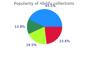
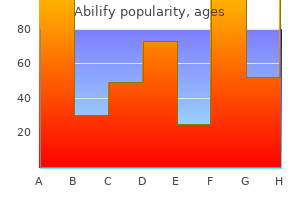
Abilify

Abilify
Abilify dosages: 20 mg, 15 mg, 10 mg
Abilify packs: 30 pills, 60 pills, 90 pills, 120 pills, 180 pills, 270 pills, 360 pills

The greatest threat is the depth of this tumor definition of depression in geography buy cheap abilify 20 mg online, which extends practically to the ventricle bipolar depression medication and weight loss 15 mg abilify generic overnight delivery. Note that this tumor seems to have a denser area (which is cheap to count on could be noninfiltrative) depression test dysthymia abilify 20 mg order free shipping, and a less dense space (more more probably to be infiltrative). We ought to be very involved concerning the lateral part given its proximity to important tracts. The quantity of reorganization on this case was remarkable, and an aggressive resection was potential with no neurologic sequelae. You ought to swap usually during the resection to make sure you are monitoring both. As I even have repeatedly expressed, gliomas are a unique illness, and often a sensible route by way of the brain is better than a slick route which avoids transgressing the brain but supplies poor or disorienting angles, or dangerous useful preservation options. The key steps are to determine the motor planning areas and motor areas, and to work slowly to approach them and educate tumor from them. Note that the minimize proceeds vertical and parallels the course of the sensorimotor system as allowed by the mapping. The preoperative picture, as usual in these circumstances, is complex, and defining goals is crucial in a case like this. This is unfortunately not the only time I even have seen this ominous sample of spread. The aim was to make a posterior frontal sort disconnection as absolute best given the useful anatomy, and to carry out a big anatomic resection of as a lot of the T2 change as potential. Of observe, the patient had important preoperative weak point from this tumor, but was not hemiplegic, being weaker within the leg than the arm. Our goal is to attempt to save whatever operate we can on this case, recognizing that with out an aggressive turn of occasions, his motor prognosis is poor. Note in the post-resection photographs that this cut is kind of irregular and complicated, and this is due to the need to define acceptable secure paths to minimize parallel to the motor planning system, which may be fairly complex within the coronal aircraft. The motor system is splayed outward around the tumor and contibutions to the corticospinal tract are pushed quite far anteriorly. An further noteworthy feature is the truth that at first look it seems to be crossing the midline, but in reality this is merely an enlargement of the midline frontal lobe herniating beneath the falx. The coronal pictures show that the callosum is basically uninvolved but is merely pushed by the tumor bulk. One factor this case highlights the want to get these tumors underneath control if in any respect possible, as early as attainable. Despite his inability to participate with motor duties, we used direct stimulation to establish and preserve motor cortices and descending motor fibers, while resecting the enhancing portion of the tumor. His perform improved to 4/5 the next day, but we may solely protect motor operate in him for about 6 months earlier than the tumor destroyed it. As traditional, the enhancing part of the tumor underestimates the T2 adjustments, and far of the T2 change is in the motor strip and never value going after. The incision looks just like the earlier case because it had been equally resected. The tumor was clearly seen upon opening the dura, and the markers demonstrate areas the place we were capable of stimulate motor responses with a cortical stimulator. We used this technique to outline boundaries and primarily resected abnormally showing areas. The resection is proscribed to the enhancing area however with that as the goal this scan looks good. We have been able to protect his remaining operate at the repeat surgical procedure with our commonplace techniques. First, note that the enhancement is mainly in the premotor areas with extension into the deep white matter. As all the time, I attempt to work a margin around the tumor as much as I can beneath the belief that there virtually at all times are tumor cells in this T2 change across the tumor, and by taking it out, if functionally possible, I am reducing tumor burden to its lowest cheap quantity. Finally, the cortical entry path for the earlier resection begins relatively premotor, and in a conventional view of the brain where cortex comes first, this must be ok to keep away from motor problems. This is straightforward to do if the affected person is asleep for the subcortical portion, or if your subcortical angles are off. The two small anterior websites point out premotor sites, where we discovered unfavorable motor events. The extra posterior marker signifies positive motor websites for the face discovered within the anatomic location of the motor strip. Following this, we carried out a posh model of a lateral frontal disconnection. It has to parallel these sites within the coronal plane to respect the coronal orientation of the motor system. Regardless, the steps are ultimately the identical: subpialize the operculum and discover the insula for orientation, defend the arteries, make the reduce with the premotor areas ensuring that the minimize stays oriented roughly parallel with the sulcal orientation, make the superior reduce and then disconnect the deep boundary to unlock and take away the tumor. I think this will account for the more distinguished premotor mapping on this case: within the setting of this harm, more of it may be essential then normal. This tumor has undergone a lesionectomy at another facility as seen, and has recurred within three months of finishing chemoradiation. We are returning to full the resection and obtain a greater margin including the T2 changes in hopes of enhancing his probability of responding to a new agent. He has some points initiating speech, but otherwise is excessive functioning, working full time, and recurrence free at 2 years after this surgery. The relationship with the insula is complex: whereas these gyri are opercula, the Sylvian fissure is quite shallow at this posterior part, and the untrained eye can easily miss the boundary between insula and opercula. This is not to mention the reality that a lot of the arteries on this region are en passage to one important mind space or one other, together with the artery of demise. Minimize contact with the artery of demise, that is the blood supply to the semantic community and the ventral visual stream. The first order of business is to remove any opercula of the junctional area that stimulate unfavorable. This defines the insula, finds the arteries early, and makes the deep tumor into a surface problem. If not realistic, coming into the atrium is kind of helpful at defining the location of deep and harmful anatomy, and should be undertaken early if the optic radiations are gone. Not only is this an easy place to meet critical white matter, but lack of sagittal plane orientation is a straightforward method to enter the thalamus or inner capsule. Obviously, these tumors have a high propensity to have eloquence within part of them, so these figures are idealized variations of the particular surgery. This simplifies this complicated area, especially as you approach the important problem of the deep white matter. Depending on what the functional anatomy allows, you should use a modified anterior occipital, medial parietal, and even posterior temporal disconnections to free the tumor from the lateral white matter system, or observe it into the deep junctional anatomy, if necessary. The cyst and nodule look of this tumor would possibly fool you into considering that this is a pilocytic tumor, or some other benign tumor. The in depth and thick T2 changes alongside the cyst wall let you know this unfortunately probably untrue. As acknowledged in other instances, at minimum, we ought to always attempt to ensure that the enhancement is dealt with first and foremost, and try to handle as a lot of the T2 change as attainable. It is noteworthy, that draining the cyst, and removing its lateral wall, solely addresses a part of the tumor, because the walls of the cyst doubtless are tumor involved brain. Sometimes, mapping these cases requires two levels of subcortical disconnection, one to disconnect and take away the lateral wall, and another to handle tumor in the deep elements of the wall. The proper image exhibits how a lot the mind can fall inward from merely draining a cyst. A less apparent query worth asking in a case like that is how the visual info will attain the semantic areas. If your resection cuts the semantic network from each side, anomia is the outcome. The resection completely removed the enhancement, but there are T2 changes remaining.

This exposes the micro organism to oxygen anxiety 9 months after baby abilify 10 mg generic with visa, which is especially dangerous to the anaerobes during their first 48 hours of growth depression and loneliness test abilify 20 mg order with mastercard. For this cause depression self help order abilify 20 mg mastercard, an acceptable holding system ought to ideally be used along side anaerobic jars. Plates can be removed from the anaerobic jar, placed in an oxygen-free holding system, eliminated one by one for rapid microscopic examination of colonies, after which quickly returned to the holding system. One or two inoculated plates are positioned right into a bag, an oxygen elimination system is activated, and the bag is sealed and incubated. However, with a number of the products, a water vapor film on the inner floor of the bag or the lid of the plate can sometimes obscure imaginative and prescient. In such instances, the plates have to be removed from the bag to observe them for development, and a brand new bag and oxygen removal system have to be used every time further incubation is required. As with the anaerobic jar, plates must be faraway from the luggage to work with the colonies on the bench. An anaerobic holding system should due to this fact be used along side any of the anaerobic baggage. For instance, a biopsy specimen can first be positioned into a sterile screw-capped tube containing sterile saline, which is then positioned in considered one of these Procedures for Identifying Anaerobic Isolates Various methods are available to medical microbiologists for figuring out anaerobic isolates (Table 22. The methodology and level of identification often depend upon the size and capabilities of the laboratory and whether the organism is from a mixed infection or from a sterile website such because the blood or different sterile physique fluid-for instance, synovial, peritoneal, or pleural fluid. Presumptive identifications of microorganisms have turn into extra in style lately primarily due to elevated emphasis on pace and value discount. Several anaerobic bacteria can be recognized in the laboratory utilizing presumptive identification standards, thereby providing physicians with well timed data concerning the presence of anaerobes in scientific specimens. The following sections on identification techniques are based mostly on progressively extra complex procedures that finally can present the genus and species of an anaerobic bacterium. Communication with the physician is important to decide how far to go together with an identification and whether susceptibility testing is required for proper affected person management. Preliminary Procedures When to Examine Primary Plates Because many anaerobic infections consist of a combination of cardio and anaerobic organisms, probably the most actionable data that can be supplied to the physician is a rapid reply about whether or not the specimen contains anaerobes. The doctor can then select the most acceptable empiric antimicrobial agents to deal with the an infection. Plates incubated in an anaerobic chamber may be examined at any time without exposing the colonies to oxygen. However, these incubated in anaerobic jars, baggage, or pouches might be uncovered to the potentially damaging effects of oxygen every time the containers are opened. This could inhibit the growth of strict anaerobes because of the poisonous impact of oxygen. If a holding system is available on the anaerobe workstation and is used at the facet of jars, baggage, or pouches, publicity to oxygen might be minimized, and plates could be removed and examined at 24 hours and then placed immediately into the holding system. As soon as all plates have been examined, those requiring additional incubation are returned to an anaerobic system and incubated for an acceptable interval before reexamination. Anaerobic cultures are routinely held for five to 7 days to permit progress of significantly slow-growing anaerobes and a minimal of 10 days every time Actinomyces is suspected. Colony Morphology and Gram Stain Reaction using a dissecting microscope to observe the nice details of each colony morphotype and decide colonies for isolation is beneficial. Growth of a selected morphotype may be semiquantitated using terms corresponding to light, average, and heavy or a coding system. Indications of the Presence of Anaerobes in Cultures Several clues can alert the scientific microbiologist that anaerobes could also be current on the first plates. Incubating the suspected isolate in aerobic and anaerobic environments determines the precise atmospheric necessities of the organism. By using this method, many alternative organisms may be tested for aerotolerance on a single plate. Presumptive Identification of Clinically Significant Anaerobes A presumptive identification of a bacterium is derived from easy colony and Gram stain observations and the results of several comparatively speedy and inexpensive exams. The subsequent part describes tests, some speedy (results in about 1 hour or less), which would possibly be of value in presumptively figuring out anaerobes commonly encountered in clinical specimens. The heavy mixture of sizes and types of colonies makes the task of isolating and figuring out colonies very troublesome with out the enhancement obtained with a dissection microscope. It is weaker than the fluorescence produced by pigmented species of Porphyromonas and Prevotella and fades utterly if the colonies are exposed to air for five to 10 minutes. To perform a catalase take a look at, a plastic, disposable inoculating loop or picket applicator stick is used to place a variety of the colony onto a small area of a glass microscope slide. A drop of 15% hydrogen peroxide is added; the manufacturing of bubbles of oxygen gasoline is a optimistic result. Among different uses, the catalase test is valuable in differentiating aerotolerant strains of Clostridium (catalase negative) from Bacillus (catalase positive). The spot indole take a look at makes use of a small piece of filter paper saturated with p-dimethylamino cinnamaldehyde. An inoculating loop or wood applicator stick is used to transfer some progress from the culture plate onto the saturated filter paper. Rapid development of a blue or green shade signifies a constructive test end result (production of indole from the amino acid tryptophan), whereas a pink or orange shade indicates a negative take a look at outcome. A constructive response can happen inside 15 minutes, however the tube should be held for up to 6 hours before a real unfavorable reaction is determined. Motility could also be decided by wet mount using either very younger (4 to 6 hours old) broth cultures or 24- to 48-hour-old colonies on agar. Using a wooden applicator stick, a small portion of a colony should be touched to a drop of saline on a microscope slide. A coverslip is positioned over the drop, and the slide is examined with a light-weight microscope. Although the Gram stain is helpful in the preliminary identification of an anaerobic isolate, certain Clostridium spp. To decide the true Gram stain reaction of the isolate, particular potency disks can be used. The disks are positioned on the closely inoculated area of the plate, often the primary quadrant of the subculture plate. Disks of the correct efficiency must be pressed firmly to the surface of the plate to ensure uniform diffusion of the agent into the medium. Clostridium-like organisms are large, unbranched, gram-positive rods, with or without spores. Not all clostridia are massive, and never all clostridia will stain as gram-positive rods. The nitrate reduction disk take a look at is a miniaturized model of the traditional nitrate discount check. The bile disk could also be added to the anaerobic subculture plate whenever the Gram stain reveals the isolate to be a gram-negative bacillus. A biletolerant, gram-negative anaerobic bacillus indicates that the isolate is most likely going a member of the B. Lecithinase cleaves lecithin found in egg yolk, releasing insoluble fat (diglyceride) that produces an opaque zone across the colony. This multicolored sheen can also appear on the surface of the agar in a slim zone across the colony. Organisms that produce proteolytic enzymes (proteases) have a completely clear zone, often quite narrow, around their colonies. Presumptive Identification of Gram-Positive Anaerobes Some gram-positive anaerobes can additionally be presumptively recognized with simple procedures (Table 22. Probably a member of the Bacteroides fragilis group, but could presumably be a Prevotella sp. A quickly rising colony exhibiting smooth swarming (as against the waves noticed with Proteus) and marking as thin rods with subterminal spores is prone to be C. Small, peaked, circular colonies appearing after 24 hours that stain as gram-positive cocci can be thought-about Peptostreptococcus spp. Small, opaque colonies which would possibly be catalase and indole optimistic and stain as coryneform rods may be recognized as C. The presence of huge gram-positive bacilli with colonies exhibiting a double zone of -hemolysis in the absence of organisms recovered on aerobic cultures of the specimen is enough to report a presumptive identification of C. Presumptive Identification of Gram-Negative Anaerobes Using Gram stain outcomes, development characteristics on main plating media. A number of strategies can be used to make a definitive identification (see Table 22.
Establishment of diagnostic cutoff points for levels of serum antibodies to pertussis toxin depression anxiety test 10 mg abilify order visa, filamentous hemagglutinin mood disorder nos 311 buy 15 mg abilify with mastercard, and fimbriae in adolescents and adults within the United States depression test accurate discount abilify 10 mg with amex. Surveillance for travel-associated legionnaires disease-United States, 2005-2006. Outbreaks of respiratory diseases mistakenly attributed to pertussis-New Hampshire, Massachusetts, and Tennessee, 2004-6. Recommended immunization schedule for children and adolescents aged 18 years or youthful, United States, 2017. Surveillance of vaccination coverage amongst adult populations-United States, 2014. National, regional, state, and chosen local space vaccination coverage among adolescents aged 13�17 years-United States, 2015. Infectious Diseases Society of America/ American Thoracic Society consensus tips on the administration of community-acquired pneumonia in adults. Current and emerging Legionella diagnostics for laboratory and outbreak investigations. List the general traits of organisms that belong to the household Enterobacteriaceae. Describe the antigenic constructions of the household Enterobacteriaceae, and explain how these antigens are used for identification. Compare the virulence components of the Escherichia coli strains pathogenic for the gastrointestinal tract and the E. Compare the pathogenesis of the three species of Yersinia most often recovered from humans. Describe the pathogenesis of the clinically relevant family members Enterobacteriaceae. Given the necessary thing reactions for identification, place an unknown organism in its proper tribe, genus, and species. Develop an algorithm for the identification of the clinically significant Enterobacteriaceae. Case in Point A 71-year-old man with diabetes who was hospitalized for diabetic ketoacidosis complained of flank pain and painful urination. However, scientific isolates in acute-care facilities consist primarily of Escherichia coli, Klebsiella pneumoniae, and Proteus mirabilis. It is nonetheless necessary to pay consideration to the opposite species as a end result of in addition they cause vital infectious ailments. This article is split into three major areas: (1) clinically significant enteric species that trigger opportunistic infections, (2) main intestinal pathogens and their related human infections, and (3) methods of identification of these organisms. Among the various organisms in the family Enterobacteriaceae, this chapter focuses solely on the members which were related to human ailments. T General Characteristics the family Enterobacteriaceae, usually referred to as enterics, consists of quite a few various organisms. Unlike other members of the family Enterobacteriaceae, Plesiomonas is oxidase-positive. Currently, a number of traits are still used to place an organism within the family Enterobacteriaceae (Box 19. These media include a number of carbohydrates, similar to lactose and sucrose, which show the power of the species to ferment particular carbohydrates. Fermentation is indicated by a shade change on the medium, which ends from a decrease in pH detected by a pH indicator incorporated into the medium. Nonfermenting species are differentiated by lack of color change, and colonies retain the original colour of the medium. These features have been used initially to differentiate and characterize sure genera. Definitive identification is decided by the biochemical reactions and serologic antigenic structures demonstrated by the actual species. Classification Members of the family are additionally subcategorized into quite a few tribes based mostly on biochemical traits. The use of tribes in classifying the members in this household was proposed by Ewing in 1963 and has been continued and extended. In classifying species into tribes, Ewing grouped bacterial species with similar biochemical traits. The idea of using tribes within the classification of micro organism has been an efficient method of inserting species into groups based Microscopic and Colony Morphology Members of the family Enterobacteriaceae are gram-negative, non� spore-forming, facultatively anaerobic bacilli. Many members of this family possess antigens that can be used in the identification of different serologic teams. These antigens embrace the following: � O antigen, or somatic antigen- this may be a heat-stable antigen positioned on the cell wall. Clinical Significance Members of the household Enterobacteriaceae are ubiquitous in nature. Except for Salmonella, Shigella, and Yersinia, they can be resident microbiota if confined to their natural setting. Some species exist as free-living organisms in water, soil, or sewage, and a few are plant pathogens. Based on the scientific infections that they produce, family members Enterobacteriaceae may be divided into two broad categories: (1) opportunistic pathogens and (2) main pathogens. The opportunistic pathogens are often a part of the same old intestinal microbiota of both humans and animals. However, outdoors their regular body websites, these organisms can produce critical extraintestinal, opportunistic infections. Other organisms could be equally devastating in immunocompromised hosts or when introduced to wounds from contaminated soil or water. These organisms produce infections resulting from ingestion of contaminated food or water or from other sources. Screening of hospitalized patients is beneficial by many public well being organizations to help contain the spread of carbapenemaseproducing bacteria. Serotyping for each O and H antigens is often useful in identification of strains, particularly strains associated with critical enteric illness. The K antigen usually masks the O antigen during bacterial agglutination testing with particular antiserum. They belong to a quantity of serotypes and are immune to the antibacterial exercise of human serum. Conversely, isolates from immunocompromised hosts consist of all kinds of strains. Cytolysins, also typically characterized as hemolysins, can kill immune effector cells and inhibit phagocytosis and chemotaxis of certain white blood cells. Aerobactin permits the bacterial cell to chelate iron; free iron is generally unavailable within the host for use by micro organism. Based on virulence components, medical manifestation, epidemiology, and completely different O and H serotypes, there are five main categories of diarrheagenic E. The serotypes associated with these classes and the features related to the intestinal infections produced by these strains are summarized in Table 19. Poor hygiene, lowered availability of sources of potable water, and inadequate sanitation are major contributing factors within the unfold and transmission of this disease. A excessive infective dose (106 to 1010 organisms) is critical to initiate disease in an immunocompetent host. Protective mechanisms corresponding to abdomen acidity have been described as inhibiting colonization and initiation of disease; achlorhydria, deficiency of hydrochloric acid inside the stomach, appears to be a excessive threat factor. Immunologic assays to detect the two toxins from culture supernatants are commercially obtainable. Enteroinvasive strains produce dysentery with direct penetration, invasion, and destruction of the intestinal mucosa. The clinical infection is characterised by fever, severe abdominal cramps, malaise, and watery diarrhea. The organisms could be simply misidentified because of their similarity to shigellae. The stool incorporates no leukocytes, which distinguishes it from dysentery attributable to Shigella spp.
The organism is proof against depression trigger definition abilify 20 mg discount on-line drying and may be recovered from swabs a number of hours after collection anxiety xr 10 mg abilify fast delivery. Incubation should be at 35� C both in ambient air or beneath anaerobic conditions anxiety job buy abilify 15 mg with amex. If none are found, incubation ought to continue for an additional 24 hours earlier than the culture is reported as negative. False-negative outcomes can occur from overgrowth of regular microbiota and lack of -hemolysis. Suspect colonies could be Lancefield-typed utilizing serologic methods, which gives a definitive, speedy identification, or biochemical tests may be performed. The correlation between presumptive identification using biochemical methods and the speedy definitive serologic technique is excessive. The reactions of some catalase-negative, gram-positive cocci in numerous biochemical exams are outlined in Table 15. Sialic acid appears to be probably the most major factor of the capsule and a crucial virulence determinant. No proof exists that any of these merchandise play a role in the virulence of this organism. It was not until Lancefield defined streptococcal classification in the Nineteen Forties that their role in human disease was recognized. Early-onset disease accounts for about 80% of the scientific circumstances in newborns and is caused by vertical transmission of the organism from the mom. Most infections of infants happen in the first three days after birth, usually within 24 hours. This infection is commonly associated with obstetric issues, extended rupture of membranes, and untimely birth. Early-onset infection often manifests itself as pneumonia and sepsis, and late-onset infection manifests itself as meningitis and sepsis. Late-onset an infection happens between 1 week and 3 months after start and often manifests itself as meningitis. One sort is a young, previously healthy girl who turns into ill after childbirth or abortion; endometritis and wound infections are commonest. In addition, tricuspid valve endocarditis is typically noticed in younger girls undergoing obstetric procedures. The second sort of patient is an aged person with a serious underlying disease or immunodeficiency. The clinical response to antimicrobial therapy is usually poor despite the heavy doses given. These organisms are gram-positive cocci that type quick chains in medical specimens and longer chains in tradition. Presumptive identification is based on antigen detection or biochemical reactions. These exams enable the organism to be readily differentiated from different -hemolytic streptococcal isolates. The definitive identification can be made by extracting the group antigen and demonstrating agglutination with particular anti�group B antisera. Numerous nucleic acid amplification checks for nonenriched scientific specimens and culture-enriched specimens can be found. The largecolony�forming isolates with group C and G and generally group A and L antigens are classified with the pyogenic streptococci. The large-colony�forming -hemolytic isolates with group A, C, G, or L antigens belong to S. The small-colony�forming -hemolytic isolates with group A, C, F, or G antigens belong to the S. Because several antigenic groups are included throughout the species, serotyping of S. It has been related to circumstances of glomerulonephritis and rheumatic fever after infections. Isolates from certain sources-for instance, circumstances of lobar pneumonia-show a predominance of specific capsular types. The capsule is antigenic and could be identified with applicable antisera within the Neufield check. In the presence of specific anticapsular serum, the capsule swells (Quellung reaction). Several toxins are produced, together with a hemolysin, an immunoglobulin A protease, neuraminidase, and hyaluronidase. It is a vital human pathogen that causes pneumonia, sinusitis, otitis media, bacteremia, and meningitis. Of the greater than ninety capsular serotypes, about a dozen account for most pneumococcal pneumonia cases. For a person to contract pneumococcal pneumonia, the organism should be present in the nasopharynx, and the person have to be deficient within the specific circulating antibody towards the capsular kind of the colonizing strain of S. After initial colonization, the micro organism can persist for weeks or months with out causing disease. In some individuals, invasive illness occurs that results in community-acquired pneumonia. Predisposing situations, such as alcoholism, anesthesia, malnutrition, and viral infections of the higher respiratory tract, can lead to pneumococcal illness within the type of lobar pneumonia. The infection begins with aspiration of respiratory secretions, which frequently include pneumococci. The infecting organisms within the alveoli stimulate an outpouring of fluid, which serves to facilitate the spread of the organism to adjacent alveoli. The process stops when the fluid reaches fibrous septa that separate the main lung lobes. Most isolates from pneumococcal lobar pneumonia are capsular serotypes 1, 2, and 3. Pneumococcal pneumonia is characterized by sudden onset of chills, dyspnea, and cough. An contaminated effusion accommodates many white blood cells and pneumococci, that are visible on Gram stain. Even with antimicrobial therapy, mortality is comparatively high (5% to 10%); nevertheless, with out therapy, the mortality fee approaches 50%. Direct smears of the cerebrospinal fluid usually reveal leukocytes and quite a few gram-positive cocci in pairs. Pneumococci may also be concerned in other infections, such as endocarditis, peritonitis, and bacteremia. Consequently, samples for blood tradition are often taken simultaneously with sputum or a fluid aspirate. This vaccine is part of the routine pediatric immunization schedule encompassing 4 doses beginning at 2 months of age. Vaccination is beneficial for these older than 65 years or individuals with long-term health drawback. The vaccine has been successful in lowering the incidence and severity of pneumococcal illness. Epidemiologic surveys report an increase in infections due to serotypes not coated by the vaccine serotypes. Laboratory Diagnosis the cells characteristically seen on Gram stain seem as grampositive cocci in pairs (diplococci). As the culture ages, the Gram stain reaction becomes variable, and gram-negative cells are seen. Clinical isolates and inventory cultures require frequent subculturing (every 1 to 2 days) to guarantee viability. The colonies might intently resemble colonies of the viridans streptococci, and the greatest concern within the laboratory prognosis is distinguishing S. Optochin susceptibility and bile solubility exams are used to accomplish this; the optochin susceptibility check is the extra generally used. Viridans Streptococci Viridans streptococci are constituents of the normal microbiota of the higher respiratory tract, the feminine genital tract, and the gastrointestinal tract. The term viridans means "green," referring to the -hemolysis many species exhibit.

Evaluate the function of the indigenous biota in host protection towards infectious illnesses mood disorder 10 discount 15 mg abilify with amex. Differentiate the mechanisms of infections attributable to true pathogens from infections brought on by opportunistic pathogens anxiety ecards 10 mg abilify cheap otc. Discuss the situations that have to be current or occasions that must occur for a microorganism to cause illness depression test dass purchase abilify 15 mg mastercard. Describe the characteristics of infectious brokers that allow them to cause illness within the host. Describe the elements and mechanisms by which the human host is protected from microbial invasion. Discuss the sequence of occasions in the phagocytosis and killing of an infectious agent. Name the routes of transmission that microorganisms use to provoke an infection in a host, and provides examples of every. Case in Point A 71-year-old man was treated for right decrease extremity cellulitis with a 10-day course of the antibiotic cephalexin. A few days after finishing the course of antibiotics, he started having loose, watery diarrhea. The affected person described having many episodes of diarrhea per day; after 3 days of diarrhea he came to the emergency division. He additionally mentioned that he had developed proper quadrant belly pain over the past 24 hours. While in the emergency department the affected person had five episodes of loose, watery diarrhea. A bacterial tradition of the stool was negative for Salmonella, Shigella, Campylobacter, Yersinia, and Vibrio species, but a polymerase chain reaction assay for the Clostridium difficile toxin B gene was optimistic. It presents the position of the microbial biota at every physique site in the host immune protection and as a supply of opportunistic infections. Factors that determine the composition of the microbiota at completely different physique sites are described. Lastly, this chapter describes factors that may make the host extra vulnerable to infections and how microbes are transmitted. Origin of Microbial Biota Key Terms Adaptive immunity Adhesin Anamnestic immune response Antigen Antibody Bactericidal impact Bacteriocin Carrier Carrier state Cell-mediated immune response Chemotactic agent Chemotaxis Colonization Commensalism Complement Diapedesis Dissemination Endotoxin Exotoxin Fimbriae Fomites Glycolysis Humoral immune response Iatrogenic an infection Immune response Immune system Immunogen Immunoglobulin A (IgA) Immunoglobulin E (IgE) Immunoglobulin G (IgG) Immunoglobulin M (IgM) Immunoglobulins Indigenous microbiota Innate immunity Interferon Lactoferrin Leukocidin Lymphocyte Lymphokine Lysozyme Opportunists Opsonin Opsonization Panton-Valentine Parasite Parasitism Pathogen Pathogenicity Pattern recognition receptor Phagocyte Phagocytosis Pili Receptor Resident microbial biota Respiratory burst Symbiosis Toll-like receptor Transient microbial biota True pathogen Virulence Zoonoses the fetus is in a sterile environment till start. During supply and the first few days of life, the new child is introduced to the various and diversified microorganisms current in the surroundings. Each organism has the opportunity to find an area on or in the toddler into which it can adapt. Microorganisms that discover their area of interest colonize numerous anatomic websites and turn into the predominant organisms. Colonization is development of microbiota in or on a body website with out the manufacturing of injury or notable symptoms. As the infant grows, the microbial biota ultimately turns into much like the microbiota seen in older people. Once established onto or into a particular body web site of the host, microorganisms develop a specific relationship with that host. The host-microbe relationship, relying on the circumstance, could also be one of symbiosis, commensalism, or parasitism. Symbiosis as a organic relationship between two or extra organisms the place both (host and organism) profit from each other may be described as mutualism. Lactobacilli within the urogenital tract of ladies provide a mutual association: the lactobacilli present the host protection by preventing colonization of pathogenic species at that web site whereas they derive nutrients from the host. The parasite Entamoeba histolytica is a pathogenic intestinal ameba that derives vitamins from the host at the expense of the host, inflicting intestinal ulcers and amebic dysentery. Characteristics of Indigenous Microbial Biota Microorganisms which are commonly discovered on or in physique websites of wholesome persons are called normal or indigenous microbiota. The different body websites might have the identical or totally different microbiota, depending on circumstances. Local conditions choose for organisms which are suited to growth in a specific space. For instance, the surroundings found on the dry pores and skin surface is different from the environment found on the moist surfaces within the oral cavity, and so the microbiota is completely different on the two sites. Microorganisms that colonize an space for months or years represent resident microbiota, whereas microorganisms which might be current at a website temporarily symbolize transient microbiota. These The consequence from the interactions between host and pathogen is influenced by numerous factors. To recognize and perceive the concepts involved in the pathogenesis of infectious ailments, knowledge and understanding of the host-pathogen relationship is necessary. This article describes the interactions between the host and infectious brokers within the pathogenesis of illness. Some pathogenic organisms may establish themselves in a host with out manifesting signs. The carrier state may be acute (short-lived or transient) or chronic (lasting for months, years, or permanently). An instance of a persistent service state is present in post�Salmonella Typhi infection. This organism can establish itself within the bile duct and could be excreted in the stool over years. In contrast, Neisseria meningitidis may be discovered in the nasopharynx of asymptomatic people during an outbreak of meningitis. After a number of days or perhaps weeks at most, these people could not harbor the organism, in which case the carrier state would be termed acute. The organisms colonizing different body sites play a big function in offering host resistance to infections. The effectivity of the microbial biota in providing protection to the human host is indicated by the relatively small variety of infections brought on by these organisms in immunocompetent individuals. Nevertheless, these organisms might cause significant, usually critical, infections or may exacerbate existing infections in people missing a fully responsive immune system. Knowing the advantage of the normal microbiota, individuals can ingest probiotics, a suspension of live bacteria that usually colonize the gastrointestinal tract, to reestablish the microbiota. In areas of the body that have a low oxidation-reduction potential, the setting supports solely organisms able to fermentation, corresponding to is seen within the gingival crevices colonized with Bacteroides and Fusobacterium. The environmental circumstances described right here could change with age, nutritional status, illness states, and drug or antimicrobial therapy use. These modifications can predispose an individual to infection by the indigenous biota, a type of infection referred to as an opportunistic infection. For example, two teams at increased threat for gram-negative bacillus pneumonia are diabetics and alcoholics. Antibiotics could cut back a selected population of bacteria, allowing the proliferation of other organisms. An improve in age brings with it a decrease within the effectiveness of the immune response. As a result, the incidence of an infection brought on by opportunistic organisms will increase. As described within the text, antibiotic use may alter the usual biota of nearly any body website. In this case antibiotic use altered the normal bacterial biota of the gastrointestinal tract and allowed Clostridium difficile to trigger an infection. Composition of Microbial Biota at Different Body Sites Human microbiome research that use molecular sequencing strategies to decide which organisms reside in or on the human body have shown that the human host is colonized by a lot of different species of microorganisms. For instance, within the oral cavity alone, roughly 500 different species have been characterised. The effectiveness of the various host defenses is evidenced by the relatively low incidence of an infection in immunocompetent people by members of the similar old or indigenous microbiota. However, infections attributable to members of the microbial biota are regularly encountered amongst immunocompromised sufferers. The scientific microbiologist must be succesful of acknowledge and determine the kinds of microorganisms discovered at the various body websites.

Analysis of eight different strategies for the detection of Helicobacter pylori an infection in patients with dyspepsia depression and symptoms abilify 20 mg discount visa. Validation of a rapid diagnostic strategy for willpower of serious bacterial counts in bronchoalveolar lavage samples depression legere definition purchase abilify 20 mg without a prescription. Evaluation of the Q score and Q234 systems for cost-effective and clinically relevant interpretation of wound cultures depression severe definition buy abilify 15 mg lowest price. Detection of Pneumocystis jirovecii in respiratory specimens by four staining methods. Points to Remember Direct microscopy of materials submitted to the laboratory for identification of infectious organisms offers the primary, final, and greatest opportunity to: Confirm possible infection. Provide a pathway to verify the suspected explanation for an infection Learning Assessment Questions 1. Direct smear examination of medical samples is a rapid means to identify presumptively the etiologic brokers of infectious illness. The presence of an infectious disease course of could be assessed on a direct smear based on which of the following Which of the next stains is greatest used to detect mycobacterial organisms in clinical samples Calcofluor white is a colorless dye that binds with which of the next constructions Detection of Campylobacter species in faecal samples by direct Gram stain microscopy. A fastidiously collected pattern of lower respiratory tree material should be submitted. The presence of purulence (neutrophils or "polys") indicates a process suspicious for infection. The absence of organisms in this usually sterile fluid is a crucial statement. Squamous epithelial cells are local to this specimen sort and make sure that the pattern is amniotic fluid. The bolus of sputum, consisting of mucus with entrapped alveolar macrophages, confirms that decrease respiratory tree materials is present. The sputum is closely coated by contaminating materials from the oropharynx or mouth. The alveolar macrophages and mucus (pink-stained background) are the local supplies from the tracheobronchial tree. Routine tradition of this specimen can grow insignificant oral biota as a outcome of culture is extra sensitive than direct examination. Acridine orange stain may be helpful in clinical settings by which micro organism are low in quantity and gram-negative. Cytocentrifuged sediments commonly have a focus of organisms sufficient for routine microscopy (105/mL). This spiral might manifest in varied sizes relying on the size of the bronchus involved. Spirals may be notably distinguished after an asthmatic episode with bronchial constriction. The gentle protein background and the pigmentcontaining cell are regular material native to the vitreous of the eye. The ability to see this brown pigment cell in smear material is related to the eye trauma. This dimension and type of carbon particle is usually seen in respiratory samples in small quantities and is usually not famous in a smear report. This small carbon particle seen prominently in respiratory samples could be related to smoking crack cocaine. This sort of carbon debris is commonly associated with smoke inhalation from house or other forms of fires. The golden macrophage present contains yellow material related to cigarette smoking. Local materials, alveolar macrophages containing very small, light to golden yellow, refractile but not polarizing particles (arrow). These small, fine particles could be hemosiderin, typically deposited as small particles or other fantastic particles from the setting. This type of large-particle hemosiderin deposition within lung phagocytes is usually related to blood within the lung as seen in heart failure or aspirated blood from large-volume nosebleeds. The presence of gram-positive diplococci, intracellular morphology suggests an antibiotic effect. This is a typical smear presentation of a handled but unresolved pneumococcal pneumonia. Neutrophils cowl the field, the diplococci are largely intracellular and partially digested, and the background amorphous material is gone. Routine bacterial tradition of this sample may be adverse for typical colonies of S. A few colonies could also be found by a careful search among the many contaminating normal biota colonies. Initial antibiotic therapy may be directed toward streptococci and staphylococci (Stomatococcus). Routine bacterial tradition isolated a pure growth of an encapsulated pressure of Streptococcus pyogenes. The heavy amorphous background is protein-rich edema fluid from the capillary mattress damaged by S. The patient subsequently died of the infection regardless of an accurate diagnosis and acceptable antimicrobial remedy. The presence of typical chains of Streptococcus on a background showing purulence with poorly preserved "polys" and amorphous debris is suggestive of hemolytic streptococci with tissue cytotoxicity. Smear morphology is typical for staphylococci, however no staphylococcal colonies had been present on the tradition plates. Careful correlation between direct and culture examinations demonstrated this organism to be the possible reason for an infection. The presumptive report implying or suggesting staphylococci followed by a negative culture report with out rationalization raises doubts about the competence of the laboratory. There was tradition isolation of Staphylococcus aureus from this same abscess 5 days previously. The progress fee of this organism is speedy, and each viable (grampositive, arrow 1) and nonviable (gram-negative) bacilli may be current within the smear materials (arrows 2). Oil faraway from smear, decolorized with acid alcohol, and instantly restained with ZiehlNeelsen acid-fast stain. The affected person had been positioned in respiratory isolation after bodily examination and historical past taking and antituberculosis remedy was instantly begun after receipt of the direct examination report. Gram-positive bacilli, filamentous, beaded, branched, partial acid-fast stain�negative. Grampositive bacilli, filamentous, beaded, branched, partial acid-fast stain�negative. Gram-positive bacilli, filamentous, beaded, branched, partial acid-fast stain�negative (arrows). Gram-negative bacilli, small, pleomorphic, intracellular, extracellular (arrow 1). Ciliated columnar epithelial cells with quite a few small bacilli adherent to cilia (arrows). The smear is consistent with a bacterial density of one hundred and five colony-forming items per milliliter of urine. Morphotype suggests combined infection with streptococci and anaerobic gram-negative coccobacilli. Care have to be taken to not mistake these conidia for streptococci or for yeasts (see Plate 87). Morphology consistent with trophozoites (tachyzoites) of Toxoplasma gondii (see Plate 95). The measurement is taken to separate this oocyst from the 8- to 10-�m oocysts of Cyclospora spp.
Salmonella: investigation update: human Salmonella typhimurium infections associated with exposure to medical and educating microbiology laboratories anxiety keeps me awake abilify 10 mg purchase with mastercard. Department of Health and Human Services mood disorder hormonal imbalance buy cheap abilify 15 mg on-line, Centers for Disease Control and Prevention anxiety jacket for dogs buy discount abilify 15 mg on line. Guidelines for protected work practices in human and animal medical diagnostic laboratories. Hexachlorophene as a element in drug and beauty merchandise for human use: last rule. Safety and effectiveness of well being care antiseptics; topical antimicrobial drug merchandise for over-the-counter human use; proposed modification of the tentative ultimate monograph; reopening of administrative record; proposed rule. Safety and effectiveness of consumer antiseptics; topical antimicrobial drug products for overthe-counter human use; last rule. Evidence-based biosafety: a evaluate of the principles and effectiveness of microbiological containment measures. Sterilization, decontamination, and disinfection procedures for the microbiology laboratory. Prospective, randomized in vivo comparison of a dual-active waterless antiseptic versus two alcohol-only waterless antiseptic for surgical hand antisepsis. A critical review of the literature concerning using povidone iodine chlorhexidine gluconate for preoperative surgical pores and skin preparation. Laboratory security practices associated with potential brokers of biocrime or bioterrorism. Food and Drug Administration perspective on topical antiseptic drug product development. Role of the hospital-based microbiology laboratory in preparation for and response to a bioterrorism event. Occupational exposure to bloodborne pathogens; needlestick and other sharps injuries; final rule-66:5317. Define and differentiate analytic sensitivity and specificity and scientific sensitivity and specificity. The doctor requested that a bunch A streptococcal direct antigen check be performed; the take a look at was adverse for group A streptococci. Laboratories have at all times taken measures to control the testing performed on affected person specimens. It is important to understand that preanalytic, analytic, and postanalytic actions all have an effect on quality. This process involves monitoring all of the components of a system or process (preanalytic, analytic, and postanalytic) and implementing adjustments when suboptimal efficiency is identified. Quality assurance is measured by affected person consequence; hence patient care might be adversely affected before problems are recognized. Individual disciplines and departments were changed with capabilities crucial to affected person care. In the ever-changing health care enviornment, the pursuit of quality continues with ideas that integrate all features of health care. Corrective action should even be recorded when any measurement falls exterior a tolerance limit. This helps prevent temperature fluctuations when the door is opened to read the thermometer. The most efficient methodology is to examine a big batch of thermometers on the identical time and on the temperature ranges likely to be used. A widespread practice in medical microbiology is to check all thermometers at -20� C, 2� to 8� C, 37� C, and 56� C. These correction elements are applied to all values obtained with particular person laboratory thermometers. For most routine work, thermometers that differ by 1� C or extra from the reference thermometer are discarded. A preventive upkeep program have to be established as an additional control measure. Preventive maintenance carried out on tools generally involves duties similar to oiling and cleaning, changing filters, and recalibrating instruments. Keeping an instrument in prime form and functioning at the proper level will enhance its lifetime and help control the quality of the results. Media Quality Control Each batch of ready media must be high quality managed to document sterility and performance. Documentation should show that the media help the expansion of acceptable microorganisms, and if applicable, inhibit progress of particular microorganisms or produce the correct biochemical response. This certificate should be retained for so long as the laboratory makes use of the desired media. Only certain forms of media must be retested by the consumer, often because of complexity or historical past of failure rate. Examples of media that require retesting are chocolate agar, selective media for pathogenic Neisseria, and Campylobacter media. Media not high quality controlled by the laboratory should still endure observation for moisture, sterility, breakage, and look with every lot or shipment received: � Moisture: Plates must be free of moisture before use but ought to never show signs of drying around the edges. Thermometer Calibration Thermometers are calibrated by batch on arrival in the laboratory. Certificates of calibration must be kept for the lifetime of the thermometer or until the expiration date on the certificates; after that date, the thermometer can be recalibrated or discarded. Gas bubbles could be eradicated by centrifugation or by putting the thermometer at a high or low temperature. Equipment Quality Control Equipment used within the scientific microbiology laboratory should be tested for correct performance at intervals applicable for every item. This could be documented on a separate document generally recognized as a media failures log (Table 5. Primary plating media ought to be tested with dilute suspensions of organisms, whereas biochemical media may be examined with undiluted organisms. Reagent Quality Control With few exceptions, reagents ought to be examined on each day of use with each positive and unfavorable controls. Reagents which are documented to have constant and reliable outcomes could additionally be tested less regularly. Reagents which may be opened and used repeatedly, similar to albumin, should be checked day by day for sterility. In any susceptibility system, many variables can affect the accuracy of outcomes, including the following: � Antimicrobial agent efficiency � Agar depth (Bauer-Kirby test) � Evaporation (microtiter dilution) � Cation content � pH � Thymidine content � Instrument failure � Inoculum concentration � Temperature � Moisture (Bauer-Kirby test) � Difficulty in figuring out end points Careful storage of degradable supplies and precision within the implementation of really helpful procedures are obligatory to obtain accurate and reproducible susceptibility results. All outcomes from the 20- or 30-day evaluation should be kept as long as the antimicrobial agent is used or for a minimum of 2 years after discontinuation of use of the agent. Personnel Competency Personnel competency, the power of a person to carry out a task accurately and successfully, is set by use of a wide selection of methods, such as direct statement, evaluation of labor sheets, or written examination. A in style technique used to determine competency is proficiency testing, by which carefully designed samples are given to laboratory scientists as unknowns for the aim of figuring out them. Proficiency testing because it pertains to the laboratory is mentioned later on this chapter. Proficiency samples for demonstrating competency could additionally be purchased commercially or prepared internally. The management organism results have to be evaluated earlier than finish factors are determined on patient isolates. A particular person could also be qualified to put together a slide for staining but might not be able to stain it, or an individual may be certified to prepare and stain a slide however to not read or interpret the smear take a look at results. Personnel should meet certain educational necessities before being permitted to perform at every stage of complexity. The many agencies concerned in accreditation and inspection have completely different necessities and interpretations of competency verification, making this a complicated task for all laboratories. These applications might educate theory or new strategies, current case research, or just provide coaching on new instrumentation. The manual have to be reviewed, signed at least yearly, and revised as wanted by a supervisor. To effectively improve high quality, all staff must perceive the plan and take energetic roles. Vision and Mission Statements Creating a short vision or mission assertion for all staff to be taught may be an efficient software for uniting everybody behind the same cause. Problems are to not be considered as actual issues however as opportunities for enhancements and an opportunity to excel.
In most combined cases mood disorder questionnaire children abilify 10 mg cheap line, I do the temporal facet first depression test legit 15 mg abilify purchase with mastercard, as a result of it makes plenty of room rapidly mood disorder youth 15 mg abilify buy, and this exhibits me where the insula is which assists with the other portions. There can be a deep reduce which separates the tumor from the basal ganglia and the posterior limb of the interior capsule which is totally laterally exposed in the posterior portion. Stimulation mapping with steady subcortical monitoring will permit you to determine what elements of the inferior insula may be safely resected and when to stop. If the tumor is following the uncinate fasciculus into the frontal lobe, you will want to stay within the insula as you head anterior-superior into the orbitofrontal area. You can even enter this by removing the pars triangularis, but this risks disorientation, as having the frontal lobe cortices in place is an effective secure guard towards taking broad angles into important white matter tracts. But there are some aspects of insular resection that are uniform, and never altered by brain mapping, which are mentioned below. This is a typical query I am asked, which should have an apparent reply by now, if you have been paying shut consideration. Thus, I resect the insula until the lateral facet of the resection cavity is roughly parallel with the hippocampus within the sagittal plane. The presence of the hippocampus prevents this from occurring, and serves as a depth gauge. It is easy to become disoriented in the insula, and as soon as you start eradicating tumor, you start to destroy the anatomy, making it very hard to re-orient your self. This might be unnerving the first time you do it, but that is what resecting the insula means. You should discipline your self to not do so, and to follow rule 2, even when which means working in a less comfortable window. Bifurcations are nice locations to overstretch arteries, as often one branch is commonly leaving the fissure and is tethered to the operculum. You will usually have to work inside a decent bifurcation, but you should strongly consider solely doing so long enough to detach the mind instantly within the "V" of this bifurcation, after which use this new mobility to work on either side of the "V" which is often roomier. A temporal lobectomy was carried out as part of a transopercular resection of the insula. The lateral part of the anterior temporal lobe has been eliminated but the insula is sort of massive, the hippocampus is stuffed with tumor, and the tumor extends into the premotor areas. Of observe, we utilized the earlier incision, which was bigger than I would normally do. I decided not to (it was earlier in my series; right now I most likely would try beneath the concept it might be difficult). Spatiotemporal mapping limited the posterior cut, slightly but despite this, this was a complete resection. The obvious method in a case like that is to approach this from the temporal lobe facet as a lobectomy is warranted. The sagittal and coronal pictures show a C-shape of this tumor which is following the uncinate into the orbitofrontal cortex. This should often be followed upward and anterior into the frontal lobe via the insula. Note that with steady subcortical monitoring, we took this resection proper as much as the internal without deficit. I truly have seen this no much less than 15 times in numerous insular gliomas and have by no means seen this space progress with out remedy as soon as, even when adopted for a few years. I suspect that that is some type of Wallerian degeneration involving some facet of the posterior thalamic peduncle as I even have followed the sign change on a quantity of cuts in these circumstances and have never seen one which immediately connects the insula and pulvinar with sign change and have never seen one of these combos cross the inner capsule to get there. It may all the time be unfold on a microscopic degree, I acknowledge; however, till I see one progress in the thalamus, I will proceed to leave the thalamic part alone in these circumstances. This tumor is so large temporally that tumor has herniated over the tentorium and is compressing the midbrain. Most of this is temporal, and the correct method is clearly from the temporal side, but the insula must be accounted for. Despite the massive query mark incision from a earlier debulking done outdoors, we ignored this and put a linear incision in the course of it which healed nice. Generally, as much as attainable, I try to do my surgical procedure as if the last surgery never happened. Thus, I almost never reopen the entire earlier incision, or take away the old bone flap. I put my incision and bone flap where I assume it needs to be until completely pressured. The reduce needs to be angled as far posteriorly underneath the speech networks as possible. Note that we discovered an anomia website which restricted our posterior extent of the reduce, however we angled beneath this and turned the mattress to be able to work way again to possible. From this, we anticipate to be capable of put our minimize posteriorly enough to attain again and remove the medial temporal lobe fairly well. Invasion of the basal ganglia is uncommon in insular tumors; however, in my expertise, resecting the insula and temporal lobe, and treating the basal ganglia part with chemoradiation is commonly capable of make the basal ganglia portion regress. This tumor is centered in the medial temporal lobe and insula, but concerningly in this case, it seems to be in the subthalamic regions, and has the anterior commissure operating via parts of it. The temporal lobe is removed beneath the bone flap and the insula (whose anterior edge is kind of posterior to the temporal tip) is centered under this flap. This 1 year post-treatment picture demonstrates regression of the tumor from the deep buildings. I am cautious medially with this high threat artery scenario, but resected the remaining, leaving some tumor medially. This tumor is filling the temporal lobe resection cavity, filling the insula, and following the uncinate fasciculus. The coronal images counsel that this tumor may be on prime of and behind the M1, and the T2 pictures suggest that there are small arteries throughout the anteromedial elements of the tumor. The giant question mark incision of the earlier surgical procedure, left us unable to totally ignore it and make a vascularly acceptable skin incision. The temporal lobe minimize was a type of J-cut, as described in Chapter 15, to reestablish a posterior temporal division airplane. This continues to be a very good cytoreductive surgery, despite not being 100 percent complete resection. The resection involved a temporoinsular kind strategy, using the anterior resection cavity as a information to know once we are anterior enough. This patient had previously undergone two surgical procedures with Professor Yasargil for a frontal low grade glioma, which explains the massive resection cavity. The plan is for a temporal insular glioma which ends within the previous resection cavity. There was a earlier pterional kind incision, which we used the bottom half of to expose the temporal lobe and insula. Interestingly, the ultimate web site of recurrence after this surgical procedure was not the edge of the resection cavity, however the diagonal band of Broca and basal forebrain. The amygdala is a pathway of spread to the opposite aspect and into unresectable areas, and this could give pause to anybody who leaves concerned amygdala behind to forestall "mild memory problems. This method concerned an anterior temporal lobe resection, however our work focused on the frontal facet to get access. Obviously the difference is that part of this tumor might be within the insula, but much of this is anterior to the insula. As seen in the operative images, we had been able to make an aggressive lateral frontal cut which made the boundaries of the tumor clear. Note that on the postoperative images that the enhancement has been fully removed. The frontal opercula have been eliminated in order that we could attain as superior as possible and to tackle as a lot of the premotor and motor region tumor that the useful anatomy would enable underneath direct visualization. It would be unwise to try to do this all from the temporal facet, as this would involve blindly sweeping tumor from the motor network blindly under the frontal opercula. This shows the boundaries of his insula and its relationship to the temporal lobe. I do this train in each case to ensure that I even have a transparent thought of which opercula need to come off to expose the round sulcus boundaries and the place the crucial opercula are so I expose them.


