Tadalafil

Tadalafil
Tadalafil dosages: 20 mg, 10 mg, 5 mg, 2.5 mg
Tadalafil packs: 10 pills, 20 pills, 30 pills, 60 pills, 90 pills, 120 pills, 180 pills, 270 pills, 360 pills

Tonsillar and other tissue homogenates can be examined for the presence of the irregular prion protein by enzyme immunoassays erectile dysfunction drugs at cvs 5 mg tadalafil order otc. These have been used in numerous studies and the development of diagnostic checks is essential not only to make a analysis but also from a public health standpoint to prevent infection erectile dysfunction doctor houston buy discount tadalafil 2.5 mg line, as transmission by blood and blood merchandise has been reported erectile dysfunction daily medication tadalafil 5 mg proven. Clinical diagnostic standards embody mind imaging and particular biomarkers in the cerebrospinal fluid. Lessons from kuru Kuru is a condition that was recognized with cannibalistic behaviour in Papua New Guinea. They are resistant to nearly all disinfectant procedures and elicit minimal immune responses. Prions can come up by mutation and hijack normal protein-folding management, producing irregular molecules that are immune to enzymes. Prions can cross from one species to one other, and have crossed from animals to humans. Genetic characteristics of potential hosts seem to play an important function in determining the course of disease after exposure. This has led to a quest to understand more fully the total range of microorganisms current in the host, collectively referred to as the microbiota and its genetic content material � the microbiome. Preceding guide chapters have focused totally on organisms which would possibly be illness brokers. Small numbers may be present in wholesome individuals, but their presence in large numbers is often associated with pathological adjustments. The first part of this chapter considers members of the microbiota found in the normal wholesome individual, in some circumstances necessary for regular functioning of the human physique however capable of cause illness under sure circumstances. Their relationship with the host makes an interesting comparability with that of species that are thought of as true parasites or pathogens discussed later on this chapter within the broader context of symbiotic relationships and the evolution of host�parasite relationships. Although bacteria are the most important contributors to the microbiome, viruses, fungi and protozoa are also often present in healthy individuals, however are far much less frequent. In addition, breast-fed infants have lactic acid streptococci and lactobacilli of their gastrointestinal tracts, whereas bottle-fed youngsters present a a lot greater variety of organisms. Thus, the human physique could be thought of as a posh of microenvironments with attribute differences in microbial composition. In this context, organisms current at a given body website of least 95% of individuals are thought-about to characterize a core microbiome whereas more minor fluctuating organisms characterize the variable microbiome. Staphylococcus epidermidis is considered one of the commonest species, making up some 90% of the aerobes and occurring in densities of 103�104 / cm2; Staph. Anaerobic diphtheroids occur beneath the pores and skin floor in hair follicles, sweat and sebaceous glands, Propionibacterium acnes being a well-known example. Changes within the pores and skin occurring throughout puberty typically lead to increased numbers of this species, which can be related to pimples. The microbiome is acquired rapidly throughout and shortly after start and modifications repeatedly all through life the organisms present at any given time are influenced by the age, diet and setting of the individual. They are rare on dry skin, however can cause infection in moist skinfolds (intertrigo). The pharynx and trachea carry their own microbiota Microorganisms in the pharynx and trachea may embody both - and -haemolytic streptococci as nicely as a variety of anaerobes, staphylococci (including Staph. The respiratory tract is often quite sterile, regardless of the regular intake of organisms by respiratory. However, substantial numbers of clinically normal people may carry the fungus Pneumocystis jirovecii (previously known as P. Both the nose and mouth may be closely colonized by bacteria the vast majority of bacteria here are anaerobes. Common species colonizing these areas include streptococci, staphylococci, diphtheroids and Gram-negative cocci. Some of the cardio micro organism found in healthy individuals are doubtlessly pathogenic. The mucous membranes of the mouth can have the same microbial density as the big intestine, numbers approaching 1011 / g wet weight of tissue. In the intestine the density of microorganisms will increase from the stomach to the large gut the abdomen normally harbours solely transient organisms, its acidic pH offering an efficient barrier. However, the gastric mucosa may be colonized by acid-tolerant lactobacilli and streptococci. The higher gut is simply frivolously colonized (104 organisms / g), but populations enhance markedly within the ileum, the place streptococci, lactobacilli, Enterobacteriaceae and Bacteroides could all be present. The vast majority (95�99%) is anaerobes, Bacteroides being especially common and a significant element of faecal material; E. Plaque is a movie of bacterial cells anchored in a polysaccharide matrix, which the organisms secrete. Entamoeba coli) and these could be thought-about as part of the variable microbiota, despite being animals. Ways during which the microbiota inhibits potential pathogens include the following: � Skinbacteriaproducefattyacids,whichdiscourageother species from invading. Gut bacteria also launch organic acids, which may have some metabolic value to the host; in addition they produce B nutritional vitamins and vitamin K in quantities which are massive enough to be valuable if the food regimen is deficient. The antigenic stimulation supplied by the intestinal flora helps to ensure the traditional development of the immune system. The urethra is frivolously colonized in both sexes, but the vagina supports an extensive presence of micro organism and fungi the urethra in each sexes is comparatively lightly colonized, though Staph. In the vagina, the composition of bacterial and fungal microbiota undergoes age-related adjustments: � Before puberty, the predominant organisms are staphylococci, streptococci, diphtheroids and E. Advantages and drawbacks of the microbiota Studies of the microbiome have confirmed the good factor about various species to the host the contribution of such species to host health is immediately shown in cases of dysbiosis, the disruption or disturbance of the microbiota. Broad-spectrum antibiotic therapy can drastically cut back the presence of helpful microbiota, and the host may then be over-run by introduced pathogens or by overgrowth of organisms normally current in small numbers. After therapy with clindamycin, overgrowth Studies of germ-free animals underscore the importance of the microbiota Germ-free animals are inclined to reside longer, presumably due to the complete absence of pathogens, and develop no caries (see Ch. However, humans acquire microbiota throughout and immediately after start, with the accompaniment of intense immunological activity. As noted earlier, overgrowth by potential pathogens can happen when the composition of the microflora changes. Under these situations, the potential pathogens benefit from the chance to enhance their inhabitants measurement or invade tissues, so changing into dangerous to the host. An account of diseases related to such opportunistic infections is given in Chapter 31. The most advanced bodies, those of birds and mammals (including humans), provide the most various environments and are essentially the most heavily colonized. Relationships between two species � interspecies associations or symbiosis � are therefore a relentless characteristic of all life. Many components influence the outcome of a specific affiliation, and organisms could also be pathogenic in a single state of affairs however harmless in another. Symbiosis has no overtones of benefit or harm and includes a broad range of relationships. Three broad classes of symbiosis � commensalism, mutualism and parasitism � can be identified on the premise of the relative profit obtained by every partner. None of these categories of association is restricted to any particular taxonomic group. Most species are independent of other species or depend on them solely temporarily for meals. May be dangerous if tissues broken (surgery), gut flora adjustments (antibiotics), or immunity reduced. Mutualism Mutualistic relationships present reciprocal advantages for the two organisms involved Frequently, the connection is compulsory for no much less than one member, and could additionally be for each. Good examples are the bacteria and protozoa residing in the stomachs of domestic ruminants, which play an essential role in the digestion and utilization of cellulose, receiving in return both the surroundings and the nutrition essential for his or her survival. Parasitism is a one-sided relationship in which the advantages go only to the parasite, the host offering parasites with their physicochemical setting, their meals, respiratory and different metabolic needs, and even the alerts that regulate their growth. Although parasites are considered essentially harmful, this is a view colored by human and veterinary clinical medication, and by the outcomes of laboratory experimentation. It may mirror selection of an increased degree of genetically determined resistance within the host inhabitants and decreased pathogenicity within the parasite (as has happened with myxomatosis in rabbits). Thus, like the opposite classes of symbiosis, parasitism is inconceivable to define completely except in the context of clear-cut and extremely pathogenic organisms. Commensalism In commensalism, one species of organism lives harmlessly in or on the body of a bigger species At its simplest, a commensal affiliation is one in which one species of organism makes use of the physique of a larger species as its physical setting and may make use of that surroundings to purchase vitamins.
The growth of scrapie in sheep shows strong genetic influences loss of erectile dysfunction causes generic 2.5 mg tadalafil otc, some breeds being rather more resistant than others intracorporeal injections erectile dysfunction tadalafil 20 mg discount, and similar genetic results have been shown in mice erectile dysfunction in early age 10 mg tadalafil buy mastercard. These mixtures of host and prion variation result in a spectrum of disease onset and severity. If the load of the latter will increase it may possibly lead to a rapid neurodegenerative phenotype. With the exception of those cases where prions arise by mutation, transmission and spread of prion illness require exposure to the infective agent. Ways by which this could happen embody eating contaminated meals material, use of contaminated medical products (blood, hormone extracts, transplants), the introduction of prions from contaminated devices during surgical procedures, as prions bind strongly to steel surfaces, and presumably mother�fetus transmission throughout pregnancy (although not one of the tons of of infants born to mothers with kuru developed the disease). In these instances, prions survive digestion and are taken up across the intestinal mucosa. Most of these infectious brokers have mutations at amino acid residue 129 of the prion protein, which are thought to trigger conversion of the protein into the pathogenic type. The fatality price fell from over 200 per yr in the late Nineteen Fifties to 6 per year in the early Nineties. A study investigating suspected kuru cases between 1996 and 2004 recognized 11 infected individuals. The minimal estimated incubation periods in this group ranged from 34 to 41 years, the range in males being from 39 to a minimal of 56 years. That quantity was a lot lower than had been predicted by mathematical modellers in the Nineties. Issues surrounding diagnostic exams include assay sensitivity and specificity, resulting in issue in comparing studies. However, little success has been reported using numerous completely different agents including antimalarials and antibiotics that had been proven to reduce the growth of irregular prion protein deposits in vitro. Humanized versions of antibodies that bind to PrP that had exercise on prion-infected nerve cells rising in vitro have been ready for a clinical trial. The speculation was that the traditional type of PrP required for prions to grow could be removed. Immunomodulation and mucosal immunization could additionally be potential therapeutic and preventative approaches, particularly because the alimentary tract is more probably to be the main route of transmission. Clinical appearances usually indicate the probable incidence of prion illness and this can be confirmed histologically submit mortem. Like all animals, people support an extensive commensal microbiota on the skin, within the mouth and in the alimentary tract. The majority of these microbes are bacteria, and their relationship with the host may be extremely specialized, with specific attachment mechanisms and precise environmental requirements. Conversely, the power of the intestinal microbiota to stop colonization by more pathogenic species could additionally be thought-about mutualistic. Some teams, similar to viruses, are exclusively parasitic (see below), but the majority embrace both parasitic and free-living representatives. We can see, then, that parasitism has been an evolutionary success; as a way of life, it must confer very appreciable benefits. At its easiest, host management is restricted to providing the cell surface molecules needed for parasite attachment and internalization. Many parasites, from viruses to protozoa, rely on the popularity of such molecular indicators for his or her entry into host cells, and this course of offers the set off for their replicative or reproductive cycles. Other parasites, primarily the eukaryotes, require extra comprehensive and sophisticated signals, typically a complex of signals, to provoke and regulate their entire developmental cycle. The complexity of the sign required for growth is probably one of the components determining the specificity of the host� parasite relationship. Where the supply of one of many indicators entails that parasite improvement can happen in just one species, host specificity is excessive. Where many host species are able to providing the required signals for a parasite, specificity is low. Parasitism has metabolic, dietary and reproductive advantages the obvious benefit of parasitism is metabolic. The parasite is offered with a big selection of metabolic necessities by the host, often at no energy price to itself, so it can devote a large proportion of its personal assets to replication or replica. This one-sided metabolic relationship reveals a broad spectrum of dependence, both within and between the various groups of parasites. Some parasites are totally dependent upon the host, whereas others are solely partly dependent. They are obligate parasites, possessing the genetic data required for production of new viruses, but not one of the mobile machinery necessary to transcribe or translate this information, to assemble new virus particles or to produce the energy for these processes. The host supplies not only the basic constructing blocks for the production of recent viruses, but additionally the synthetic equipment and the vitality required. Virusesthereforerepresent the final word parasitic condition and are qualitatively completely different from all different parasites in the nature of their relationship with the host (see Ch. The foundation for the elemental distinction between viruses and different parasites is the difference between virus group and the mobile organization of prokaryotic and eukaryotic parasites. Non-viral parasites have their very own genetic and mobile machinery, and multienzyme methods for impartial metabolic exercise and macromolecular synthesis. The diploma of reliance on the host for nutritional necessities varies significantly and follows no constant pattern between the assorted teams, nor does it comply with that smaller parasites are likely to be extra dependent. All, after all, obtain nutrition from the host but, whereas some use macromolecular material (proteins, polysaccharides) of host origin and digest it utilizing their own enzyme methods, others depend on the host for the process of digestion as nicely, being in a position to take up only low-molecular-weight materials (amino acids, monosaccharides). Nutritional dependence may embrace host provision of progress elements that the parasite is unable to synthesize itself. Disadvantages of parasitism the most obvious drawback of parasitism arises from the reality that the host controls the event of the parasite. No development is feasible with no suitable host, and many parasites will die if no host becomes out there. For this cause, several diversifications have advanced to promote prolonged survival in the outdoors world and so maximize the possibilities of profitable host contact. Nevertheless, where parasites fail to make contact with a number, their powers of survival are in the end limited. In many, parasitism likely arose as a consequence of unintended contacts between organism and host. Of many such contacts, some would have resulted in prolonged survival and, underneath beneficial nutritional circumstances, prolonged survival would have been related to enhanced replication, giving the organism a selective benefit within the surroundings. For instance, parasites of blood-feeding arthropods have ready entry to the tissues of the animals on which the arthropods feed. Where the parasite becomes specialised for the non-arthropod host it may lose the ability to be transmitted by blood feeding. Parasite development can be controlled by the host the benefit that parasitism confers in reproductive terms makes it very important to coordinate parasite development with the supply of appropriate hosts. Many strains of evidence suggest that mitochondria of modern eukaryote cells developed from micro organism that established symbiotic (mutualistic) relationships with ancestral cells. Initially, the organisms involved would have needed to be facultative parasites, able to life each within or outside host organisms (many pathogenic micro organism nonetheless have this property. Legionella, Vibrio), but selective pressures would have forced others into compulsory parasitism. Such events are after all speculative, but are supported by the close relationship of enteric micro organism such as E. Subsequent survival of the pathogen would rely upon the possession of surface or metabolic properties that prevented digestion and destruction by the host cell. The success of intracellular life can be measured not solely by the massive number of bacteria which have adopted this habit, but in addition by the extent to which some organisms have integrated their biology with that of the host cell. The pathway of virus evolution is unsure Clearly, parasitism by micro organism, which are undoubtedly historical organisms (they can be traced again 3�5 billion years in the fossil record), depended upon the evolution of upper organisms to act as hosts. Whether the identical is true of viruses is open to question, and relies upon upon whether or not viruses are considered primarily or secondarily simple. A third different is that viruses had been never something aside from fragments of the nuclear materials of other organisms and have in impact all the time been parasitic. Eukaryote parasites have developed via unintended contact the evolution of parasitism by eukaryotes is prone to have arisen much as it may have carried out in prokaryotes.
Diseases
Adduction Thumb extension and flexion happen within the frontal (coronal) airplane when standing in anatomical place erectile dysfunction diabetes permanent 2.5 mg tadalafil purchase with visa. Palmar interossei are also found between metacarpals and adduct digits two erectile dysfunction pump medicare 20 mg tadalafil buy free shipping, 4 impotence over 40 5 mg tadalafil discount mastercard, and five. The space is occupied by the median nerve and long tendons of flexor digitorum superficialis (4), flexor digitorum profundus (4), and flexor pollicis longus (1). Prior to entering the carpal tunnel, the median nerve gives off a cutaneous department to the skin of the lateral palm-palmar cutaneous V. Bands of the palmar aponeurosis associated with the fourth and fifth digits start to shorten and fibrose, causing these digits to transfer right into a flexed position. After passing through the carpal tunnel, the median nerve divides into three to 4 palmar digital nerves and the recurrent department of the median nerve to serve the thenar muscles; lumbricals (1 and 2); and the pores and skin overlying digits one, two, three, and half of four. Compromise of the median nerve inside the carpal tunnel may trigger downstream motor and sensory impairments. The ulnar nerve innervates the skin over the medial palm and dorsum of the hand and digits 5 and half of four. Green region is equipped by the median nerve, yellow region is provided by the radial nerve (superficial branch), and blue area is supplied by the ulnar nerve. Symptoms embrace numbness and tingling within the lateral three and a half digits, pain, thenar eminence atrophy, and decreased thumb power. Surgical launch of the flexor retinaculum may relieve stress on the median nerve. Transverse view m Carpal tunnel (colored blue) contents include median nerve and tendons of flexor pollicis longus and flexor digitorum superficialis and profundus. This kind interphalangeal of lesion may happen because of a distal joints humeral fracture or as an entrapment syndrome because the median nerve travels between the two heads of the pronator teres muscle ("pronator syndrome"). Clinically, a patient with a median nerve lesion at the elbow presents with the lack to flex digits one by way of three utterly ("hand of benediction"). Thenar wasting and loss of thumb opposition would even be current over time ("simian" or "ape hand"). In some instances, a lesion of the anterior interosseous department of the median nerve could happen. The other long flexors of the forearm are spared, as are the thenar muscles and median nerve cutaneous sensory distribution. A sequence of ligaments and capsular buildings assist these higher limb joints and allow a broad range of motion. Glenohumeral: this is a synovial ball-and-socket joint between the top of the humerus and shallow glenoid fossa. It is supported by a joint capsule, glenohumeral and coracohumeral ligaments, and rotator cuff muscles. Movement: this joint permits flexion, extension, abduction, adduction, medial rotation, lateral rotation, and circumduction. Overuse, as in baseball pitchers, or direct injury can drive the humeral head in an anterior path, particularly if the shoulder is extended, kidnapped, and externally rotated. Depending on the mechanism of injury and amount of pressure, dislocation on this path could be paired with a glenoid labrum tear. Posterior glenohumeral dislocations are less widespread because the posterior capsule is additional supported by rotator cuff musculature. The axillary nerve and circumflex humeral vessels are weak in this kind of injury. Anterior glenohumeral dislocation A/Pview Plain film A/P radiograph of anterior glenohumeral dislocation. Yellow arrow signifies direction of dislocation of humeral head from the glenoid fossa. Note that the acromioclavicular joint is unaffected in a glenohumeral dislocation. Blood provide: this joint is equipped by anterior and posterior circumflex humeral arteries and suprascapular, axillary, and lateral pectoral nerves. Associated bursae: these embrace the subacromial/subdeltoid and subscapular bursae. Acromioclavicular: this can be a synovial aircraft joint between the acromion and the acromial end of the clavicle. It is supported by a joint capsule, instantly by the acromioclavicular ligament and indirectly by the coracoclavicular ligament (conoid and trapezoid portions). Movement: this joint permits rotation of the acromion on the clavicle during scapular movement. Blood provide: this joint is provided by branches of the suprascapular and thoracoacromial arteries and supraclavicular, lateral pectoral, and axillary nerves. Sternoclavicular: this can be a synovial saddle joint (functionally ball and socket) between the manubrium and the sternal finish of the clavicle. It is supported by a joint capsule and anterior and posterior sternoclavicular, interclavicular, and costoclavicular ligaments. Rupture of the coracoclavicular ligaments also can occur, which will increase the severity of the shoulder separation. In this case, the load of the higher limb pulls it inferiorly, accentuating the purpose of the acromion by way of the lateral shoulder pores and skin and giving the shoulder a much less rounded, squarer look. Movement: this joint permits clavicular elevation, despair, and anterior/posterior motion throughout humeral motion. Blood supply: this joint is provided by branches of the inner thoracic and suprascapular arteries and medial supraclavicular and subclavius nerves. It allows for flexion and extension actions and is equipped by anastomoses around joint (from brachia! Humeroradial: this joint articulates between the capitulum and radial head and is supported by radial collateral and anular ligaments (also helps proximal radioulnar joint by sustaining position of radial head in radial notch of ulna). Humeroulnar: this joint articulates between the trochlea and trochlear notch and is supported by the ulnar collateral ligament (anterior, posterior, and oblique bands). Forearm Synovial pivot-type joints allow for pronation and supination of the forearm. This usually occurs in younger kids as a result of a forceful pull on the upper limb. The radial head turns into dislocated and seems as a palpable mass in the cubital fossa. The biceps brachii pulls the radial head anteriorly and superiorly as soon as dislocated. Children will commonly splint their injured arm towards their bodies and complain of ache. The supply of pain is believed to be the torn anular ligament being pinched within the joint. The joint may be lowered by "screwing" the head again in place-supinating the forearm whereas the elbow is flexed. Lateral plain film radiograph indicating anterior displacement of radial heads in reference to humerus (yellow line). This repair is often referred to as "Tommy John surgical procedure" (after a well-known main league pitcher). Postoperative changes could be seen within the epicondyle and the chic tubercle after Tommy John surgical procedure on the T1 -weighted photographs (yellow arrows). Proximal radioulnar: this joint articulates between the radial head and radial notch of the ulna and allows for rotation of the pinnacle in the notch. It is supported by the anular ligament and supplied by elbow anastomoses and musculocutaneous, median, and radial nerves. Distal radioulnar: this joint articulates between the ulnar head and ulnar notch of the radius with an articular disc. The ulna is comparatively mounted, permitting the radius to transfer throughout pronation/supination. It is supported by weak anterior and posterior ligaments and the interosseous membrane and provided by anterior and posterior interosseous arteries and nerves. At the distal radioulnar joint, the distal radius is parallel to the ulna within the supinated place and crosses anterior and medial to the ulna in the pronated position.

Saline or sodium bicarbonate instillation into the trachea followed by instant suction also helps to scale back the likelihood of such problems erectile dysfunction niacin tadalafil 5 mg trusted. Apnoea Some patients with chronic obstructive airways illness might develop apnoea following restoration of their airway erectile dysfunction pump how do they work discount tadalafil 5 mg without prescription. These sufferers want monitoring and the administration of carbon dioxide through a flowmeter via the tracheostomy if needed erectile dysfunction drugs stendra tadalafil 2.5 mg purchase otc. Speech A pocket book or erasable pad should be provided for the patient to communicate. If the larynx is still functioning, the affected person can be proven how to converse by temporarily blocking the tube while exhaling or utilizing a phonation valve. Patients with a permanent tracheostomy should, if attainable, have a fenestrated tube with a speaking valve incorporated with the inside tube. Swallowing Some sufferers with a tracheostomy experience swallowing difficulty, often because of the condition which necessitated the tracheostomy, however generally because the tracheostomy tube and cuff tethers the larynx, decreasing its elevation when swallowing. Deflation of the cuff will generally improve laryngeal elevation, however some patients could not be capable of swallow solids or liquids without aspirating and require a nasogastric tube for feeding. Replacement or cleansing of the outer tube is normally left for the first 5 days till a observe has turn out to be established, then this ought to be carried out regularly, often monthly or 2 monthly. If a cuffed tube has been used, it must be inflated with the minimal amount of air that forestalls an air leak, and it should have a low-pressure cuff to minimise the chance of tracheal stenosis. A spare tube of similar size, plus the scale under and a tracheostomy dilator must all the time be available at the bedside in case a fast change is critical. A cuffed tube should be out there in the occasion of an emergency when a cuffed tube may be needed for ventilation. The first tube change is often accomplished about 5 days after the tracheostomy and will all the time be carried out by a health care provider and ideally the surgeon who performed the procedure. Whenever the nursing employees perform subsequent tube adjustments, it must be accomplished within the neighborhood of a physician educated in tracheostomy care in case of an issue. Decannulation the tracheostomy tube must be downsized, spigotted and removed as soon as possible. A physiotherapist should assess the patient to guarantee secretions may be expectorated safely prior to removal of the tube. To test this is the case, the affected person should be able to handle with the tube spigotted for a full 24-hour interval, including a period of sleep. They even have a relatively smaller tracheal airway which can be partly blocked by granulation tissue. Surgical closure by excision of the scar tissue and the tracheocutaneous track may be required in some instances. After decannulation, the affected person ought to remain in hospital underneath surveillance for no much less than 2 days, together with in a single day statement. An occlusive dressing ought to be used and the patient educated to splint his or her dressing when talking and coughing to assist within the fistula healing; an assessment must be made to make positive the wound is therapeutic and a light-weight dressing applied. London: Hodder Arnold; 2008;three:2292�2304 Brass P, Hellmich M, Ladra A, Ladra J, Wrzosek A. Multidisciplinary guidelines for the management of tracheostomy and laryngectomy airway emergencies. Effect of early vs late tracheostomy placement on survival in patients receiving mechanical air flow: the TracMan randomized trial. The repaired tympanic membrane is positioned onto the footplate after which the promontory but evaded the spherical window creating a round window baffle or carva minorum (not common). The stapes footplate is fixed and a fenestration of the lateral semicircular canal is carried out (now a historic operation as stapedotomy is now done). The spherical window area of interest is left uncovered and the tympanic membrane reconstructed so that its inferior edge lies on the promontory above the round window, thereby creating an oval window baffle. These are typically academic however do get requested about in examinations and understanding them will give an concept of the method to think about reconstruction when undertaking an operation. Type 1 Reconstruction of the tympanic membrane with a normal ossicular chain (myringoplasty), a typical operation. The tympanic membrane is reconstructed over the malleus remnant and the lengthy strategy of the incus (not very common). The tympanic membrane is repaired to lie immediately on the stapes head (very commom). All the plastic, ceramic, carbon and lots of different artificial supplies fail within the medium term regardless of being positioned in the immune-privileged center ear and are now confined to history. The exception is the stapedotomy prosthesis, but these only work in ears that have got otosclerosis and no other disease. A myringostapediopexy or malleostapediopexy is carried out to create the tympanic membrane or malleus-to-footplate meeting. The incus physique is used with a cup for the stapes head and a strut or hook to the malleus handle just below the tensor tympani insertion. There is an incudostapedial discontinuity secondary to an absent lenticular process and/ or stapes head. An incudostapediopexy utilizing a cortical bone slither is the preferred methodology of reconstruction. Bone cement was used but has now been withdrawn because of issues of safety and poor long-term outcomes. If surgical procedure is to be accomplished, then a realistic prognosis has to be given and a detailed understanding of the course and consequence of the surgery within the regular course of occasions has to be understood by the patient. The patient should contemplate the potential complications and their effects as properly as their frequency. The patient should be warned of the following: � Failure of operation: Graft failure and no hearing enchancment, 20 to 50%, relying on the operation. The lengthy process of the incus appears to have a tenuous blood provide and is definitely eroded by the inflammatory effects of infectious illness. Homografts fair higher and have leads to the region of 70% if the stapes superstructure is current and 50% if it is absent. Complex reconstruction with plastic prostheses and Silastic bands may go better however in a number of specialist hands solely. Most perforations or ossicular deficiencies are greatest left alone and no surgical procedure done. Surgery ought to solely be carried out if the outcomes are definitively higher than the pure historical past of the illness. If the hearing loss could be aided and the ear waterproofed, then no surgery is indicated. The perforation has the sides freshened, the middle ear inspected and the graft inserted deep to the eardrum remnant. The most important thing in any tympanoplasty is having the graft tight up against the medial surface of the eardrum remnant. The most important factor in ossicular reconstruction is having the inserted bone (or artificial material) flippantly touching the stapes and the eardrum and never touching the other middle ear buildings. Probably all profitable tympanic membrane grafts work by being in effect, a scaffold which allows squamous epithelium from the freshened fringe of the tympanic membrane perforation to develop throughout. The ear may need repeated microsuction to compensate for the disrupted squamous migration and wax clearance that the surgery has temporally induced. Diving or repeated head damage (heading a football) could predispose to prosthesis displacement and must be prevented. If artificial ossicle grafts are used, the affected person should be adopted up for the lengthy run as displacement or rejection is likely and revision surgical procedure or listening to aids are commonly required. It can be used to cowl synthetic ossicles to cut back extrusion by way of the tympanic membrane. Cartilage is used in the eardrum both as palisades or often as a butterfly plug. On otoscopy, deposits present as sharply demarcated areas of white opaque, chalk-like materials. Plaques usually occur only within the pars tensa, mostly situated in the anterior or posterior segments, various in size. Middle ear Middle ear involvement is much less common, but when it occurs, it normally accompanied by a perforation of the tympanic membrane.
Combination of biochemistry (commercial kits available) and serotyping required for full identification; important to distinguish enteric fever salmonellae from others erectile dysfunction caused by lack of sleep tadalafil 2.5 mg cheap on line. Detection of circulating antibody (Widal test) could aid analysis of enteric fevers what medication causes erectile dysfunction tadalafil 2.5 mg discount on line. While serotyping (and phage typing of most essential serotypes) is useful for investigation of outbreaks impotent rage random encounter tadalafil 5 mg generic on line, molecular approaches corresponding to pulsed-field gel electrophoresis are extra definitive. Vast majority trigger diarrhoeal illness; very often invasive (particularly S. Widespread in animals; encountered in meals chain (especially in poultry, eggs, meat, milk, and cream). Acquired by ingestion of contaminated meals or person to particular person through faecal�oral route. Antibiotic resistance is an increasing downside in many nations (important implications for travellers). Prevention relies upon upon interrupting faecal�oral transmission and on eliminating opportunities for transmission through the meals chain. Characteristics Laboratory identification Diseases Transmission Pathogenesis Gram-negative rods. Full identification requires use of biochemistry (commercial kits available) and serological checks for O antigens. Intense inflammatory response involving neutrophils and macrophages characteristic. Many strains carry multiple antibiotic resistances, often on plasmids; thus susceptibility testing is necessary. Treatment and prevention Genus Pseudomonas and Related Organisms Burkholderia, Stenotrophomonas and Acinetobacter this group contains a lot of species, a couple of of which are human pathogens, some are animal pathogens and others are necessary pathogens of crops. Species additionally widely distributed and may contaminate the hospital setting and cause opportunist infections. Pseudomonas aeruginosa Characteristics Aerobic Gram-negative rod, motile via polar flagella. Able to make the most of a very big selection of carbon and energy sources and to grow over a wide temperature vary. Does not grow anaerobically (except when nitrate is provided as a terminal electron acceptor). Carriage as a part of the conventional gut flora occurs in a small share of normal healthy individuals and in a better proportion of hospital inpatients. A number of virulence elements have been identified, including endotoxin and exotoxin A, which acts as an inhibitor of elongation factor in eukaryotic protein synthesis. Laboratory identification Diseases Transmission Pathogenesis e30 Copyright � 2019, Elsevier Ltd. Combination antimicrobial chemotherapy based on susceptibility testing is required. Prevention relies upon upon good aseptic practice in hospitals, avoidance of unnecessary or prolonged broad-spectrum antibiotic treatment and prophylaxis. Curved Gram-Negative Rods There are several genera of curved Gram-negative rods containing species that occur in people as pathogens. Characteristics Laboratory identification Curved Gram-negative rods, highly motile by means of single polar flagellum. Grow in alkaline circumstances (can be chosen from other gut flora in alkaline peptone water). Biochemical tests and use of particular antisera required for complete identification. Chromosomally encoded subunit toxin produced after cells bind to intestinal epithelium enters cells and binds to ganglioside receptors activating adenyl cyclase and inflicting fluid loss, leading to huge watery diarrhoea. Prevention of cholera depends upon provision of a clear (chlorinated) water supply and enough sewage disposal. Diseases Transmission Pathogenesis Treatment and prevention Genus Campylobacter Curved Gram-negative rods once categorized as vibrios, campylobacters are primarily pathogens of animals, but a quantity of species additionally cause infections in people. Campylobacter jejuni Characteristics Laboratory identification Slender, curved (seagull-shaped) Gram-negative rods. Full identification by biochemical tests and characteristic antibiotic susceptibility sample. First-line brokers for treatment for invasive illness embody fluoroquinolones or azithromycin. Helicobacter pylori Characteristics Laboratory identification Associated with gastritis and duodenal ulcers; initially named C. Organism in endoscopic biopsy specimens; positive urease test from endoscopic biopsy specimens or labelled urea breath-test additionally very useful. Protease affects gastric mucosa; urease produces ammonia and buffers abdomen acid. Diseases Transmission Pathogenesis Treatment and prevention Gram-Negative Non-Spore-Forming Anaerobes Historically, all short Gram-negative anaerobic rods or coccobacilli have been categorised in the genus Bacteroides and longer rods with tapering ends within the genus Fusobacterium. Recent applications of new strategies to the Bacteroides have resulted in the definition of two additional genera: Porphyromonas and Prevotella. The genus Bacteroides is now restricted to species found among the normal intestine flora. The genus Porphyromonas incorporates asaccharolytic pigmented species, which kind part of the traditional mouth flora (P. Bacteroides fragilis Characteristics Laboratory identification Small pleomorphic Gram-negative rods or coccobacilli. Grows on blood agar incubated anaerobically and in other media designed for isolation of anaerobes. Full identification in the diagnostic laboratory relies on biochemical tests and antibiogram. Intra-abdominal sepsis; liver abscesses; aspiration pneumonia; mind abscesses; wound infections. Endogenous infection arising from contamination by intestine contents or faeces is most common route of acquisition. An anaerobic setting is essential and in combined infections development of cardio organisms most likely helps the expansion of Bacteroides by utilizing up obtainable oxygen. Metronidazole, imipenem, or beta-lactam�beta-lactamase inhibitor combos used in remedy. Many strains produce beta-lactamases and thus susceptibility to penicillin and ampicillin is unreliable. Prevention of endogenous infection is tough; good surgical approach and appropriate use of prophylactic antibiotics are necessary in abdominal surgery. Treatment and prevention Gram-negative Cocci Genus Neisseria this genus contains a number of kind of fastidious species of which two, N. Characteristics Laboratory identification Non-motile Gram-negative diplococci with fastidious development requirements: capnophilic; N. Gram stains of pus or cerebrospinal fluid might reveal Gram-negative kidney-shaped diplococci, often intracellular (in polymorphs). Formerly considered a commensal in the respiratory tract, it has been associated with a selection of infections, including bronchitis, bronchopneumonia, sinusitis and otitis media. Although kind b has been most regularly present in disease, this has changed with the introduction of vaccines towards the type b strains. Capsulate organisms could be agglutinated by particular antisera and detected instantly. Human pathogen unfold by airborne route from circumstances of disease (healthy carriage not documented). Several virulence components, together with tracheal cytotoxin, fimbrial antigen and endotoxin. Macrolides erythromycin or clarithromycin for cases and close contacts of whooping cough. Antibacterial therapy has little impact on medical course, however could cut back infectivity and incidence of superinfection. Vaccine administered to young children in 5 doses together with diphtheria and tetanus toxoids.
Note that the transient fall in pH accompanying the respiratory burst enhances the activity of the cationic microbicidal proteins and defensins erectile dysfunction studies purchase tadalafil 2.5 mg without a prescription. Neutrophil serine proteinases have homology to the cytotoxic granzymes released by cytotoxic T cells erectile dysfunction treatment delhi tadalafil 5 mg without a prescription. Another phagocytic cell erectile dysfunction doctor manila cheap tadalafil 10 mg otc, the eosinophil, is especially rich in cytotoxic granules (Table 15. Five distinct eosinophil cationic proteins are recognized and seem to be notably poisonous to parasitic worms, no less than in vitro. Because of the enormous distinction in size between parasitic worms and eosinophils, this sort of injury is restricted to the outer surfaces of the parasite. The eosinophilia typical of worm infections is presumably an try to deal with these large and virtually indestructible parasites. Lysozyme is an antibacterial molecule that assaults peptidoglycan in the cell wall of micro organism, which is particularly effective in opposition to Gram-positive micro organism where it has easier entry to the peptidoglycan. Activated macrophages have a greatly enhanced ability to kill both intracellular and extracellular targets. The way in which these molecules acquired their sometimes somewhat deceptive names, and the bewildering overlap of function between molecules of quite different structure, are described in detail in Chapter 12. The beneficial results could be direct or extra usually oblique via the induction of some other antimicrobial course of. The name is derived from the demonstration in 1957 that virus-infected cells secreted a molecule that interfered with viral replication in bystander cells. In many instances, this is adopted by secondary binding to different cells or molecules of the immune system. Microorganisms tend to develop extra slowly in vivo than in vitro, which reveals that the host surroundings is mostly hostile. Usually the antibody response continues as long as antigen is present, although some down-regulation may happen in very prolonged responses, presumably in an effort to restrict immunopathology (see Ch. The lifelong immunity that follows many virus infections might usually be due to common boosting by viruses locally, however typically. Such persistence of immunological reminiscence could additionally be due to the non-specific stimulation of memory B and T cells by cytokines during responses to other antigens, a course of referred to as bystander activation. A B Affinity It seems self-evident that the next antigen-binding affinity would render antibody more useful, and passive protection experiments have confirmed this. Affinity is decided by each the germline antibody gene pool and somatic mutation in individual B lymphocytes, and seems to be underneath genetic control which is separate from that controlling the entire quantity of antibody made. For example, T-independent antigens similar to some polysaccharides induce mainly IgM antibodies, T cells being required for the change to IgE and helpful for IgG switching. IgG antibodies to polysaccharides tend to be primarily IgG2, whereas IgG antibodies to protein are primarily IgG1. The poor improvement of IgG2 in kids beneath the age of about 2 years explains their lack of response to micro organism with polysaccharide capsules. Antibodies to viruses are predominantly IgG1 and IgG3, and those to helminths are IgG4 and IgE. Antigens encountered via the digestive tract induce primarily IgA, which is processed throughout its passage via epithelial cells to sIgA, the only sort of antibody that can function in this protease-rich intestinal surroundings; the gut microbiota might induce T-independent IgA class switching by way of interactions with intestinal epithelial cells. Speed, amount and length Because of the cell interactions concerned and the necessity for proliferation of a small number of specific precursor lymphocytes, a primary antibody response may be dangerously gradual in reaching protective levels. Experiments with specifically bred lines of mice recommend that the pace and size of an antibody response is underneath the management of a lot of genes, and the same is undoubtedly true in people. To help present cover while specific antibodies are produced, there are some pre-existing pure antibodies that are normally low-affinity and cross-reactive IgM antibodies. It might bodily interfere with the receptor interplay necessary for microbial entry. This is the basis of many life-saving vaccines against viruses or bacterial toxins. Such vaccines have to generate high-affinity antibodies, and T-cell assist will be wanted. Blocking of attachment and entry could be effective in opposition to all organisms that use particular attachment sites, whether viral, bacterial or protozoal (see Ch. An important exception is these organisms that parasitize the macrophage, such as the virus of dengue fever; here the presence of a low focus of IgG antibody can really improve infection by promoting attachment to Fc receptors (see Ch. A extra subtle blocking impact of antibody is interference with important floor parts of the parasite, notably if these are enzymes or transport molecules. Needless to say, the successful pathogen takes steps to shield such elements whenever potential, as described in Chapter 17. Lysis Lysis of micro organism within the presence of complement offers another handy assay for the presence of antibody (IgG and IgA). However, lysis in all probability plays a significant protecting role in solely a restricted vary of infections, notably these brought on by Neisseria and a few viruses (see Ch. Telling proof for that is the general similarity in the results on the affected person of defects in antibody, complement (up to and including C3) and phagocytic cells (see Ch. The protecting value of agglutination in vivo is difficult to assess; once clumped, most organisms are most likely quickly phagocytosed, but clumps of still-motile trypanosomes could be seen in the blood of infected animals with sufficient serum antibodies. Well A shows agglutination of group A streptococci with latex particles coated with anti-group A antibodies. The free form of IgM adopts a star-like configuration (arrow), as shown within the image obtained with low temperature atomic drive microscopy. Antibody and complement collectively accelerate the clearance of pneumococci from the blood of mice (blue line); depletion of complement permits some opsonization if antibody is present but fails to control the an infection (red line). Of course, the effectiveness of opsonization is dependent upon the phagocytic cell being capable of finishing off the ingested organism. Some examples of the significance of antibody and cell-mediated immunity in resistance to systemic infections are given in Table 15. Antibody-dependent cellular cytotoxicity In the case of larger organisms (worms being the most obvious example), phagocytosis is clearly not a risk. However, several types of cell, having made contact with the parasite through antibody and Fc receptors in the same method as phagocytes do, can inflict injury extracellularly by way of antibody-dependent mobile cytotoxicity. Indeed, the exact method in which antibody protects against infection is, within the majority of circumstances, still unknown. For instance, the enormous production of IgA within the intestine, which may quantity to half of all antibody produced in the physique, suggests the important importance of mucosal safety, and yet deficiency of IgA is relatively widespread and not significantly severe. Once once more, it have to be emphasized that the presence of antibody by no means denotes a protecting function. The greatest indication of the worth of antibody comes from antibody-deficiency syndromes (Ch. This resulted in an inflow of T cells and macrophages into the pores and skin lesions, and a reduction within the number of micro organism. Three Maltese households had kids who had been susceptible to atypical mycobacterial infection (solid symbols), two of whom died (slashed symbols). This mutation introduces a cease codon leading to a non-functional truncated protein. Those with a big skin test response are at elevated danger of growing tuberculosis, showing that robust T-cell responses could be induced during disease development and that a strongly constructive pores and skin check can point out infection somewhat than immunity. However, once more these checks might be optimistic in those with latent tuberculosis or with lively tuberculosis disease. Th17 cells play a role in immunity towards a variety of bacterial infections including Klebsiella pneumoniae, E. The granule contents are delivered into the contact zone between the cytotoxic T cell and its goal cell, and close by cells are spared. Granzymes are discovered as proenzymes in acidic granules where they bind to serglycin; as soon as cleaved by cathepsin they enter the target cell and induce apoptosis. A recent study has proven that, once inside a target cell infected with micro organism or protozoa similar to Listeria or Toxoplasma, granulysin, a third part of human lytic granules, permits granzymes to kill the micro organism and parasites in a process just like apoptosis. In some circumstances, apoptotic blebs launched by apoptotic, infected macrophages could additionally be taken up by dendritic cells. In the early levels of an infection, nonetheless, adaptive immunity can want some assistance. Often, one mechanism is responsible for restoration and one other for resistance to re-infection.
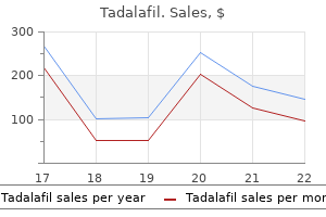
cis-1,2,3,5-trans-4,6-Cyclohexanehexol (Inositol). Tadalafil.
Source: http://www.rxlist.com/script/main/art.asp?articlekey=96321

The muscularis mucosa consists of a clean muscle layer that may not be distinguished erectile dysfunction protocol + 60 days 20 mg tadalafil generic mastercard. Perineum muscle that thickens to form the interior anal sphincter erectile dysfunction nofap purchase tadalafil 10 mg with mastercard, an outer longitudinal layer of easy muscle (may not be prominent) erectile dysfunction 35 year old male discount tadalafil 10 mg amex, and the myenteric (Auerbach) nerve plexus. Lower anal canal: the wall of the lower anal canal also consists of mucosa, submucosa, muscularis externa, and adventitia. The mucosa of the lower anal canal is characterised by a squamous zone as outlined by the type of epithelium present. Located immediately distal to the pectinate line, the squamous zone options nonkeratinized stratified squamous epithelium. The muscularis mucosa consists of a smooth muscle layer, which is most likely not prominent. The epidermis of the anal verge features keratinized stratified squamous epithelium. The dermis consists of dense, irregular connective tissue, blood vessels, lymphatics, myelinated and unmyelinated axons, sensory receptors, hair follicles with sebaceous glands, and circumanal glands (apocrine sweat glands). Boundaries: Each house is certain by the levator ani muscles and anal canal medially, obturator internus muscle and ischial tuberosity laterally, and skin and fascia of the anal area on the base. Contents: the fossae primarily contain adipose, which permits for distension during passage of fecal matter and provides additional support to the anal canal. In each fossa, this neurovascular bundle travels deep to the obturator internus fascia, although the pudenda! Pelvis and Perineum Gluteus maximus (reflected) Superomedial wall of ischioanal fossa Pudenda! To keep away from the spread of infection to adjacent buildings, abscesses on this area must be surgically drained and packed if essential. Males are more probably to develop an abscess and fistula on this area, in comparability with females. The inner iliac arteries course medially, divide into posterior and anterior divisions, and function the principle V. External and inside iliac veins unite to type common iliac veins, which merge to kind the inferior vena cava at roughly the L4-L5 level. Internal iliac posterior division Three branches arise from the posterior division of the interior iliac artery. Lateral sacral: this department can present as paired arteries and programs interiorly alongside the sacrum to enter an anterior sacral foramen. Superior gluteal: the most important of the three posterior division branches, this artery sometimes passes between the lumbosacral trunk (L4-L5) and S1 to exit the pelvis through the higher sciatic foramen. It enters the gluteal area superior to the piriformis and provides gluteus maximus, minimus, and medius and tensor fasciae latae. Internal iliac anterior division Multiple branches arise from the anterior division, which supply thigh, perinea! Obturator: this artery exits the pelvis via a small opening in the obturator membrane to enter the medial thigh and gives off a small acetabular department to the head of the femur. Umbilical: the distal finish of this artery turns into obliterated after birth to kind the medial umbilical ligament, and its proximal end offers off the artery to ductus deferens and a quantity of superior vesical branches to the bladder. Vaginal: Commonly a branch of the uterine artery, this artery provides the vaginal wall. The intimate relationship between the uterine artery and ureter at the base of the broad ligament is very important clinically. Great care should be taken to establish the ureter earlier than ligating the uterine artery to keep away from pointless damage. Inferior vesical: A homologue to the vaginal artery in females, this artery provides the bladder, seminal glands, and prostate. Pelvis and Perineum sciatic foramen, then enters the ischioanal fossa via the lesser sciatic foramen. It gives off inferior gluteal branches before supplying buildings of the perineum. Inferior gluteal: this artery exits the pelvis through the larger sciatic foramen and enters the gluteal region inferior to piriformis. Venous drainage A sequence of pelvis venous plexuses encompass pelvic viscera throughout the true (lesser) pelvis. These plexuses primarily drain into tributaries of inner iliac veins, although portacaval anastomoses are present within the rectum. Venous drainage from the gonads (testes and ovaries) terminates in the caval system by means of gonadal veins (testicular and ovarian). Right frequent iliac artery Right exterior iliac artery Right inside iliac artery Superior gluteal artery Umbilical artery (partially obliterated) 6. This course of entails transvaginal palpation of the ischial spine and injection of anesthesia on the spine where the pudenda! Nerves to levator ani and coccygeus: these nerves arise from 8 3-84 anterior rami. Additional details about contributions to the lower limb from the sacral plexus could be found in Chapter 6. Primarily, preganglionic parasympathetic and sympathetic fibers merge to kind the inferior hypogastric plexus, which is 196 5. For example, presacral neurectomy of the superior hypogastric plexus can alleviate menstrual pain, whereas accidental lesion of the hypogastric plexus can interrupt sympathetic coordination throughout ejaculation. Sympathetic: Sympathetic preganglionic fibers from lumbar splanchnic nerves travel by way of the superior hypogastric plexus and hypogastric nerves before becoming a member of the inferior hypogastric plexus. In most circumstances, these fibers synapse in ganglia inside the inferior hypogastric plexus after which distribute to viscera by way of arteries. This includes coordinated closure of inside urethral sphincter throughout ejaculation, to forestall retrograde flow of semen into the bladder. Sympathetic afferent fibers mediate sensation from the uterine fundus and body and superior floor of the bladder. Sympathetic fibers will also be part of sacral and coccygeal spinal nerves by the use of the grey rami communicantes of the sacral sympathetic trunks. Parasympathetic: Parasympathetic preganglionic fibers from pelvic splanchnic nerves (S 2-S 4) exit the spinal nerves to join the inferior hypogastric plexus. Parasympathetic stimulation drives opening of sphincters, bladder emptying, erection, and sexual excitation. Aside from the uterine fundus and body and superior bladder, autonomic afferent fibers mediate sensation from pelvic viscera. Therefore, pain sensation from viscera typically travels back to the origin of pelvic splanchnic nerves (S 2-S 4). Common iliac nodes External iliac nodes Superficial inguinal nodes Lymphatic drainage from prostate follows inferior vesical vessels � Superficial inguinal lymph nodes receive lymph from most buildings of the perineum. Three primary groups of lymph nodes drain buildings on this region-inguinal nodes, iliac nodes, and para-aortic (lumbar) nodes. Smaller collections of nodes are current within the pelvis, including sacral and pararectal. Note that lymph flow can vary in this area or move into a couple of set of nodes, but the information introduced right here represents general move patterns (see Table 5. Organized into superficial and deep inguinal teams, these nodes are organized along the inguinal ligament. A small amount of lymph from the uterus in the area of the spherical ligament drains through the superficial lymph nodes as properly. The spongy urethra in males, glans penis, and clitoris usually drain into deep inguinal nodes. Iliac nodes 197 Midsagittal views Organized into external, inner, and customary iliac teams, these nodes are organized along arteries of the identical name. The internal iliac nodes drain selective deep structures of the perineum, anal canal superior to the pectinate line, and pelvic viscera (urinary bladder fundus, cervix, superior vagina, prostate, seminal glands, prostatic, and intermediate urethra in males and the entire urethra in females). External iliac nodes drain many of the uterine physique, ductus deferens, and superior urinary bladder. Para-aortic (lumbar) nodes Detailed in Chapter 4, these nodes are organized along the stomach aorta.
Posterior Thoracic Wall one hundred and five (b) Intercalated disc: this can be a extremely specialised attachment web site that exits between neighboring cardiac muscle cells erectile dysfunction mayo clinic tadalafil 20 mg buy with mastercard. It is positioned alongside the finger-like projections on the ends of a cardiac muscle cell that interdigitate with neighboring cardiac muscle cells goal of erectile dysfunction treatment tadalafil 5 mg buy cheap on line. An intercalated disc consists of a fascia adherens erectile dysfunction doctor boca raton buy discount tadalafil 2.5 mg on line, a macula adherens (desmosome), and a gap junction (nexus). It consists of three components: mesothelium, easy squamous epithelium that traces the within of the pericardia! The coronary arteries, cardiac veins, and autonomic nerve bundles travel within this connective tissue layer. Structures of the posterior thoracic wall embody: � Branches of the thoracic (descending) aorta-posterior intercostal arteries � Tributaries of the azygos venous system- posterior intercostal veins Inferior thyroid vein (cut) Thymic vein (cut) Right inner jugular vein (cut) Right subclavian vein (cut) J Left inner jugular vein (cut) Left subclavian vein Left brachiocephalic vein Right brachiocephalic vein Left superior intercostal vein Esophageal veins (cut) Accessory hemiazygos vein Right posterior intercostal veins, Esophageal veins (cut) Ascending lumbar vein -;- - - - -;n. Thorax � Thoracic sympathetic trunks with rami communicantes � Anterior rami (intercostal nerves) � Subcostal muscles-innermost intercostal muscles that span more than one intercostal area lntercostal veins, arteries, and nerves travel in the intercostal house between the inner and innermost intercostal muscles. Bilateral thoracic sympathetic trunks run vertically just lateral to thoracic vertebral bodies T1 via T12. Gray and white rami communicantes connect the sympathetic trunk to spinal nerves at every spinal level on this region. Therefore, gaining a stable understanding of the conventional anatomy of the lungs, pleurae, coronary heart and bony structures of the thorax is essential for evaluating radiographs of this area. Interpretation of any chest radiograph requires an in-depth understanding of the anatomy of the thorax. Intravenous or oral contrast may be used to differentiate specific thoracic constructions, especially vasculature. Thorax Chapter Summary Body Cavities and Diaphragm � the intraembryonic coelom gets partitioned during growth into the definitive adult body cavities referred to as the pleural cavity, pericardia! It separates the thoracic and stomach cavities however allows for passage of large buildings just like the abdominal aorta, inferior vena cava and esophagus. Respiratory � the respiratory diverticulum is the primary sign in the development of the respiratory system. The esophagus travels posterior to the trachea via the superior and inferior mediastinal areas. A small quantity of serous fluid is found inside this space to decrease friction during respiration. Sympathetic innervation increases bronchodilation and reduces mucous secretions. Heart � Pre-cardiac mesoderm types a crescent-shaped area in the cranial area of the embryo. The proper atrium receives venous blood by way of the superior vena cava, inferior vena cava, coronary sinus and anterior cardiac veins. Thorax Chapter Summary (continued) Breast Development � the mammary ridges are epidermal (ectoderm) thickenings that kind on either side of the embryo throughout week four. In addition, the terminal ductules differentiate and form secretory alveoli, that are surrounded by myoepithelial cells. Supernumerary nipples along the midline Supernumerary breasts alongside the midline Supernumerary breasts along the mammary ridge Supernumerary nipples alongside the mammary ridge Supernumerary nipples near the clavicle. Which of the next embryonic constructions is responsible for separating these two buildings When the tracheoesophageal folds fuse within the midline, they form the tracheoesophageal septum. This septum is answerable for separating the grownup trachea ventrally from the esophagus dorsally. Laryngotracheal groove Posterior esophageal folds Laryngotracheal diverticulum Tracheoesophageal septum Bronchopulmonary phase V. Physiologic umbilical herniation Esophageal hiatal hernia Tetralogy of Fallot Congenital diaphragmatic hernia Tricuspid atresia Correct answer = D. The loss of breath and cyanosis outcome from pulmonary hypoplasia related to congenital diaphragmatic hernia. Auscultation reveals no breath sounds on the left facet because of pulmonary hypoplasia. It is characterised by a multilayered anterolateral muscular wall, which allows for varying levels of enlargement, and is lined internally by parietal and visceral peritoneum, thus making a peritoneal cavity. Nine topographic abdominal areas are sure by imaginary vertical lines via the midclavicular planes bilaterally and horizontal strains via subcostal and transtubercular planes. Quadrants are sure by an imaginary vertical line via the median airplane and horizontal line through the transumbilical aircraft. Anterolateral abdominal wall Unlike the thoracic wall, the anterolateral stomach wall is primarily made up of layers of soppy tissue, together with fascia, adipose, muscle, and peritoneum. For descriptive purposes, these constructions contribute to the lateral and anterior walls of the abdomen. External indirect: this muscle originates from the exterior surfaces of ribs 5-12 and inserts onto the anterior iliac crest, pubic tubercle, and linea alba. Internal indirect: this muscle originates from the iliac crest and thoracolumbar fascia and inserts onto the pectin pubis (by the conjoint tendon), linea alba, and ribs 10-12. To keep in mind fiber path for exterior obliques, think of putting your arms in your front pockets. Abdomen iliac crest, and thoracolumbar fascia and inserts onto the pubic crest, pectin pubis (by the conjoint tendon), and linea alba. Layers: the general association of layers from superficial to deep is offered in Table 4. The rectus sheath consists of the aponeuroses of the three flat muscle tissue and encapsulates the rectus abdominis muscle and inferior epigastric arteries and veins. In conjunction with the lateral flat muscles, rectus abdominis flexes the trunk and supplies assist to stomach viscera in addition to aids in pelvic tilt management. It is characterised by perpendicular tendinous intersections alongside its length, which anchor the muscle to the internal surface of the anterior rectus sheath. Pyramidalis is taken into account insignificant in the help of the anterior wall, though it features to tense the linea alba. Rectus abdominis: this muscle originates from the pubic crest and symphysis and inserts superiorly onto the xiphoid process and costal cartilages of ribs 5-7. Pyramidalis: this muscle originates from the anterior pubis and pubic ligament and inserts within the linea alba. Anterior rectus sheath (aponeuroses of external and internal oblique muscles) Rectus abdominis muscle Posterior rectus sheath (aponeuroses of internal indirect and transverse abdominis muscles) Transversalis fascia Extraperitoneal adipose Parietal peritoneum Internal indirect muscle Transverse abdominis muscle Transversal is fascia Extraperitoneal adipose Pa Parietal peritoneum Umbilical hernias in newborns happen on the website of an incompletely closed umbilicus. Layers: Anteriorly, the general arrangement of layers from superficial to deep is offered in Table 4. This arrangement adjustments at the midway point between the umbilicus and pubic symphysis, because of the transition of aponeuroses. At this level, all three aponeuroses travel anterior to the rectus abdominis muscle, leaving only the transversalis fascia to line the posterior surface of the muscle. This defect permits for abdominal viscera-typically the small intestine-to protrude into the amniotic cavity in utero. Therefore, the surface of the viscera is in direct contact with the amniotic fluids, which may cause tissue damage, dilation, and wall thickening. Postnatal surgical reduction of eviscerated bowel and closure of belly wall defect are indicated. C, Left rectus abdominis muscle reflected to right to reveal posterior rectus sheath and related buildings. Skin Superficial fascia External oblique Internal oblique Transversus abdominis Transversalis fascia Extraperitoneal fat Parietal peritoneum At this stage, transition of all three lateral belly wall aponeuroses into the anterior rectus sheath allows for entry of interior epigastric vessels posterior to rectus abdominis. It also units up a weakness within the anterior belly wall and potential website for a direct inguinal hernia. Abdomen Paracentesis entails passing a needle by way of the constructions of the stomach wall to entry the peritoneal house for excess fluid removing. This procedure is performed within the midline both anteriorly by way of the linea alba or laterally through the muscular wall. Innervation: these muscular tissues and the overlying pores and skin and fascia receive innervation from segmental anterior rami from thoracic and upper lumber spinal nerves (T 6-T12, L1-L2)4. Blood provide and lymphatics: the abdominal wall has in depth venous, arterial, and lymphatic networks.
Most strains are vulnerable to penicillin; nevertheless erectile dysfunction toys 10 mg tadalafil buy visa, reasonable to high resistance has additionally been observed erectile dysfunction age 16 tadalafil 20 mg purchase free shipping. Moderately resistant isolates could additionally be treated with penicillin plus an aminoglycoside whereas highly resistant strains require a broad-spectrum cephalosporin or vancomycin ginkgo biloba erectile dysfunction treatment 20 mg tadalafil fast delivery. Genus Enterococcus (Faecal Streptococci) Formerly categorised in the genus Streptococcus, with which they share many traits; there are more than 30 species of enterococci. Characteristics Laboratory identification Gram-positive cocci, cells usually in pairs and chains; more ovate look than streptococci. Resistant to 40% bile salts and Optochin; relatively heat tolerant (grow at 45�C), and salt tolerant (grow in 6. Urinary tract an infection; endocarditis; infrequent, but severe septicaemia after surgical procedure and within the immunocompromised. Most infections thought to be endogenously acquired, however cross-infection might happen in hospitalized patients. Patients with known coronary heart defects must be given prophylactic antibiotics to forestall endocarditis before dentistry or surgery on intestine or urinary tract. Diseases Transmission Pathogenesis Treatment and prevention Gram-positive Rods Genus Corynebacterium this genus contains many species, is extensively distributed in nature. This and other pathogens throughout the genus must be distinguished from commensal corynebacteria. Pathogen parade Corynebacterium diphtheriae Characteristics Gram-positive, non-capsulate, non-spore-forming, non-motile rods, 2�6 �m in size. Species identification is established on the premise of biochemical checks or species-specific sequencing. It is important to demonstrate toxigenicity to confirm diphtheria analysis, but non-toxigenic strains can also be associated with illness. Disease is due to production of diphtheria toxin controlled by the tox gene, which is integrated into the bacterial chromosome on a lysogenic phage. When concentration of exogenous inorganic iron (Fe3+) could be very low, exotoxin production is maximal; the selective advantage to the organism is unknown. The mode of motion of the toxin is to block protein synthesis of the host cells by inactivating an elongation issue. Immunization effective in prevention of diphtheria; in areas the place immunization charges reach 85%, herd immunity sufficient to shield complete inhabitants. Circulating antibody after immunization neutralizes check dose of standardized toxin (Schick test). Laboratory identification Diseases Transmission Pathogenesis Treatment and prevention Other Corynebacteria C. It produces two toxins, one of which is neutralized by diphtheria antitoxin, the other is much like that produced by C. It is usually detected by its relative resistance to antibiotics apart from glycopeptides such as vancomycin. These, and different associated genera corresponding to Brevibacterium and Rhodococcus, are lipophilic and require lipids for optimum progress. Genus Bacillus this genus incorporates more than 70 species, most of that are soil organisms. Bacillus anthracis Characteristics Laboratory identification Large (4�10 �m) Gram-positive spore-forming encapsulated rods. In smears of body fluids, the capsule could be stained with polychrome methylene blue McFadyen reaction or direct fluorescent antibody is diagnostic of B. Characteristic colonies (Medusa head) are probably associated to chaining of the long rods. It is a zoonosis and humans are often infected by contact with contaminated hides or bones. However, the doubtless deadly impact of anthrax infections has particularly attracted interest as an aspect of organic warfare. The carcasses of animals dying with anthrax are buried 6 toes deep to prevent organisms being carried to the floor. Humans are accidental hosts, and an infection is usually acquired when spores enter abrasions on the skin or are inhaled. Pasteur used warmth attenuation to produce a virulent pressure that could presumably be used as an attenuated vaccine. Ciprofloxacin is the drug of alternative but (depending on susceptibility and particularly within the case of inhalation anthrax) may be combined with other antibiotics. Prevention includes management measures such as formalin disinfection of hides, strict management of infected home animals, and the immunization of veterinarians and laboratory staff in danger. Transmission Pathogenesis Treatment and prevention Bacillus cereus Characteristics Laboratory identification Diseases Large Gram-positive spore-forming rod. Lecithinase production and inability to make the most of mannitol are used as distinguishing features on a specifically designed selective medium. The organism is also a uncommon explanation for bacteraemia, especially in immunocompromised hosts. Some strains produce heat-stable toxin in meals related to spore germination; this gives rise to a syndrome of vomiting within 1�5 h of ingestion. Others produce a heat-labile enterotoxin after ingestion, which causes diarrhoea within 10�15 h. Cooked meals must be saved in a refrigerator and reheated completely earlier than serving. Transmission Pathogenesis Treatment and prevention Genus Listeria these organisms have been included with the genus Corynebacterium in older classifications. Pathogen parade Listeria monocytogenes Characteristics Short Gram-positive rods, usually coccobacillary in clinical materials (must keep away from confusion with streptococci in chains); regularly Gram variable. Selective medium aids restoration of these organisms, particularly from food samples (fish, rooster and cheeses). Cold enrichment at +4�C for a quantity of weeks can be an effective selective method. On translucent, non-blood-containing agar, colonies seem green-blue in oblique light. Catalase constructive, nitrate reduction negative; coupled with motility at room temperature these results are useful figuring out features. Infections within the immunocompromised (particularly meningitis) and in pregnant women. Reaches food chain through silage in addition to more instantly through for example greens. While thirteen totally different serotypes exist, pulsed-field gel electrophoresis and other molecular methods are routinely used to examine outbreaks. Virulent strains produce internalins (cell attachment factors), haemolysins, and a motility protein; organism can survive in phagocytes. Widespread distribution of organism in nature makes prevention of acquisition tough. Pregnant ladies have been suggested against consuming uncooked meals thought to be of specific danger. Laboratory identification Diseases Transmission Pathogenesis Treatment and prevention Genus Clostridium this genus incorporates many species of Gram-positive anaerobic spore-forming rods; a number of are aerotolerant. The major ailments associated with species of the genus are gangrene, tetanus, botulism, meals poisoning and pseudomembranous colitis. In each of those, the production of potent protein exotoxins is a crucial explanation for pathology, and in a number of species the genes encoding toxins are carried by plasmids or bacteriophages. Clostridium perfringens Characteristics Laboratory identification Anaerobic Gram-positive rods; spore forming, but spores rarely seen in contaminated materials. Germination of heat-resistant spores (with subsequent toxin production) may be responsible for food poisoning. Food poisoning following ingestion of meals contaminated with enterotoxin-producing strains. Spores and vegetative organisms widespread in soil and normal flora of people and animals. In ischaemic wounds, manufacturing of quite a few toxins and tissue-destroying enzymes allows organism to set up itself and multiply in wound.
Collectively shakeology erectile dysfunction tadalafil 2.5 mg cheap line, the infrahyoid musclessternohyoid top rated erectile dysfunction pills purchase tadalafil 2.5 mg otc, sternothyroid erectile dysfunction at the age of 18 buy tadalafil 20 mg, thyrohyoid, and omohyoid (omo = scapula)-attach to the constructions of their names. The omohyoid has superior and inferior bellies, which function muscular borders in the neck. Specific functions embody: (a) Sternohyoid: this muscle depresses the hyoid bone from an elevated position during swallowing. The right and left lobes of the thyroid are related anteriorly by an isthmus at approximately the second or third tracheal ring stage. It eventually courses into the larynx to mediate sensation to the mucosa beneath the vocal folds as nicely as to provide branchiomotor innervation to the remaining intrinsic laryngeal muscular tissues. The recurrent laryngeal nerve can also be referred to because the inferior laryngeal nerve as soon as it enters the larynx. Submental lymph nodes lie superficial to the mylohyoid muscle throughout the boundaries of the triangle. These buildings are contained within the visceral layer of the deep cervical fascia and have endocrine, respiratory, and digestive capabilities, respectively. Thyroid gland the thyroid gland is a bilobar endocrine organ responsible for manufacturing of thyroid hormone and calcitonin. The thyroid gland is located anteriorly within the muscular triangle simply inferior to the thyroid cartilage. Its midline connection-isthmus-is located at roughly the second or third tracheal ring stage. Vasculature: the thyroid gland receives blood supply from the superior and inferior thyroid arteries, which arise from the exterior carotid and thyrocervical trunk, respectively. Bilaterally, these vessels pierce the visceral fascia (pretracheal) and anastomose throughout the gland to ensure sufficient collateral circulation. Superior and middle thyroid veins are tributaries of the interior jugular vein, whereas the inferior thyroid vein drains instantly into the brachiocephalic veins. Care ought to be taken with any surgical process in the anterior thyroid region as a outcome of harm to this artery can cause bleeding. Thyroidectomy is partial or complete surgical removing of the thyroid gland to deal with thyroid disorders-both neoplastic (cancer and goiter) or non-neoplastic (hyperthyroidism). Additionally, care should be taken to avoid damaging the recurrent laryngeal nerves and parathyroid glands throughout this procedure. Following full removing of the thyroid gland, supplemental thyroid hormone must be administered daily. Additional postoperative problems can include thyroid storm or hypothyroidism, which makes medical administration of those patients much more necessary. Lymphatics: Lymphatic drainage of the thyroid gland begins at associated lymph nodes, which include pretracheal, paratracheal, and prelaryngeal teams. These fibers distribute alongside the vasculature and function solely to regulate vasoconstriction. Neck Anterior view Mandible Internal laryngeal nerve Superior thyroid artery and vein - -. Inferior - -l== thyroid artery Cricothyroid muscle Left recurrent laryngeal nerve Lesion of recurrent laryngeal nerve would lead to hoarseness of voice and vocal fatigue. Pharyngeal pouches form from endoderm situated on the lateral aspects of the pharynx. Parafollicular cells (or "C cells") are derived from neural crest cells from the ultimopharyngeal body. Embryology: At week 4, the thyroid primordium, a small mass of endoderm, forms in the mid line of the ground of the pharynx close to the foramen cecum of the developing tongue. The thyroid primordium migrates caudally down the midline, passing ventral to the hyoid bone and the laryngeal cartilages to its adult location. During this migration, the thyroid primordium stays linked to the tongue by the thyroglossal duct. The endoderm of the thyroid primordium ultimately differentiates into the thyroid follicular cells. Viscera 427 A fragment of the thyroid gland could turn into detached during its descent and kind ectopic thyroid tissue anyplace alongside its midline descent within the neck. Most commonly, extra thyroid tissue called the "pyramidal lobe" could also be discovered near the superior floor of the thyroid gland. Histology: the normal grownup thyroid gland is pink brown in shade and butterfly formed with two bulky lateral lobes related by a skinny isthmus. Thyroglobulin is the inactive storage type of the thyroid hormones (triiodothyronine [T3] and thyroxin [T41). The wall of the thyroid follicle is fashioned by follicular cells and parafollicular cells. Follicular cells include tough endoplasmic reticulum, polyribosomes, a Golgi advanced, mitochondria, endocytotic vesicles, juxtaluminal zonula occludens (tight junctions), apical microvilli, lysosomes, and lipid droplets. They synthesize iodinated thyroglobulin and secrete T3 and T4, each of which are lysosomal degradation merchandise of iodinated thyroglobulin. Thyroid is the one endocrine gland with follicular cells that first synthesize colloid, which is stored, then endocytose and degrade colloid in lysosomes to type triiodothyronine (T3) and thyroxine (T4), which are then released into close by capillaries. Spherical thyroid follicles comprise colloid, which is synthesized by thyroid follicular cells and is the storage type of the thyroid hormones. Hypothyroidism throughout embryologic development is characterised by intellectual incapacity, deafness, muscle hypertonia, and dwarfism. Colloid � [3] Parafollicular cell: the parafollicular cell is a spherical cell with a spherical, centrally located nucleus. It accommodates tough endoplasmic reticulum, polyribosomes, a Golgi complex, mitochondria, and quite a few secretory granules. Parafollicular cells could lie within the basal lamina that surrounds the thyroid follicle or as a cluster of cells within the unfastened connective tissue between follicles. Calcitonin lowers blood Ca2+ ranges by appearing directly on osteoclasts to decrease their bone resorption activity. Clinical features of hypothyroidism include myxedema; chilly intolerance; weight acquire; low pitch of voice; mental slowing; increased menstrual bleeding; constipation; edema of face, eyelids, and palms; and brittle, dry hair that falls out. Clinical features of hyperthyroidism include restlessness, irritability, and fatigue; resting tremor within the palms; heat intolerance; sweating with warm, moist pores and skin; tachycardia with palpitations; fat and muscle loss despite elevated caloric intake; and thinning of hair; diarrhea; and amenorrhea. Vasculature and lymphatics: the main blood provide to the parathyroid glands is by the use of the inferior thyroid artery, although surrounding arterial provide can also contribute depending on the exact location. Parathyroid veins drain the glands, and lymphatic drainage communicates with that of the thyroid gland. Histology: the parathyroid glands are yellow to orange tan in colour (depending on the amount of stromal fat). Most people have four parathyroid glands, but the number might differ from 1 to 12. The form of the parathyroid glands varies because the glands are molded by adjoining anatomical buildings. Each parathyroid gland is surrounded by a connective tissue capsule that sends connective tissue septae into the parathyroid gland for help. They include rough endoplasmic reticulum, polyribosomes, a Golgi advanced, mitochondria, secretory granules, lipid, and glycogen. It may be brought on by the presence of an adenoma, hyperplasia, or in affiliation with multiple endocrine neoplasia syndromes. Clinical features include osteitis fibrosa cystica (bone softening and painful fractures), urinary calculi, belly ache (due to constipation, pancreatitis, or biliary stones), depression/lethargy, and cardiac arrhythmias. Oxyphil cell: Oxyphil cells seem after puberty and have a definite eosinophilic cytoplasm due to numerous mitochondria. It may be caused by accidental surgical removal, DiGeorge syndrome, or autoimmune destruction. Clinical features include carpopedal spasm, laryngospasm, Chvostek signal (tapping facial nerve elicits spasm of facial muscles), Trousseau phenomenon (elated blood pressure cuff on arm elicits carpel tunnel spasm), calcification of basal ganglia, cataracts, and tetany. Weight: the burden of the parathyroid glands is a vital parameter in histopathologic assessment.


