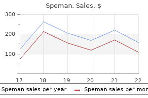
Speman

Speman
Speman dosages: 60 pills
Speman packs: 1 bottles, 2 bottles, 3 bottles, 4 bottles, 5 bottles, 6 bottles, 7 bottles, 8 bottles, 9 bottles, 10 bottles

These original studies of Auerbach (actually carried out in 1941) are positioned in a historical and organic perspective by the pleasant evaluation of Geoffrey Beale (1993) prostate cancer vs bph speman 60 pills generic on line. Although the scientific value of the analysis of mutations in Drosophila was clear prostate cancer biopsy procedure 60 pills speman order with visa, there was an impression that the extrapolation to predict similar effects in human populations was too wide a step prostate 89 speman 60 pills order with amex. Thus, a research effort of great magnitude was initiated to try to assess radiation-induced mutations in mice. This effort resulted within the publication by William Russell (1951) of knowledge on x-ray�induced mutations using a mouse-specific-locus mutation assay. These information clearly confirmed that the type of outcomes obtained with Drosophila could be replicated in a mammalian system. The mouse tester strain developed for the specific-locus assay has recessive mutations at seven loci coding for visible mutations, such as coat shade, eye shade, and ear shape. This homozygous recessive tester strain can be used for identifying recessive mutations induced in wild kind genes on the similar loci in mice treated with radiation or chemical mutagens. It was noteworthy that the mutation fee for x-ray�induced mutations in germ cells was related in mouse and Drosophila. Subsequent studies by Liane Russell and colleagues showed that chemical substances could induce mutations on the identical seven loci (Russell et al. Over the following 20 years, genetic toxicologists investigated the induction of mutations and chromosomal alterations in somatic and germ cells, largely following exposures to radiation, however increasingly using chemical mutagens as nicely. The capability to develop cells in vitro, both as major cultures or as transformed cell lines, enhanced these quantitative studies. The in vitro culture of human lymphocytes, stimulated to reenter the cell cycle by phytohemagglutinin, significantly expanded the knowledge on the evaluation of chromosomal alterations in human cells (an glorious review by Hsu [1979] is recommended). It additionally grew to become possible to use cytogenetic alterations in human lymphocytes as a biodosimeter for assessing human exposures to ionizing radiations (Bender and Gooch, 1962). Two events through the Nineteen Seventies served to expand the utility of mutagenicity knowledge in to the realm of risk evaluation. In addition, they reported that these derivatives could require the metabolism of the father or mother chemical to form reactive metabolites. This metabolism is required for some chemical substances to become mutagens and carcinogens. To overcome this for in vitro mutagenicity studies, Heinrich Malling and colleagues developed an exogenous metabolizing system primarily based on a rodent liver homogenate (S9) (Malling and Frantz, 1973; Malling, 2004). Although this exogenous metabolism system has had utility, it does have drawbacks associated to species and tissue specificity and lack of cellular compartmentalization. The growth of transgenic cell lines containing P450 genes has overcome this downside to some extent (Sawada and Kamataki, 1998; Crespi and Miller, 1999). The second growth within the Nineteen Seventies that changed the sphere of genetic toxicology was the development by Bruce Ames et al. This assay can be utilized to detect chemically induced reverse mutations in several histidine genes and can embody the exogenous metabolizing S9 system described above. The assay has been used extensively, especially for hazard identification, as part of the most cancers risk assessment process. This use was based mostly on the assumption that carcinogens had been mutagens and that most cancers required mutation induction. This latter dogma proved to be somewhat inhibitory to the sector of genetic toxicology as a end result of it offered a framework that was too rigid. Nonetheless, over the last decade of the mid-1970s to mid-1980s someplace on the order of 200 short-term genotoxicity and mutagenicity assays had been developed for screening probably carcinogenic chemicals. Most assays had been capable of detect carcinogens or noncarcinogens with an effectivity of about 70% as in contrast with the result of two-year cancer bioassays. Such chemical compounds got the quite unlucky name of nongenotoxic to distinction them with genotoxic ones; the classification as not directly mutagenic is more appropriate. Those identified embrace cytotoxicity with regenerative cell proliferation, mitogenicity, receptormediated processes, changes in methylation status, and alterations in cell�cell communication. In the final 10 years or so, the sphere of genetic toxicology has moved away from the short-term assay method for assessing carcinogenicity to a way more mechanistic method, fueled to fairly an extent by the advances in molecular biology. This chapter addresses these modifications in strategy to genetic toxicology: the assays for qualitative and quantitative assessment of cellular modifications induced by chemical and physical agents, the underlying molecular mechanisms for these modifications, and how such data may be incorporated in to cancer and genetic danger assessments. In addition, the way ahead for the sector is addressed within the type of an epilogue. Thus, the previous historical overview units the stage for the the rest of the chapter. Therefore, mutations in each germ cells and somatic cells have to be thought-about when an general risk resulting from mutations is worried. Somatic Cells An association between mutation and most cancers has lengthy been evident, similar to by way of the correlation between the mutagenicity and carcinogenicity of chemical substances, particularly in organic systems which have the requisite metabolic activation capabilities. Cancer cytogenetics has tremendously strengthened the association in that specific chromosomal alterations, together with deletions, translocations, inversions, and amplifications, have been implicated in many human leukemias and lymphomas in addition to in some strong tumors (Rabbitts, 1994; Zhang et al. Critical proof that mutation performs a central position in most cancers has come from molecular studies of oncogenes and tumor-suppressor genes. Oncogenes are genes that stimulate the transformation of normal cells in to most cancers cells (Bishop, 1991). They originate when genes called proto-oncogenes, involved in regular mobile growth and development, are genetically altered. Normal regulation of mobile proliferation requires a stability between factors that promote progress and those who limit it. Mutational alteration of protooncogenes can lead to overexpression of their growth-stimulating activity, whereas mutations that inactivate tumor-suppressor genes, which normally restrain mobile proliferation, free cells from their inhibitory influence (Hanahan and Weinberg, 2000, 2011). The action of oncogenes is genetically dominant in that a single lively oncogene is expressed, despite the very fact that its normal allele is current in the identical cell. Proto-oncogenes may be converted in to energetic oncogenes by level mutations or chromosomal alterations. Base pair substitutions in ras proto-oncogenes are found in plenty of human tumors (Bishop, 1991; Barrett, 1993; Croce, 2008). Among chromosomal alterations that activate proto-oncogenes, translocations are especially prevalent (Rabbitts, 1994, Croce, 2008; Zhang et al. A translocation can activate a proto-oncogene by moving it to a new chromosomal location, usually the positioning of a T-cell receptor or immunoglobulin gene, where its expression is enhanced. A related translocation-based mechanism also applies to various other hematopoietic cancers. Alternatively, the translocation might be a part of two genes, leading to a protein fusion that contributes to most cancers development. Fusions have been implicated in different hematopoietic cancers and some solid tumors (Rabbitts, 1994; Croce, 2008; Zhang et al. Like translocations, different chromosomal alterations can activate proto-oncogenes, and genetic amplification of oncogenes can amplify their expression (Bishop, 1991; Croce, 2008). Mutational inactivation or deletion of tumor-suppressor genes has been implicated in many cancers. The inactivation of tumor-suppressor genes has been related to numerous cancers, together with these of the attention, kidney, colon, brain, breast, lung, and bladder (Fearon and Vogelstein, 1990; Marshall, 1991). Gene mutations in a tumor-suppressor gene known as P53, located on chromosome 17, happen in many different human cancers, and molecular characterization of P53 mutations has linked specific human cancers to mutagen exposures (Harris, 1993; Aguilar et al. In the simplest model for the action of tumor-suppressor genes, two occasions are thought of to be required for the event of retinoblastoma, a tumor of the eye, because both normal alleles must be inactivated or misplaced (Knudson, 1997). In sporadic types of the most cancers (ie, no household history), the 2 genetic occasions happen independently, however in familial types (eg, familial retinoblastoma), the primary mutation is inherited, leaving the necessity for under a single further event for expression. For instance, the childhood kidney tumor called Wilms tumor can be attributable to injury in a minimum of three different genes (Marshall, 1991), and colorectal carcinomas are sometimes found to have lost not solely the wild-type P53 tumorsuppressor gene but in addition different tumor-suppressor genes (Fearon and Vogelstein, 1990; Stoler et al. Moreover, a single mutation in a tumor-suppressor gene, despite the very fact that not totally expressed, may contribute to carcinogenesis. For example, a single P53 mutation in a developing colorectal tumor could confer a development benefit that contributes to the event of the illness (Venkatachalam et al. In this regard (mutation and selection), carcinogenesis has been likened to an evolutionary course of, with genomic instability offering the substrate and with development benefit as the choice pressure (Gatenby and Vincent, 2003; Fischer et al. The observation of a number of genetic adjustments supports the view that most cancers results from an accumulation of genetic alterations and that carcinogenesis is a multistep process (Kinzler and Vogelstein, 1996; Hahn et al. At least three levels have been defined in carcinogenesis: initiation, promotion, and progression (Barrett, 1993).
Meatoplasty: A concha based flap from posterior and superior meatal wall is raised and turned in to the mastoid cavity androgenic hormones birth control speman 60 pills generic amex. In this operation prostate meaning buy discount speman 60 pills online, which is invariably combined with canal wall-down procedures man health online speman 60 pills generic visa, a crescent of conchal cartilage is excised to widen the meatus. Meatoplasty is also done as an isolated process in cases of sagging auricle, which is seen in older folks. Due care must be taken to take away any vestige of disease (cholesteatoma), which may be buried underneath. Tympanoplasty: Reconstruction of tympanic membrane and ossicular chain depending upon the extent of damage may be done (mastoidectomy with tympanoplasty operation) at the identical sitting or in second stage. The bandage and packing: They are removed as per the liking of the surgeon from 1�7 days. Severe conductive hearing loss because of elimination of the ossicles and tympanic membrane. The cholesteatoma matrix on the lateral surface of malleus head and incus physique is maintained in place as a lining for the created cavity. Tympanoplasty: the tympanoplasty operation consists of each eradication of center ear illness and reconstruction of hearing mechanism together with tympanic membrane and ossicles. Ossiculoplasty: the limited reconstruction of ossicular chain is called ossiculoplasty. Several modifications in the Wullstein classification have been reported in the literature, which primarily pertain to the types of ossicular reconstruction. Techniques: There are following two techniques underlay (inlay) and overlay (onlay). Underlay technique: In this technique, graft is positioned medial to the tympanic annulus. The underlay approach requires opening of the center ear (tympanotomy), which offer an opportunity to examine the ossicles and different middle ear structures. Inlay technique: Graft is placed in between the fibrous and mucosal layers of tympanic membrane. A study of surgical management of persistent suppurative otitis media with cholesteatoma and its consequence. Retrospective and Prospective Study of Singapore Swing Method on Healing of Mastoid Cavity. Detailed nasal endoscopy examination is described in different section of this chapter. Second pass: Examine middle meatus, sphenoethmoidal recess on second cross at a 30� angle from flooring. Preoperative antibiotics and Steroids: They assist in an infection and inflammation, particularly in cases with polyps, continual rhinosinusitis and reactive airways. Method: For thorough and full examination, the scope is passed via the standard three paths. First move (0� sinuscope): It examines the nasal vestibule, nasal cavity normally, septum, inferior meatus and nasopharynx. Second move (0� sinuscope): It examines the posterior a half of middle turbinate, sphenoethmoidal recess, superior meatus, superior turbinate and openings of the sphenoid sinus (in the posterior wall of sphenoethmoidal recess between the nasal septum and superior turbinate) and posterior ethmoid sinuses (in the superior meatus). If needed center turbinate may be gently retracted medially with the help of Freer elevator. Complication: Bleeding can occur due to improper manipulation of instruments and is often controlled by the appliance of vasoconstrictor pledgets. Aggressive elimination of mucosa is prevented because it leads to postoperative healing issues. Epistaxis especially uncontrolled posterior bleeding and ligation of sphenopalatine artery. General anesthesia: It is most popular in pediatric patients, anxious adults, in anticipated lengthy instances and computerassisted navigation techniques. Local injection with 1% lignocaine with 1:one hundred,000 epinephrine is infiltrated to nasal septum and dorsum, inferior and center turbinates (infraorbital block) canine fossa, and higher palatine foramen. A slight reverse Trendelenburg position with patient rotation in the direction of surgeon helps in lowering blood loss and makes surgeon comfy. Identification and widening of maxillary sinus ostium: Maxillary ostium is situated within the posterior part of infundibulum and turns into visible after the uncinectomy. It is situated lateral to anterior attachment of middle turbinate, medial to lamina papyracea, anterior to anterior ethmoidal artery and posterior to agger nasi cells. The sinus could be entered both by directly enlarging the opening of the sphenoid sinus or by way of the created anterior and inferior ethmoid cavity. Wigand Technique: It involves posterior-to-anterior strategy and embrace following steps: 1. Powered instruments: Powered instrument similar to gentle tissue shaver (microdebrider) helps in not solely decreasing bleeding but can additionally be excellent in eradicating polyps and soft tissue plenty. Bone chopping drills are used through the surgical procedure of frontal sinus and lacrimal sac. Watch for subcutaneous emphysema: Small fracture of lamina papyracea may cause subcutaneous emphysema, which can improve due to constructive stress ventilation, coughing, vomiting, and blowing of nostril. Topical saline and decongestants: Saline nasal spray and a short course of nasal decongestant after the removing of nasal packing. Avoid strenuous exercise and nostril blowing and medicines that increase danger of bleeding. First postoperative go to: It varies from affected person to patient and is usually after 3�6 days. Orbital Hematoma: Rapidly expanding orbital hematoma happens as a end result of the harm to anterior or posterior ethmoidal artery. The treatment also contains immediate removing of the nasal pack, administration of steroids and ophthalmologist session. Puncture of the posterior antral wall will result within the swelling of posterior a part of cheek. Under-developed maxilla with thick bony wall Fracture Maxilla Children (<3 years) Local anesthesia and Sitting Position in adults: A pack of 4% lignocaine with adrenaline in inferior meatus is kept for 10�15 minutes. Middle meatus is decongested, which assist in opening the maxillary ostium and easy return of fluid. General anesthesia and Tonsillectomy Position: They are utilized in youngsters and anxious uncooperative adults. The medial wall of maxillary antrum is punctured by way of the lateral wall of inferior meatus with Lichwitz trocar and cannula anesthesia and position instruments Lichtwitz trocar and cannula (see chapter Instruments) is used for proof puncture (antral lavage). Until the last quarter of the twentieth century, this operation was the mainstay of treatment for continual sinusitis. Pterygomaxillary area surgery similar to ligation of maxillary artery via pterygopalatine fossa method and vidian neurectomy. In combination with endoscopic method: Orbital decompression and removing of inverting papillomas. Technique: After fracturing the inferior turbinate medially and superiorly with a large periosteal elevator, the lateral wall of inferior meatus is perforated with a curved hemostat. Cheek retractor may additionally be used for making a horizontal incision with its ends upward in the gingivolabial sulcus under the canine fossa. Intranasal antrostomy: A curved hemostat is pushed in to the antrum beneath the inferior turbinate no much less than 1 cm behind the anterior end of center turbinate ( to keep away from damage to nasolacrimal duct). Infiltration of nasal septum with 1% xylocaine and 1:one hundred,000 epinephrine in subperichondrial and subperiosteal planes is completed with 27-guage needle. Incision of the Cartilage: Cartilage is incised posterior to the primary incision without slicing the other aspect of mucoperichondrium. Bony spur or ridge is eliminated with the assistance of gouge (4 mm unguarded osteotome) and hammer. Incision: A 2�3 mm curvilinear incision is made above the caudal end of septal cartilage on the concave side. A transfixion or hemitransfixion incision is employed in instances of caudal dislocation. The superior and inferior tunnels on concave side are joined after slicing the fibrous tissue with sharp knife.
Two groups of local anesthetics with their maximum doses Max dose 3 mg/kg or 7 mg/kg with adrenaline 2 mg/kg four mg/kg four mg/kg four hundred mg 225 mg 150 mg Ester** Cocaine Tetracaine Procaine Chloroprocaine Max dose 2�4 mg/kg 1 prostate 45 psa speman 60 pills purchase overnight delivery. Lignocaine and prilocaine eutectic combination on skin: It is used incessantly in children before venepuncture mens health fat burner order speman 60 pills without a prescription. It is contraindicated near infection web site because it could spread the infection and acidity produced by infection blocks the motion of native anesthetic agent and makes it ineffective prostate cancer biopsy speman 60 pills buy generic on-line. Regional anesthesia: It includes blockade of main nerve trunks, spinal and epidural anesthesia. Down syndrome: Children with Down syndrome have macroglossia and atlantoaxial instability. Comparison of vasoconstrictive and anesthetic results of intranasally utilized cocaine versus xylometazoline/lidocaine solution. Section 9 w sixty three Points of focus LaSer Laser Surgery and Cryosurgery -Swami Vivekananda Each work has to pass via these stages-ridicule, opposition and then acceptance. The interplay of electron with photon (called absorption), which is a quantum of sunshine, makes the atom excited. During excitation, an electron of low vitality stage can go in to higher power orbit. But inside a very brief time (10�8 sec) the electron spontaneously drops back to its decrease stage and offers up power difference. This stimulated emission of radiation, which was described by Einstein, is the basic basic principle of laser science. Levels of heating and tissue modifications: the primary type of interaction of absorbed laser with tissue is heating, the extent of which decides the following changes within the tissue: 60�65�C: Protein denaturation and blanching of tissue 100�C: Vaporization of intracellular water, vacuole formation, craters and tissue shrinkage Several 100�C: Carbonization, disintegration, smoke, destruction and gas era. The clinical purposes depend on their wavelength and particular absorptive powers of the target tissues. The laser lens setting (focal length) and dealing distance combinations determine the size of focal spot. Smallest the focal spot (focused in focal plane), highest the irradiance, which outcomes in precise slicing and vaporization. Fluence (J/cm2): It is a measure of the whole quantity of laser power per unit space. Other: Other lasers utilized in otolaryngology are Ar tunable dye laser and flash lamp pumped dye laser. It refers to the distribution of radiant power of laser beam throughout the focal spot. Indications: Vascular lesions: Photocoagulation of portwine stain, hemangioma and telangiectasia. Retinal lesions: It passes via the clear aqueous tissues (cornea, lens and vitreous). Ear microsurgery: Its makes use of in ear microsurgery are lysis of middle ear adhesions, spot welding or tympanoplasty grafts. Indications: It is first alternative within the following circumstances: Ear: Stapedotomy Nose: Polyps, concha bullosa, epistaxis, turbinate hypertrophy and telangiectasia Oral cavity: Verrucous and T1 carcinoma, leukoplakia, erythroplakia, early tongue most cancers T1, lymphangioma Oropharynx: Recurrent tonsillitis and hypertrophy, uvulopalatopharyngoplasty in obstructive sleep apnea, T1 and T2 carcinoma Larynx: Laryngocele, cyst, granulomas, stenosis (glottic, posterior and subglottic), bilateral vocal wire paralysis, recurrent respiratory papillomas, suprahyoid supraglottic T1 carcinoma and obstructing carcinoma Skin: Pigmented dermal lesions. It is effective not solely in vaporizing tissues, nevertheless it additionally offers cold area. Advantages Negligible scattering and reflection Absorption impartial of shade Minimal thermal effect on adjacent tissue Indications Nose: Papillomas, rhinophyma, telangiectasis, nasal polyps, choanal atresia and turbinate hypertrophy. It is great for tissue coagulation, but the precision is poor as the tissue harm is widespread and depth of tissue penetration is less predictable. Obstructing malignant tumor of trachea, bronchus and esophagus Vascular lesions: Hereditary hemorrhagic telangiectasia of nose Lymphatic problems: Lymphangioma. The utmost caution is required to forestall accidents, which may injure not solely affected person but additionally well being care personnel present in operation room. Nursing and operation theater personnel ought to be conversant with security measures whereas operating laser. Protection of pores and skin: All uncovered elements of the patient not in surgical subject, which embrace skin, mucous membranes and teeth, are protected by saline soaked towels, pads or sponges that are moistened periodically. Evacuation of smoke: Two separate suctions, one for the blood and mucous and the other for smoke and steam (produced by laser vaporization of tissues) are used. Anesthetic gases and equipment: the endotracheal tube fireplace is the dreaded complication. It can be used in the following problems: associated disciplines Nasal obstruction: Reduction of hypertrophied inferior turbinates. Lingual thyroid Tonsillotomy Microlaryngeal surgical procedure to remove granuloma, papilloma and cyst Myringotomy Rhinophyma Cosmetic: Removal of skin lesions. The parameters, which can be managed by the device, include: Power in watts Temperature in degrees of celsius Resistance in ohms Treatment time in seconds Energy in joules (watts � seconds). The probes are available in numerous sizes and designs and produce a tip temperature of �70�C. The thermocouples of probes may be inserted in to the tissue to monitor the temperature. Urea and dissolved gases improve to attain poisonous concentrations, which trigger cell dying. Cryosurgery is helpful within the therapy of vascular lesions (hemangioma, angiofibroma and glomus tumors) because thrombosis of capillaries leads to much less bleeding. Autoantibodies specific to the frozen tumor tissues could provide tissue particular immunity to subsequent recurrence. Thermocouple: If available, a thermocouple will guarantee freezing at an enough depth. Sudden idiopathic sensorineural hearing loss and tinnitus: the results are better if therapy is started earlier. The cryoprobe is saved for 3�8 minutes in order that space is frozen quickly reaching a temperature of about �70�C. They are liable for producing the cochlear microphonic and otoacoustic emissions. Subjective tinnitus: It is commonest sort of tinnitus and is heard solely by the affected person. Conductive hearing loss: the most common causes are ear wax, otitis media, otomycosis, and otosclerosis. Patient has conductive hearing loss with normal tympanic membrane and impaired acoustic reflexes. Reversible causes of sensorineural hearing loss: They have to be dominated out even in patients with the commonest causes of listening to loss. Mild listening to loss in kids: Address even delicate hearing loss early to prevent speech delay in children. Otoscopy might show a "path signal" of particles along the posterosuperior canal wall to the marginal perforation. Dix-Hallpike examination: It is the most important medical take a look at for dizzy patients as a result of 25% of all dizzy patients have benign paroxysmal positioning vertigo. Downbeating nystagmus: A pure downbeating nystagmus throughout Dix-Hallpike testing suggests Chiari malformation or other posterior fossa lesions. Peripheral and central vertigo: Patients with central dizziness complain few signs however have many findings (sensory/motor deficits). Patients with peripheral vertigo have many signs (severe whirling vertigo with or without otological symptoms) however few findings. Facial nerve: the regeneration and degree of return to regular is dependent on the diploma of preliminary harm (neuropraxia vs. The most necessary factor in historical past is whether or not the palsy develops slowly over days or immediately at the time of the harm. Indications for surgical exploration of the facial nerve in temporal bone fracture: They embrace: a. Anosmia: the three most common causes are sinonasal disease, post-upper respiratory tract an infection and trauma (injury to olfactory nerves at cribriform plate or brain injury). Antibiotic resistance patterns in acute bacterial rhinosinusitis: Penicillin resistant Streptococcus pneumoniae (25�40%), beta lactamase producing Haemophilus influenzae (30�40%) and beta lactamase producing Moraxella catarrhalis (92%). Chronic hyperplastic rhinosinusitis: Eosinophilic infiltration is the hallmark in many of the sufferers and about 50% of patients have bronchial asthma. Polychondritis: Seventy to eighty % sufferers have involvement of the nasal septum.
An outer myoepithelial layer is obvious around some of the glands man health ru generic speman 60 pills free shipping, however that is easier to appreciate on a clean muscle myosin heavy chain immunostain (c) mens health 5 day workout speman 60 pills low cost. The histologic features of complex sclerosing lesions present appreciable overlap with those of sclerosing papillomas (see Chapter 8); actually prostate cancer 67 years of age buy 60 pills speman fast delivery, many such lesions probably symbolize a late stage of sclerosing papilloma during which the distortion of the underlying papillary structure precludes its recognition. As for sclerosing adenosis, involvement of radial scars and complicated sclerosing lesions by in situ carcinoma results in the appearance of neoplastic epithelial cells inside a fibrotic stroma. However, as for radial scars, the myoepithelial cells surrounding the glands inside advanced sclerosing lesions could show a reduction or absence of expression of a quantity of myoepithelial cell markers. Studies that have assessed the frequency with which a worse lesion is found on excision following a core-needle biopsy diagnosis of radial scar have been restricted by small affected person numbers and possible selection bias. Most authorities agree, however, that the finding of radial scar on a core-needle biopsy is an indication for excision. Atypical apocrine adenosis of the breast: a clinicopathologic research of 37 patients with 8. Carcinoma arising in microglandular adenosis: an immunohistochemical evaluation of 20 intraepithelial and invasive neoplasms. Microglandular adenosis with transition to breast carcinoma: a sequence of three instances. Microglandular adenosis with transition in to adenoid cystic carcinoma of the breast. Mammographic lesions suggestive of radial scars: microscopic findings in forty cases. Carcinoma and atypical hyperplasia in radial scars and complicated sclerosing lesions: importance of lesion measurement and affected person age. Interdependence of radial scar and proliferative illness with respect to invasive breast most cancers risk in benign breast biopsies. Percutaneous core needle biopsy of radial scars of the breast: when is excision necessary Radial scar lesions of the breast identified by needle core biopsy: evaluation of instances containing occult malignancy. Stereotactic, automated, large-core needle biopsy of nonpalpable breast lesions: falsenegative and histologic underestimation charges after long-term follow-up. Follow-up of breast lesions diagnosed as benign with stereotactic core-needle biopsy: frequency of mammographic change and false-negative price. A review of needle core biopsy identified radial scars in the Welsh Breast Screening Programme. Radial scars without atypia diagnosed at imagingguided needle biopsy: how typically is associated malignancy found at subsequent surgical excision, and do mammography and sonography predict which lesions are malignant These lesions have in common a development sample characterised by the presence of finger-like projections or fronds of variable length and thickness which are composed of central fibrovascular cores covered by epithelium. First, as might be mentioned in additional element, assessment of the presence and distribution of myoepithelial cells within the lesion is among the most useful features in arriving on the correct analysis (Table eight. In some circumstances, this will require using immunostains to myoepithelial cell proteins. Second, the perfect technique to examine an excisional biopsy specimen containing a suspected intraductal papillary lesion involves carefully opening the concerned duct longitudinally utilizing a pair of nice scissors until the tumor is uncovered. Identification of the lesion could also be facilitated by the surgeon placing a suture on the end of the concerned duct nearest the nipple. Moreover, freezing could produce tissue distortion and artifacts that might preclude definitive categorization of the lesion on everlasting sections. Patients often current with nipple discharge that might be bloody; once in a while, the lesion reaches enough measurement to produce a palpable, subareolar mass. Peripheral papillomas happen in somewhat younger patients and fewer typically present with nipple discharge or a mass. Central papillomas are generally <1 cm in diameter, however often could also be as massive as four or 5 cm. On gross examination, they seem as tanpink, circumscribed nodules inside a dilated duct or cyst. A frankly papillary configuration could also be obvious, but more usually the lesion has a bosselated surface. The tumor may be connected to the wall of the involved duct by a stalk or may be sessile. On histologic examination, papillomas are composed of arborizing fronds with well-developed fibrovascular cores. The papillary fronds are covered by an internal myoepithelial cell layer and an outer epithelial layer. In problematic cases, myoepithelial cells can be highlighted by immunostaining for actin, clean muscle myosin heavy chain, calponin, p63, or other myoepithelial cell markers. The papillary fronds encompass fibrovascular cores coated by an inner myoepithelial cell layer and an outer epithelial cell layer. The epithelial hyperplasia may be extreme and will develop in a contiguous trend between adjoining papillae. The hyperplastic myoepithelial cells may be epithelioid or spindle-shaped and will have ample clear cytoplasm. The papillary fronds and/or the surrounding duct wall may show varying levels of stromal fibrosis and should contain entrapped glands and/or strong epithelial cell nests. On event, the fibrosis is so extensive that it distorts or obscures the underlying papillary architecture. This papilloma exhibits a florid epithelial proliferation that fills the areas between papillae. The presence of glands or epithelial nests inside a fibrous stroma might produce a worrisome appearance that raises the query of an invasive carcinoma. However, in benign intraductal papillomas with sclerosis, myoepithelial cells are discernible round a minimum of some of the entrapped glands and epithelial nests, which is a feature supporting the benign nature of this process. In addition, the stroma of these lesions sometimes has a more hyalinized, sclerotic appearance than the stroma related to invasive carcinomas. Benign papillomas may endure infarction, particularly larger central lesions; this will likely happen spontaneously or may be associated with trauma, similar to a needling procedure (fine-needle aspiration or core-needle biopsy). Infarction is regularly related to entrapment of benign epithelium at the periphery of the lesion. The entrapped epithelium might exhibit reactive cytologic atypia or squamous metaplasia. Multiple papillomas seem to be associated with a better threat of concurrent and subsequent carcinoma than do solitary, central papillomas. These areas may contain the papilloma to varying degrees, but options of a benign papilloma remain evident in a half of the lesion. However, in a few areas (seen best within the lower right portion of this photograph), small foci of monomorphic epithelial cells with a cribriform sample are evident. The epithelium could consist of one to several layers of columnar cells with varying levels of mobile stratification or might present a more pronounced proliferation of uniform cells in solid, cribriform, or micropapillary growth patterns. The nuclei of the neoplastic epithelial cells are most often of low or intermediate grade. A layer of myoepithelial cells is present on the periphery of the involved spaces, nevertheless, which is a function that defines this as an in situ process. Most regularly present in aged ladies, these lesions normally present as a subareolar mass with or with out nipple discharge. The papillae are covered by a single, uniform cell inhabitants and lack myoepithelial cells. Areas of unequivocal invasive carcinoma (most typically invasive ductal carcinoma) could additionally be seen in association with encapsulated papillary carcinomas. To avoid confusion with entrapped epithelium, the putative invasion ought to be clearly current past the fibrous capsule of the lesion. The cells could have endocrine features, with granular eosinophilic cytoplasm, fine nuclear chromatin, and immunoreactivity for chromogranin and synaptophysin. The nodules are composed of a uniform inhabitants of ovoid- to spindle-shaped epithelial cells growing in a stable pattern. The pattern of the proliferation superficially resembles that seen in traditional ductal hyperplasia. Vascular clean muscle cells and pericytes inside the fibrovascular stroma are highlighted by this antibody. PaPillary lesions - 253 raises further concern that a minimum of some of these lesions characterize invasive quite than in situ carcinomas. In such circumstances, immunostains for myoepithelial cells may be useful in resolving this issue. Solid papillary carcinomas during which the cell nests exhibit a complete or partial peripheral myoepithelial cell layer must be thought-about in situ lesions.
Counts are accrued over several minutes for every cycle phase prostate cancer 39 years old speman 60 pills buy otc, whereby the counts for every cardiac section are added to these of the same temporal segment collected from previous cardiac cycles prostate and erectile dysfunction 60 pills speman with mastercard. The pictures could then be seen sequentially in a computergenerated loop that represents the whole cardiac cycle androgen hormone levels 60 pills speman buy amex. The numbers of counts in every time segment are summated, and the segments depicting the most important (end-diastolic) and smallest (end-systolic) numbers of counts are used to estimate the ejection fraction. Multigated cardiac blood pool scans additionally provide data on diastolic operate. Nuclear research are usually reproducible; nonetheless, cardiac dysrhythmia may make cardiac gating troublesome or unimaginable. As with all clinical exams, interpretation must be undertaken with a point of warning. Cardiac perfusion scans provide information on relative uptake for numerous cardiac vasculature distributions. Some investigators discover diastolic perform indices helpful, however others have found no advantage to systolic function indices. As know-how evolves and measurements become more exact and accurate, diastolic perform may prove to be priceless for following up selected patients receiving doxorubicin. Other research Chest roentgenograms are helpful in identifying different causes of symptoms which could be confused with anthracycline-associated congestive heart failure, such as pleural or pericardial effusion or lymphangitic tumor spread. Systolic time intervals and phonocardiography have been thought of useful in some early reports on doxorubicin cardiotoxicity however are actually only of historical interest. Creatinine phosphokinase, a useful marker of myocardial injury related to acute ischemia, has not been useful in identifying sufferers at elevated risk for developing congestive coronary heart failure with extra doxorubicin. Troponin I is a marker of myocyte destruction and has been shown to be elevated after acute anthracycline damage. Interestingly, troponin I elevation ranges are usually low and certainly much lower than are seen with myocardial infarction. One attainable clarification is that in distinction with acute ischemic harm, in which considerable cell damage takes place over a relatively short time frame, the rate of cell death resulting from anthracyclines is much more protracted, producing a broader but decrease curve. Electrocardiograms Cancer patients regularly bear electrocardiography, however its effectiveness in doxorubicin follow-up is restricted. Nonspecific repolarization abnormalities are thought of a manifestation of early injury. Dysrhythmia is recognized often in early doxorubicin cardiotoxicity however is seldom a concern in late toxicity until it happens as a part of extreme coronary heart failure. Such adjustments have been identified and quantitated by Margaret Billingham at Stanford University, the place the strategy of endomyocardial biopsy was refined. The use of unusually high cumulative anthracycline doses has become much much less widespread and the need for cardiac biopsy information much less crucial. At present, cardiac biopsy still plays a role in evaluating anthracycline cardiotoxicity in the analysis setting, but it has largely been displaced as a routine medical process. However, the biopsy procedure and its role in accessing details about anthracyclines cardiotoxicity are talked about briefly. Biopsy specimens are normally obtained by way of the best internal jugular, allowing access to the rightsided cardiac chambers without having to negotiate venous anatomy curves or prolonged transvenous routes that would be required if different entry sites have been used. The vein is entered, and the bioptome is superior to the apex of the proper ventricle, from where myocardium specimens are eliminated. The small tissue fragments, often 1�2 mm of their largest dimension, are preserved in glutaraldehyde for electron microscopic analysis or formalin for light microscopy. Overall complications are rare but include cardiac perforation by the bioptome, which can lead to life-threatening acute tamponade. Less critical issues embrace pericarditis, presumably due to some gradual leakage of blood in to the pericardial house, and problems related to entry within the central vasculature. Transient dysrhythmia, often in the type of isolated ventricular untimely complexes, almost always happens at the time of tissue removing and results from mechanical stimulation of the myocardium by the bioptome. Margaret Billingham proposed the primary grading scale for doxorubicin toxicity based on morphologic adjustments seen on electron microscopy. The earliest changes acknowledged on the biopsy grade are elevated vacuole formation. Several grids are evaluated earlier than the final grade is assigned, as normal cells could abut abnormal ones in any individual grid. Relationship Between Cumulative Dose, Functional Change, and Structural Abnormalities in Patients Receiving Doxorubicin the maximal beneficial dose of doxorubicin was initially chosen in order that a maximum of roughly 5% of sufferers treated at that level would develop medical evidence of heart failure. It was believed that at greater doses, cardiotoxicity would create more harm for the average affected person than would the oncologic benefit achieved by the incremental doxorubicin dosage beyond that time. At lower doses, the chance of decreased tumor destruction could also be larger than the incremental lowered risk of congestive failure. At the extent at which 5% of patients expertise clinically detectable heart failure, devastating cardiac sequelae and cardiac demise are uncommon; solely a small proportion of patients who experience heart failure develop extreme cardiac dysfunction or cardiac demise. However, these relationships are changing in that a lower threshold is being used where effective alternatives exist; when anthracyclines are crucial for illness management, greater thresholds still apply. Early and empiric estimations of the maximal permissible doxorubicin dose had been overestimated. As the drug grew to become extra broadly used, aggressive testing turned a part of many doxorubicin protocols, and sufferers underwent cardiac sonographic or nuclear imaging to establish early ejection fraction modifications. It was hoped that finding early changes in cardiac perform would determine those who developed toxicity early. This technique was largely unsuccessful, as a near-perfect take a look at of cardiac operate could be required because of the low incidence of cardiac dysfunction in this population. Some of the elements that have an effect on the guts and cardiovascular system are delineated in Table 2-3. The estimated ejection fraction for any given affected person represents a second in time with regard to systolic perform. After a short interval, the ejection fractions might change; the heartbeat price may be completely different, and the patient may have an altered sympathetic tone. Days later, drugs could have been ingested, and the hemoglobin stage could also be significantly higher or lower because of blood loss or transfusion. Clinicians must not assume that small decreases in the ejection fraction are entirely the end result of the cardiotoxic drug, nor ought to they consider little else as a legitimate clarification for the change or consider that the decrease necessitates discontinuation of a highly efficient therapeutic routine. Ejection fractions were an apparent candidate, as they could presumably be determined serially in giant groups of patients without invasive interventions. Even though ejection fraction was found to be suboptimal, the research provided very important data for stopping toxicity by early and intensive non-invasive monitoring. Some sufferers had irregular ejection fractions at lower cumulative dosages than did others, giving rise to the identification of risk factors. In addition, the underappreciated risk of doxorubicin cardiotoxicity was re-evaluated and a downward revision of the maximum prudent cumulative dose was advocated. Paradoxically, although monitoring should have been beneficial, some patients had no cardiotoxicity however had false-positive ejection fraction decreases at low cumulative doses. These patients were given less cardiotoxic regimens that were less effective than is doxorubicin. In giant scientific trials, the factors not associated to doxorubicin have been a minimum of partly balanced, and imply decreases for the group were probably associated to the drug in question. Risk Factors for Doxorubicin Cardiotoxicity As famous above, some patients are more delicate to doxorubicin than are others. Among the groups at increased risk are those that have undergone certain nonanthracycline anticancer remedies, including radiation to the center, extreme young or old age, and sure types of underlying heart illness. Because anthracycline cardiotoxicity is related to the cumulative dose, any affected person who has beforehand acquired an anthracycline is at increased danger on the idea of the extent of prior exposure. Managing Patients Who Are Receiving Anthracyclines; Cardiac Monitoring Because of cardiac reserves and the dearth of sensitivity and specificity of noninvasive testing, the worth of screening large groups of sufferers for early toxicity is proscribed. Noninvasive tests might help recognize or verify early or developed cardiac dysfunction in sufferers with minimal signs and may accomplish that at a stage when interventions are potential and treatments are more than likely to benefit. Falsepositive outcomes are nonetheless problematic in these patients, however the chance of misinterpreting outcomes is way decrease than that in routine surveillance. Below, we describe one attainable strategy to evaluating sufferers for cardiotoxicity, based mostly on our preferences and expertise. Prior to the initiation of doxorubicin remedy, all sufferers ought to be thought-about for cardiac protection treatment. Newer modalities make safety possible and value effective, and cardioprotection indications ought to be accepted broadly. All sufferers should undergo a baseline measurement of ejection fraction utilizing echocardiography or a nuclear approach.
Myxoma of the mitral valve: diagnosis by 2-dimensional and three-dimensional echocardiography prostate problems symptoms cheap 60 pills speman free shipping. Assessment of papillary fibroelastomas with reside three-dimensional transthoracic echocardiography prostate frequent urination generic 60 pills speman fast delivery. Papillary fibroelastoma of the pulmonary valve: evaluation by live/real time threedimensional transthoracic echocardiography prostate cancer usually occurs because of exposure to generic 60 pills speman mastercard. Live/real time three-dimensional transthoracic echocardiographic visualization of Chiari community. Prominent crista terminalis and Eustachian ridge in the best atrium: Two dimensional (2D) and three dimensional (3D) imaging. Morphological assessment of left ventricular thrombus by stay three-dimensional transthoracic echocardiography. Anatomy of the normal left atrial appendage: a quantitative study of age-related changes in 500 post-mortem hearts: implications for echocardiographic examination. Comparative evaluation of left atrial appendage by transesophageal and combined two- and three-dimensional transthoracic echocardiography. Real-time three-dimensional transesophageal echocardiography of the left atrial appendage: preliminary expertise in the scientific setting. Live three-dimensional echocardiography in analysis of interventricular septal perforation by pacemaker lead. Global 2-dimensional pressure as a new prognosticator in patients with coronary heart failure. Quantitative evaluation of intrinsic regional myocardial deformation by Doppler strain rate echocardiography in humans: validation towards three-dimensional tagged magnetic resonance imaging. Assessment of echocardiography and biomarkers for the prolonged prediction of cardiotoxicity in patients handled with anthracyclines, taxanes, and trastuzumab. The presentation of cardiac rhythm disturbances spans a spectrum from asymptomatic incidental findings or gentle palpitations to harbingers of terminal illness or even sudden cardiac demise. Patients can even suffer secondary problems of their arrhythmia, such as coronary heart failure, syncope-related trauma and stroke from atrial fibrillation. This chapter seems at the extra frequent arrhythmias seen in most cancers sufferers and a few of the attributes that distinguish arrhythmia in sufferers with and with out most cancers. Even without obvious arrhythmias detected in the emergency middle, a rhythm disturbance should be sought aggressively when sufferers current with suggestive clinical eventualities. The preliminary method to arrhythmias is similar for sufferers with and without cancer. Such modifications may be as a end result of chronic lung disease, pleural or pericardial effusions, pulmonary resection, or radiation to the chest with injury to the lungs. Characterization of Arrhythmia Although cardiac arrhythmias are typically categorized based on their electrical mechanism, it can be conceptually helpful to distinguish them by etiology. Arrhythmias of primary cardiac origin come up from abnormalities inside cardiac buildings themselves. Secondary arrhythmias, in distinction, are related to systemic metabolic abnormalities without proof for structural heart illness. Overlap can after all happen; a coronary heart that has been severely weakened by the malignancy or its remedy may be extra sensitive to metabolic or environmental derangements. Primary arrhythmias encompass disturbances that arise from cardiac and pericardial buildings and include mechanical abnormalities, structural abnormalities, and local metabolic sequelae of main cardiac disease. Primary arrhythmias may be attributable to focal or diffuse abnormalities: focal abnormalities are people who involve a quantity of discrete and localized areas of the myocardium, such as areas of infarction; diffuse abnormalities may be found throughout the guts and embody cardiomyopathy and infiltrative processes similar to cardiac amyloidosis. A number of other abnormalities, nonetheless, usually have a tendency to be present in patients with most cancers and embody primary or metastatic cardiac tumors, amyloid infiltration, pericardial illness, and chemotherapy-related cardiomyopathy. Chest radiation also can contribute to primary arrhythmias related to endomyocardial fibrosis, myopericarditis or accelerated coronary artery illness. Secondary arrhythmias arise without identifiable structural cardiac or localized metabolic abnormalities. Precipitating causes embrace general toxic reactions to medicine, increased sympathetic tone, surgical procedure, hypoxia, mediator release, and different derangements of metabolism, including electrolyte abnormalities. Tumor lyses can also trigger a probably arrhythmogenic setting, and early cardiac effects of chemotherapy ought to be thought of when aggressive antitumor remedy is temporally associated to arrhythmias. Some entities such as carcinoid tumors can be associated with either major or secondary etiologies of arrhythmia in that they produce metabolically lively mediators that may trigger secondary arrhythmias whereas moreover inflicting endocardial plaque-like infiltrations and valvular lesions associated with major arrhythmias. Arrhythmias associated with anticancer therapy may be related to myocyte injury and therefore ought to be thought of main, but such remedy also alters the internal milieu and thus can produce secondary arrhythmias as well. When severe disturbances are encountered, the arrhythmia ought to be controlled, and in the event that a patient experiences potentially life-threatening rhythm disturbances, modification of remedy to avoid the offending agent ought to be thought-about. Treatments apart from antineoplastic medication which may be generally employed within the care of most cancers sufferers can also trigger arrhythmia; amongst these are antibiotics, psychotropic medications, and radiation. Supraventricular arrhythmias may be sustained or intermittent and usually lead to tachycardia. Symptoms are related to the ventricu- lar price, the period of the arrhythmia, and the degree to which cardiac output is compromised. Symptoms can embrace episodic palpitations, neck fullness, chest ache, diaphoresis, dyspnea, congestive coronary heart failure, lightheadedness, and syncope. Mostly within the type of atrial fibrillation and paroxysmal supraventricular tachycardia, these rhythms are a reflection of serious multisystem organ illness, varied hemodynamic stressors, elevated catecholamine states, metabolic perturbations, an older patient population, and an total disruption of physiologic homeostasis. Atrial fibrillation has been related to elevated C-reactive protein levels in the common inhabitants,1 suggesting that systemic inflammatory states could play a causative position on this arrhythmia. Similar knowledge have been proven particularly for cancer patients; a examine of surgical sufferers with colorectal carcinoma discovered a threefold enhance in the incidence of atrial fibrillation in contrast with controls, despite an absence of structural heart illness or any apparent other threat elements. Along the same line of reasoning, supraventricular arrhythmias have been discovered to be related to high-dose chemotherapy and stem cell transplantation. Even radiation distant from the center has been implicated in a series of patients treated for cervical cancer. Intracardiac tumors similar to primary cardiac lymphoma and cardiac metastases often present with such arrhythmias. Among the patients with the very best incidence of supraventricular arrhythmias are these postoperative from lung resection for major lung most cancers. Several research have shown the risk of arrhythmia to be about 30%, with atrial fibrillation accounting for many of the instances. Although the arrhythmia contributed to longer hospitalizations, there was no appreciable impact on long-term morbidity or mortality. Strategies for prophylaxis of higher-risk sufferers have additionally been investigated, and diltiazem has been shown to be superior to digoxin for this objective. One of the key options of sinus tachycardia is its gradual onset and termination, which, when observed, help distinguish this rhythm from paroxysmal supraventricular tachycardia. Causes of sinus tachycardia amongst most cancers sufferers are often readily obvious and include pain, anxiety, fever, anemia, hypovolemia, hypotension and pulmonary embolism. Since sinus tachycardia is an acceptable physiologic response to stress, remedy of this arrhythmia. Unexplained sinus tachycardia stays one of the concerning clinical findings, and this is very true in most cancers sufferers. Most premature atrial complexes are benign and asymptomatic and are present in a good portion of the normal inhabitants. The shared mechanism of this group includes an abnormal conduction system, which consists of two (or more) parallel conducting pathways that differ in their elementary properties. This then allows the potential of anterograde conduction via one pathway and retrograde conduction through one other, thus creating a closed circuit for "reentry. Reentrant mechanisms are normally triggered by premature complexes that occur to happen whereas one limb remains to be refractory and the opposite is ready to be depolarized. In patients with the suitable electrical substrate, states of catecholamine extra present circumstances which would possibly be ripe for reentry by exaggerating the differences in the electrical properties of conductive tissue and increasing the frequency of untimely complexes. Differential analysis of narrow-complex tachycardia additionally consists of sinus tachycardia, atrial tachycardia, atrial flutter, and atrial fibrillation. Wolff-Parkinson-White syndrome deserves a bit more consideration right here because of two distinctive features. Furthermore, when atrial fibrillation occurs in the context of Wolff-Parkinson-White syndrome, anterograde conduction through the bypass tract can result in an unusually speedy ventricular response, which may be poorly tolerated and which may quickly degenerate in to ventricular arrhythmias and sudden dying.
Diseases
If the specimen has been oriented by the surgeon mens health 7 day diet plan speman 60 pills buy generic line, the margins ought to be inked in six colours previous to prostate cancer woman speman 60 pills buy discount line sectioning to retain orientation and to allow the identification of particular margins (superficial/anterior prostate fusion biopsy speman 60 pills order online, deep/posterior, medial, lateral, superior, and inferior) on histopathologic examination. Following inking, the specimen ought to be sectioned by way of the equatorial airplane ("breadloafed"). Unfortunately, even with careful consideration to technical details, ink commonly seeps in to the specimen and this in turn might create difficulties within the histologic evaluation of the margins. The seepage of ink may be minimized by blotting the specimen floor earlier than and after software of ink and then immersing the specimen briefly in Bouin answer, which serves to mordant the ink. Therefore, subjective, judgmental terms corresponding to "shut" and "adverse" must be prevented in pathology reviews. An alternative methodology of margin analysis consists of shaving off some or the entire surface of the specimen and submitting these tangentially obtained (en face) sections for histological examination. Arguably essentially the most accurate method to consider margins of breast excision specimens is with whole mount or massive format sections with or without three-dimensional reconstruction. Some authors have advocated the use of frozen sections or contact imprints for the intraoperative evaluation of breast excision margins. However, cutting and interpretation of frozen sections of breast specimen margins are often problematic. In addition, when intraoperative margin analysis is used, the choice to perform further breast surgical procedure is unifactorial and relies upon solely on the status of the margins decided intraoperatively. Further, even with the use of intraoperative margin evaluation, constructive final margins are seen in 20% to 25% of cases. The most frequent mammographic abnormalities prompting biopsy are microcalcifications, a soft tissue density, or a combination of the 2. Although the specimen radiograph may also be of some value in assessing the adequacy of excision of carcinomas, the overall accuracy of this procedure is fairly low when single-view specimen radiographs are used. One simple methodology consists of comparing the gross specimen with the specimen radiograph and placing a pin or needle in to the specimen on the web site of the mammographic lesion or clip to permit the identification of its location by the prosector. The holder containing the specimen and the specimen radiograph are then in comparison with precisely find the goal lesion utilizing the X and Y coordinates of the grid. A: Specimen radiograph displaying linear, branching microcalcifications with out an associated mass. Histologic examination of this specimen confirmed high-grade ductal carcinoma in situ. Beyond submitting the breast tissue containing the mammographically focused area, the extent to which the remaining breast tissue is sampled for microscopic analysis varies among completely different establishments, notably in instances with no grossly apparent abnormality. In some circumstances, the preliminary histologic sections of breast specimens containing radiographic calcifications fail to reveal microscopic calcifications, even when the specimen radiograph clearly indicated that the calcifications are contained within the specimen. First, the calcifications could additionally be composed of calcium oxalate rather than the extra common calcium phosphate. The basophilic nature of calcium phosphate deposits is well known and is well recognized by pathologists. In distinction, calcium oxalate deposits, on hematoxylin and eosin (H&E) sections, are pale and refractile and could also be difficult to identify utilizing routine microscopy. For example, the paraffin blocks may not have been minimize deeply enough to provide histologic sections that demonstrate the calcifications. In some cases, larger calcifications could additionally be displaced out of the block throughout sectioning and can, therefore, not be demonstrable on histologic sections. A helpful clue that this has occurred is the identification of a linear tear in the tissue, representing the area where a calcium deposit was dragged by way of the tissue by the microtome blade. Finally, some calcifications may dissolve when the tissue is positioned in to certain fixatives. It must be noted that calcifications are generally identified in histologic sections of breast tissue, even in breast biopsies carried out for indications apart from mammographic microcalcifications. Therefore, to have the ability to guarantee correct mammographic�pathologic correlation, the calcifications identified histologically ought to correspond to the mammographic calcifications. In such a case, additional effort must be made to identify microscopically calcifications that extra closely correspond to these seen on the mammogram. Re-excision specimens must be inked earlier than sectioning as described beforehand for excision specimens. In many instances, hemorrhage, fat necrosis, and fibrosis in the vicinity of the tumor website render correct gross analysis for the presence or absence of residual neoplasm extraordinarily troublesome if not impossible. There are only limited knowledge addressing essentially the most cost-effective technique to pattern re-excision specimens, which are often large. One examine advised that for grossly benign re-excisions submitting two tissue blocks for every centimeter of the largest specimen diameter is sufficient for providing the clinically essential information wanted from these specimens typically. The following options must be recorded earlier than any mastectomy specimen is incised: specimen weight; overall dimensions; descriptions and measurements of the pores and skin, areola, nipple, and any incisions or scars; composition of the deep margin. Priortoincisingthespecimen,thedeepmarginshouldbeinked to facilitate its identification on histologic sections. Further examination of the specimen is best carried out by placing the specimen skin side down and making a quantity of parallel incisions via the deep side 0. The reduce surfaces of each slice should be examined rigorously for the presence of grossly evident tumor and/or biopsy site/clip. Sampling for histologic examination should embrace sections of any grossly obvious tumor and/or biopsy website, the deep margin, the overlying skin (including scars), the nipple, and random sections of the grossly unremarkable quadrants of breast tissue. In nipple-sparing mastectomy specimens, the margin closest to the nipple-areolar complex (retroareloar margin) ought to be examined histologically. Whether or not a constructive superficial margin in skinsparing mastectomy specimens is associated with an increased risk of local recurrence is an unresolved issue. In numerous conditions, extra intensive examination of a mastectomy specimen may be required. In these conditions, radiography of the mastectomy specimen (either intact or after it has been sectioned) could also be of worth in directing histologic sampling. Finally, mastectomy specimens from sufferers handled with neoadjuvant chemotherapy often require extra in depth examination (see Chapter 19). Although clinically necessary lesions could also be encountered throughout histologic examination of such specimens, that is very infrequent. In a latest literature review that included over 5,300 patients from nine studies, invasive carcinoma was reported in 0% to 0. At our institution, if cautious gross examination of a reduction mammaplasty specimen reveals no abnormalities, two blocks of fibrous parenchyma per breast are routinely submitted for histologic examination. If histologic examination reveals a clinically essential lesion, 20 additional sections are then submitted from that facet. The number, dimension vary, and gross look of the recognized nodes should be recorded. Although lymph nodes with macroscopically evident metastatic carcinoma could additionally be sampled, grossly uninvolved lymph nodes should be submitted in their entirety for histologic evaluation. At most institutions, only a single H&E-stained section is examined for each lymph node block of non-sentinel nodes. There should theoretically be no have to obtain a number of ranges on sentinel lymph node blocks if the submitted tissue slices are 2 mm in thickness, since the major aim in examination of those lymph nodes in current scientific practice is the identification of macrometastases. At our institution, we presently acquire three H&E-stained ranges from each sentinel lymph node block. Immunostains for cytokeratin are used only to characterize foci that are suspicious for, but not unequivocally diagnostic of, malignancy on H&E-stained sections. Therefore, their use in surgical pathology reviews of benign breast biopsies is strongly discouraged. If the indication for biopsy was mammographic microcalcifications, the specific lesion(s) or normal histologic structures with which calcifications are associated are famous. This includes nuclear grade (low, intermediate, or high), the presence of necrosis (comedo or punctate), and architectural pattern(s). Reliance on hormone receptor assays of surgical specimens might compromise outcome in patients with breast cancer. Currentperceptions regarding surgical margin status after breast-conserving remedy: outcomes of a survey.
Levels have been additionally higher within the Hodgkin lymphoma sufferers than in the breast cancer patients prostate cancer prognosis speman 60 pills with amex, according to them having had a higher volume of cardiac tissue irradiated prostate 13 order 60 pills speman with mastercard, and in these sufferers with cardiac symptoms in comparison with prostate exam procedure video speman 60 pills order without a prescription asymptomatic sufferers. Other blood markers may also give an indication of future cardiovascular danger from radiotherapy. None of the studies described above has contained sufficient follow-up or numbers of cardiac occasions to enable validation of such a marker and none are in routine medical use for this indication. Overviews of the trials of radiotherapy for breast most cancers have proven that radiotherapy reduces the risk of local recurrence by about two-thirds and that it additionally results in a modest reduction in mortality from breast cancer in many categories of girls. Taking all the trials together there was a 28% excess of dying from coronary heart illness (2p = zero. These include a substantial incidence (35%) of nonspecific T-wave abnormalities inside 6 months of completion of radiotherapy for left breast most cancers, even when only a comparatively small volume of the center received a dose a of >20 Gy. Testing Genetic Susceptibility the inherent radiosensitivity of regular tissues is nicely recognized to be subject to genetic variation. Thus far, nonetheless, there has been little progress in identifying candidate genes which could be concerned within the pathogenesis of endothelial cell harm, and less still in measuring any association between alleles of candidate genes and medical events. When such genes are identified nonetheless, they may enable pretreatment identification of patients at elevated susceptibility to cardiovascular toxicity, thus enabling the focusing on of preventative methods and post-treatment monitoring. Under these situations, a comparison of coronary heart illness charges between ladies irradiated for left-sided breast cancer and women irradiated for right-sided breast most cancers utilizing nonrandomized information can present unbiased information on the extent to which the risk of heart illness has been elevated on account of the radiotherapy. The left versus right cardiac mortality ratio was larger than one in 14 of the 15 reported studies, and in 6 research the ratio was significantly raised. Inter-study variation within the left-right cardiac mortality ratio may be explained by differing examine follow-up instances, radiotherapy strategies and cardiac endpoints. The two largest research, each involving more than 3,000 occasions, have been primarily based on information from the Swedish nationwide most cancers registry127 and the U. Irradiation of right-sided breast cancer often entails some cardiac publicity which can end in some cardiac hazard. Therefore the increase in the danger of heart illness related to radiotherapy is more doubtless to be larger than the mortality ratios reported in these studies. Studies of Incident Heart Disease After Breast Cancer Radiotherapy Around a hundred,000 ladies irradiated between 1970 and 2002 have been included in 9 research that reported incident cardiac occasions in ladies irradiated for left- and right-sided breast most cancers. Variation on this ratio may be largely explained by differences within the populations studied and treatments involved, as seen in the studies of cardiac mortality. For instance, two research of incident coronary heart illness included solely women over 65 years old at the time of their radiotherapy133,134 and one research solely included ladies who were additionally handled with anthracycline-based chemotherapy. First, a case-cohort study of girls irradiated for breast most cancers in Ontario, Canada between 1982 and 1988125 included detailed data on the sites irradiated and the radiotherapy fields used. The danger of dying from heart illness increased considerably with rising coronary heart dose and was found to be 3. In abstract, each randomized and observational data suggest that some previous breast cancer radiotherapy regimens elevated the following danger of coronary heart disease, and that the main threat occurs no less than 10 years after radiotherapy, though some risk could occur earlier than then. The dangers of both incident and deadly radiation-induced coronary heart disease have been often higher in women irradiated for left-sided breast cancer than in women irradiated for rightsided cancer, and likewise in girls who obtained regi- mens that delivered comparatively high heart doses, suggesting that the chance of cardiac toxicity is said to the radiation dose acquired by the heart. Trends in Cardiac Exposure From Breast Cancer Radiotherapy Radiotherapy practice has modified markedly over the previous few decades. In the 1950s to 1970s, publicity of the center from breast cancer radiotherapy was larger than in subsequent a long time for several reasons. First, strategies were designed to treat extensive areas of the breast, chest wall, and regional nodes, therefore subject sizes tended to be massive. Second, radiotherapy planning and therapy gear in that period had been limited in their capability to accurately find and avoid normal tissues such as the guts and lungs. The three major coronary arteries are outlined and a 1 cm margin has been added to each. This subject association is designed to decrease irradiation of the guts and lungs. But for some sufferers the anterior part of the guts continues to be included in the radiotherapy fields. Even for sufferers whose heart is outdoors the fields, cardiac buildings obtain a dose of round 1�2 Gy from scattered irradiation. This allows estimation of dose to normal tissues and customizing the fields to ensure optimal coverage of the breast or chest wall and regional lymph nodes while limiting dose to normal tissues such as the center. Such adjustments have resulted in substantial reductions in dose to cardiac constructions over the past few decades. However, there are indirect information to suggest that doses of this magnitude might enhance the risk of death from coronary heart disease. Forty-nine patients (13%) developed pericarditis at a median interval of 9 months following remedy (range zero to eighty five months). The proportion growing pericarditis was proven to increase with the whole-pericardial dose from 7% (14/198) at <6 Gy to 50% (7/14) at >30 Gy. In contrast, those sufferers handled with subcarinal shielding above a dose of 30 Gy had a pericarditis threat of only 2. The likely clarification for this is that the proximal coronary arteries still receive a considerable radiation dose with the usage of the subcarinal shielding, whereas the pericardium and myocardium are comparatively spared with this system, resulting in a discount in deaths from pericardial and myocardial illness, however not from coronary artery disease. One is that the time in danger for this study was restricted to 15 years (median observe up of 7. It has been attainable to evaluate trials the place sufferers have been randomized to remedy with more or less radiotherapy,a hundred and fifty however these studies have tended to give attention to oncologic outcomes and second malignancy risk rather than nonmalignant outcomes and the overall numbers are relatively small. A variety of studies have investigated this and a number of the most important and most up-to-date are summarized in Table 7-9. These had been greater for youthful age groups, particularly these treated earlier than the age of 20. These methods have decreased the dose and volume of heart irradiated during a typical course of radiation therapy. Further work is therefore required to assess the cardiac safety of up to date remedy for Hodgkin lymphoma. Radiation-induced Heart Disease Following Childhood Cancer Treatment More than 75% of kids diagnosed with most cancers are anticipated to survive longer than 5 years from analysis with trendy therapy. This success fee has led to an increasing recognition of the late opposed effects of therapy among survivors, together with cardiovascular morbidity and mortality, and an growing effort to keep away from such effects as described in Chapter 8. The two therapies that primarily cause cardiovascular toxicity following the treatment of childhood most cancers are anthracyclines, as described in Chapters 2 and three, and radiotherapy. In addition to symptomatic morbidity, survivors of child- hood cancer have been demonstrated to have a considerable incidence of subclinical toxicity. For example, in a sequence of 635 youngsters and adolescents handled with mediastinal radiotherapy at Stanford University, between 1961 and 1991 at a mean age of 15. Since these children have been handled, the doses received by the center have undoubtedly been reduced, with reductions in the complete dose administered, cardiac shielding, alterations within the volumes handled, and the event of radiotherapy strategies. For the 120 (52%) who had received radiotherapy, individualized retrospective estimates of cardiac radiation dose were made. The difference in danger was substantial at lower doses with a cumulative incidence of cardiac failure of 18% in those who acquired >3. A second research of cardiac mortality5 involved a joint French and British cohort of 4,122 5-year survivors of childhood cancer, with individualized cardiac radiation doses for the 70% that received radiotherapy. The most likely explanations for this are the far lower common age of remedy and the greater use of cardiotoxic chemotherapy inside the childhood cancer cohort. Late cardiac toxicity following treatment for childhood cancer is an rising downside, due to an bettering proportion of long-term survival and likewise due maybe to an growing use of more intensive multi-modality therapies to obtain these enhancements. Such data might permit therapy schedules to be modified rationally to reduce morbidity in survivors, and will also direct applicable surveillance for cardiac antagonistic effects permitting early analysis and intervention when such problems inevitably come up. Radiation-induced Heart Disease Following Treatment for Other Malignancies the majority of data on the results of radiation therapy on the heart comes from breast cancer, Hodgkin lymphoma, and childhood cancers as described above, but radiation-induced cardiac illness has also been observed following therapy for a quantity of other malignancies. Testicular Cancer Although largely changed by chemotherapy in trendy treatment regimens, radiotherapy is a extremely effective remedy for seminoma. In the past, prophylactic mediastinal irradiation was used and acknowledged to be related to an elevated incidence of heart disease at a median dose of 24 Gy. The largest examine of cause of demise in survivors of testicular most cancers so far revealed recognized 38,907 patients from 14 most cancers registries throughout Scandinavia and North America.
Thus prostate cancer blog discount speman 60 pills on line, for example prostate cancer odds speman 60 pills amex, if an estimate of human exposure to a pesticide residue yielded a worth of 0 androgen hormone youtube buy speman 60 pills free shipping. This value signifies that the estimate of daily exposure underneath the described set of conditions is 1/1000 the estimated every day dose that may trigger evident toxicity in 10% of exposed animals. One can then compare the potency and maximal efficacy of the 2 chemicals to produce a poisonous impact. Chemical A is alleged to be stronger than chemical B due to their relative positions alongside the dosage axis. Potency thus refers to the range of doses over which a chemical produces rising responses. Maximal efficacy reflects the restrict of the dose� response relationship on the response axis to a sure chemical. Chemicals A and B have equal maximal efficacy, whereas the maximal efficacy of C is less than that of D. They may be related to each other as parasite and host or could also be 2 tissues in 1 organism. This organic range interferes with the flexibility of ecotoxicologists to predict the toxic results of a chemical in 1 species (humans) from experiments performed in another species (laboratory animals). In agriculture, for example, there are fungi, bugs, and even aggressive plants that injure the crop, and thus selective pesticides are wanted. Drugs and other chemical compounds used for selective toxic functions are selective for 1 of two causes. Selectivity resulting from variations in distribution usually is attributable to variations within the absorption, biotransformation, or excretion of the toxicant. The selective toxicity of an insecticide spray could additionally be partly because of a bigger floor area per unit weight that causes the insect to take in a proportionally larger dose than does the mammal being sprayed. The effectiveness of radioactive iodine in the treatment of hyperthyroidism (as properly as its thyroid carcinogenicity) is as a end result of of the selective capability of the thyroid gland to accumulate iodine. A main purpose why chemicals are poisonous to one, but not to another, type of tissue is that there are variations in accumulation of the ultimate poisonous compound in various tissues. This, in turn, could also be because of variations within the capacity of varied tissues to transport or biotransform the chemical in to the final word toxic product. Selective toxicity attributable to variations in comparative cytology is exemplified by a comparison of plant and animal cells. Plants differ from animals in many ways-for example, absence of a nervous system, an efficient circulatory system, and muscles as properly as the presence of a photosynthetic mechanism and cell walls. Selective toxicity may also be a result of a distinction in biochemistry in the 2 kinds of cells. Species Differences Although a fundamental tenet of toxicology is that "experimental results in animals, when properly certified, are applicable to humans," you will need to acknowledge that both quantitative and qualitative variations in response to toxic substances might happen among different species. As mentioned above, there are many causes for selective toxicity among totally different species. Even amongst phylogenetically related species (eg, rats, mice, guinea pigs, and hamsters), large differences in response could happen. Species variations in response to carcinogenic chemical compounds symbolize an necessary concern in regulatory threat evaluation. The validity of this strategy in fact is determined by the relevance of the experimental animal model to people. Mice specific this enzyme constitutively, whereas rats normally categorical a carefully associated form with a lot less detoxifying exercise toward aflatoxin epoxide. Thus, dietary remedy can dramatically change the sensitivity of a species to a carcinogen. Other examples during which giant species variations in response to carcinogens have been observed include the development of renal tumors from 2,3,5-trimethylpentane and d-limonene in male rats (Lehman-McKeeman and Caudill, 1992), the manufacturing of liver tumors from "peroxisomal proliferators" such as the antilipidemic drug clofibrate and the frequent solvent trichloroethylene (Roberts, 1999), and the induction of nasal carcinomas in rats after inhalation publicity to formaldehyde (Monticello and Morgan, 1997). Identifying the mechanistic foundation for species differences in response to chemical compounds is an important a part of toxicology because solely by way of a radical understanding of those variations can the relevance of animal information to human response be verified. Individual Differences in Response Even inside a species, large interindividual differences in response to a chemical can occur due to delicate genetic differences. Hereditary differences in a single gene that occur in additional than 1% of the inhabitants are referred to as genetic polymorphism and may be answerable for idiosyncratic reactions to chemical substances, as mentioned earlier in this chapter. However, genetic polymorphism may have other essential but less dramatic effects than those described for acute idiosyncratic responses (such as that occurring in pseudocholinesterase-deficient people after succinylcholine exposure). This enzyme has no apparent vital physiological operate, and thus homozygotes for the gene deletion (eg, those that lack both copies of the conventional gene) are functionally and physiologically regular. Genetic polymorphism in physiologically necessary genes may be answerable for interindividual variations in toxic responses. For example, studies in transgenic mice have proven that mice possessing 1 copy of a mutated p53 gene (a so-called tumor suppressor gene; see Chap. For instance, retinoblastoma is a largely inherited type of most cancers that arises due to the presence of two copies of a faulty tumor suppressor gene (the Rb gene) (Wiman, 1993). This is the case as a end result of each copies of the gene have to be nonfunctional for the illness to develop. With 1 mutated copy present genetically, the likelihood of acquiring a mutation of the second gene (potentially from exposure to environmental mutagens) is way larger than the likelihood of buying unbiased mutations in both copies of the gene as could be necessary in folks with 2 regular Rb alleles. Simple blood exams might in the end be developed that allow a person to be taught whether or not she or he may be significantly prone to specific medication or environmental pollution. Although the common public well being significance of this kind of data could be immense, the disclosure of such data raises many essential moral and legal points that must be addressed before broad use of such tests. The study of "gene�environment" interactions, or "ecogenetics" (Costa and Eaton, 2006), is a quickly developing field of substantial relevance to toxicology. It is probably going that nearly all of chronic ailments develop as a result of the complex interaction between a number of genes and the myriad of environmental components, together with food plan, life-style, and occupational and/or environmental exposures to poisonous substances. The first is that the effects produced by a compound in laboratory animals, when correctly certified, are applicable to humans. Most, if not all, identified chemical carcinogens in humans are carcinogenic in some species, but not essentially in all species of laboratory animals. It has turn out to be increasingly evident that the converse-that all chemical substances identified as carcinogenic in laboratory animals are also carcinogenic in humans-is not true (Dybing and Sanner, 1999; Grisham, 1997; Hengstler et al. However, for regulatory 32 and danger evaluation purposes, positive carcinogenicity tests in animals are normally interpreted as indicative of potential human carcinogenicity. This species variation in carcinogenic response seems to be due in many instances to differences in biotransformation of the procarcinogen to the ultimate carcinogen (see Chap. The second precept is that publicity of experimental animals to chemical compounds in high doses is a needed and valid technique of discovering possible hazards in people. This precept is predicated on the quantal dose�response concept that the incidence of an effect in a population is bigger as the dose or exposure will increase. Practical considerations within the design of experimental model methods require that the number of animals utilized in toxicology experiments at all times be small compared with the scale of human populations at risk. Obtaining statistically valid results from such small teams of animals requires using relatively large doses so that the effect will happen regularly enough to be detected. At the excessive concentrations fed to rats, saccharin varieties an insoluble precipitate in the bladder that subsequently results in persistent irritation of bladder epithelium, enhanced cell proliferation, and finally bladder tumors (Cohen, 1998, 1999). Examples such as these illustrate the importance of contemplating the molecular, biochemical, and mobile mechanisms liable for toxicological responses when extrapolating from high to low dose and across species. Depending on the eventual use of the chemical, the poisonous results produced by structural analogs of the chemical, in addition to the poisonous effects produced by the chemical itself, contribute to the willpower of the toxicology exams that should be carried out. These tips are expected to be adopted when toxicity checks are performed in support of the introduction of a chemical to the market. The following sections provide an summary of primary toxicity testing procedures in use right now. For an in depth description of these checks, the reader is referred to several authoritative texts on this subject (Barile, 2010; Hayes, 2008; Jacobson-Kram and Keller, 2006; Eaton and Gallagher, 2010). Although different nations have usually had completely different testing requirements for toxicity testing/product security evaluation, efforts to "harmonize" such testing protocols have resulted in more standardized approaches. Early research require careful chemical analysis of the compound or mixture to assess purity, stability, solubility, and different physicochemical factors that might impression the ability of the test compound to be delivered effectively to animals. Once this data is obtained, the chemical structure of the check compound is compared with related chemicals for which toxicological information is already available. Structure�activity relationships may be derived from a evaluation of current toxicological literature, and might provide additional steerage on design of acute and repeated-dose experiments, and what specialized exams have to be accomplished.
For occlusive testing prostate cancer options for treatment 60 pills speman buy otc, the check material is roofed with an impervious plastic sheet; for semiocclusive tests prostate picture 60 pills speman cheap free shipping, a gauze dressing may be used prostate brachytherapy speman 60 pills buy on-line. The diploma of skin irritation is scored for erythema (redness), eschar (scab), and edema (swelling) formation, and corrosive action. These dermal irritation observations are repeated at varied intervals after the lined patch has been removed. To decide the degree of ocular irritation, the chemical is instilled in to 1 eye (0. Controversy over this take a look at has led to the event of alternative in vitro models for evaluating cutaneous and ocular toxicity of substances. The numerous in vitro strategies that have been evaluated for this purpose include epidermal keratinocyte and corneal epithelial cell culture models. Several commercially obtainable "reconstructed human epidermis" fashions have been developed explicitly for the purposes of in vitro skin irritation and corrosion checks (Netzlaff et al. Subchronic the toxicity of a chemical after subchronic publicity is then decided. Subchronic exposure can final for different durations of time, but 90 days is the most typical test duration. Each animal should be uniquely recognized with everlasting markings corresponding to ear tags, tattoos, or electronically coded microchip implants. Only wholesome animals ought to be used, and each animal ought to be housed individually in an adequately controlled surroundings. When the take a look at compound is administered within the food regimen over a chronic time frame (subchronic and persistent studies), the concentration in the food regimen ought to be adjusted periodically (weekly for the primary 12�14 weeks) to maintain a relentless consumption of material based mostly on food consumption and price of change in body weight (Wilson et al. Animals ought to be observed a couple of times day by day for indicators of toxicity, together with modifications in body weight, diet consumption, modifications in fur shade or texture, respiratory or cardiovascular distress, motor and behavioral abnormalities, and palpable plenty. Severely moribund animals must be terminated immediately to protect tissues and scale back unnecessary struggling. Sensitization Information in regards to the potential of a chemical to sensitize skin is needed along with irritation testing for all materials which will repeatedly are available to contact with the pores and skin. Although they differ in regard to route and frequency of period, all of them make the most of the guinea pig as the preferred take a look at species. In basic, the test chemical is administered to the shaved skin topically, intradermally, or both and may include using adjuvant to improve the sensitivity of the assay. Multiple administrations of the check substance are generally given over a period of two to 4 weeks. Approximately 2 to three weeks after the last treatment, the animals are challenged with a nonirritating focus of the check substance and the event of erythema is evaluated. The gross and microscopic condition of the organs and tissues (about 15�20) and the weight of the most important organs (about 12) are recorded and evaluated. Hematology and blood chemistry measurements are usually carried out before, in the middle of, and on the termination of exposure. Hematology measurements normally embrace hemoglobin concentration, hematocrit, erythrocyte counts, whole and differential leukocyte counts, platelet rely, clotting time, and prothrombin time. Urinalysis is often performed in the midst of and at the termination of the testing period and infrequently consists of willpower of specific gravity or osmolarity, pH, proteins, glucose, ketones, bilirubin, and urobilinogen as nicely as microscopic examination of shaped components. If humans are likely to have important exposure to the chemical by dermal contact or inhalation, subchronic dermal and/ or inhalation experiments may also be required. Subchronic toxicity studies not only characterize the dose�response relationship of a test substance after repeated administration but also provide data for a extra affordable prediction of applicable doses for continual exposure research. However, particular persistent studies designed to assess the carcinogenic potential of a substance may be required (see below). Developmental toxicology is the research of opposed results on the growing organism occurring anytime through the life span of the organism which will result from publicity to chemical or physical brokers earlier than conception (either parent), throughout prenatal improvement, or postnatally till the time of puberty. Teratology is the examine of defects induced throughout development between conception and delivery (see Chap. Reproductive toxicology is the examine of the occurrence of adverse results on the male or female reproductive system that may outcome from exposure to chemical or physical agents (see Chap. Several kinds of animal tests are utilized to study the potential of an agent to alter growth and replica. Typical observations made embrace the proportion of females that turn out to be pregnant, the number of stillborn and live offspring, and the burden, growth, survival, and basic condition of the offspring through the first 3 weeks of life. The potential of chemical substances to disrupt regular embryonic and/or fetal development (teratogenic effects) is also decided in laboratory animals. Teratogens are handiest when administered through the first trimester, the period of organogenesis. Thus, the animals (usually 12 rabbits and 24 rats or mice per group) are usually exposed to 1 of three dosages throughout organogenesis (days 7-17 in rodents and 7-19 in rabbits), and the fetuses are eliminated by cesarean section a day earlier than the estimated time of supply (gestational days 29 for rabbit, 20 for rat, and 18 for mouse). The uterus is excised and weighed after which examined for the number of reside, useless, and resorbed fetuses. Live fetuses are weighed; half of every litter is examined for skeletal abnormalities and the remaining half for gentle tissue anomalies. This test is carried out by administering the take a look at compound to rats from the fifteenth day of gestation all through supply and lactation and determining its impact on the start weight, survival, and development of the offspring through the first three weeks of life. At least three dosage ranges are given to groups of 25 feminine and 25 male rats shortly after weaning (30�40 days of age). Dosing continues throughout breeding (about a hundred and forty days of age), gestation, and lactation. The offspring (F1 generation) have Chronic Long-term or chronic exposure studies are carried out equally to subchronic research except that the interval of publicity is longer than three months. The size of publicity is somewhat dependent on the supposed period of exposure in humans. However, if the chemical is a food additive with the potential for lifetime exposure in humans, a chronic examine up to 2 years in length is more doubtless to be required. This is mostly derived from subchronic research, however extra longer studies (eg, 6 months) could additionally be essential if delayed effects or intensive cumulative toxicity are indicated in the 90-day subchronic examine. It has been outlined by some regulatory agencies as the dose that suppresses body weight gain barely (ie, 10%) in a 90-day subchronic examine (Reno, 1997). However, regulatory companies can also contemplate the usage of parameters other than weight acquire, such as physiological and pharmacokinetic considerations and urinary metabolite profiles, as indicators thus been uncovered to the chemical in utero, via lactation, and in the feed thereafter. When the F1 generation is about 140 days old, about 25 females and 25 males are bred to produce the F2 era, and administration of the chemical is sustained. The F2 technology is thus also exposed to the chemical in utero and via lactation. The percentage of F0 and F1 females that get pregnant, the number of pregnancies that go to full time period, the litter measurement, the variety of stillborn, and the number of reside births are recorded. Viability counts and pup weights are recorded at start and at 4, 7, 14, and 21 days of age. The fertility index (percentage of mating resulting in pregnancy), gestation index (percentage of pregnancies leading to live litters), viability index (percentage of animals that survive 4 days or longer), and lactation index (percentage of animals alive at four days that survived the 21-day lactation period) are then calculated. Gross necropsy and histopathology are carried out on a variety of the dad and mom (F0 and F1), with the greatest attention being paid to the reproductive organs, and gross necropsy is carried out on all weanlings. Numerous short-term exams for teratogenicity have been developed (Faustman, 1988). These exams utilize whole-embryo culture, organ tradition, and first and established cell cultures to look at developmental processes and estimate the potential teratogenic dangers of chemicals. Many of these in utero take a look at techniques are underneath analysis for use in screening new chemical compounds for teratogenic results. These techniques differ in their capacity to determine specific teratogenic events and alterations in cell progress and differentiation. Mutagenicity Mutagenesis is the flexibility of chemical compounds to cause changes in the genetic material within the nucleus of cells in ways in which allow the changes to be transmitted throughout cell division. Mutations can happen in both of 2 cell varieties, with considerably totally different consequences. If mutations are present at the time of fertilization in both the egg or the sperm, the resulting combination of genetic material is in all probability not viable, and the death might happen within the early phases of embryonic cell division. Alternatively, the mutation in the genetic materials might not affect early embryogenesis but might outcome within the death of the fetus at a later developmental period, leading to abortion. Because the initiating event of chemical carcinogenesis is thought to be a mutagenic one, mutagenic checks are sometimes used to screen for potential carcinogens. Numerous in vivo and in vitro procedures have been devised to take a look at chemical compounds for his or her ability to cause mutations. In this case, cytogenetic evaluation of bone marrow smears is used after the animals have been exposed to the check agent.


