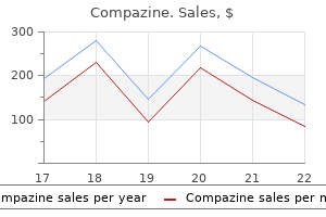
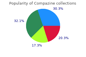
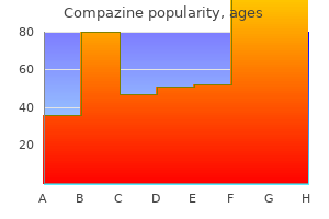
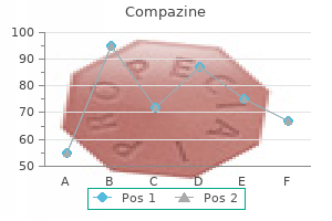
Compazine

Compazine
Compazine dosages: 5 mg
Compazine packs: 90 pills, 180 pills, 270 pills, 360 pills

This uncommon structure is expelled following uptake of virus particles into host cells by phagocytosis to permit fusion of the viral membrane with that of the mobile vacuole symptoms 10 days before period generic compazine 5 mg free shipping. Unprecedented assemblies specialised for release of the viral genome in host cells might prove to be a characteristic property of the very large viruses medications for adhd 5 mg compazine safe. The gold beads are electron dense and seem as darkish spots in the electron micrograph symptoms 6 weeks pregnant generic 5 mg compazine free shipping. They are present at a single vertex in each nucleocapsid, which due to this fact incorporates one portal. Tegument proteins that bind to hexons plus pentons and to triplexes are shown in blue and pink, respectively. The virus Pithovirus sibericum was isolated following culture of a suspension of soil from a pattern of permafrost collected in 2000 in Siberia with the ameba Acanthamoeba castellani. Shown is an electron micrograph of a particle noticed in infected ameba late in the infectious cycle following ultrathin sectioning of fastened cells and negative staining. However, many include additional viral proteins or other parts, that are typically current at much decrease concentrations however are important or essential for establishing an environment friendly infectious cycle (Table 4. Reovirus Reovirus kind 1 2 three Enzymes Many kinds of virus particles contain enzymes needed for synthesis of viral nucleic acids. Other forms of enzymes found in virus particles embody integrase, cap-dependent endonuclease, and proteases. Among the most effective characterised are the protein primers for viral genome replication which are covalently linked to the genomes of picornaviruses corresponding to Structure 117 poliovirus and adenoviruses. The cores of vaccinia virus additionally include proteins which would possibly be essential for transcription of viral genes, as they allow recognition of viral early promoters. Retroviruses with advanced genomes, corresponding to human immunodeficiency virus kind 1, comprise additional proteins required for efficient viral replica in certain cell sorts, for instance, Nef and Vpr. Nongenomic Viral Nucleic Acid the presence of a viral nucleic acid genome has lengthy been acknowledged as a definitive function of virions. It is subsequently difficult to exclude the chance that their presence is a functionally irrelevant and secondary consequence of nonspecific nucleic acid binding by viral structural proteins. For instance, mobile glycoproteins is most likely not excluded from the membrane from which the viral envelope is derived. Furthermore, as a bud enlarges and pinches off throughout virus assembly, inside cellular parts may be trapped within it. As a outcome, preparations of those viruses could additionally be contaminated with vesicles fashioned from cellular membranes. Indeed, analysis by the sensitive proteomic strategies offered by mass spectrometry has identified from 50 to a hundred mobile proteins in purified, enveloped particles of assorted herpesviruses, filoviruses, and rhabdoviruses. Consequently, it can be difficult to distinguish mobile components specifically included into enveloped virus particles from those trapped randomly or copurifying with the virus. The cellular elements captured in retrovirus particles have been significantly properly characterised. One of the most unusual properties of human immunodeficiency virus sort 1 is the presence of cellular cyclophilin A, a chaperone that assists or catalyzes protein folding. This protein is the major cytoplasmic member of a ubiquitous family of peptidyl-prolyl isomerases. Cellular membrane proteins, similar to Icam-1 and Lfa1 (see Chapter 5), can be integrated within the viral envelope and might contribute to attachment and entry of retroviral particles. It is obvious from these examples that virus particles contain a surprisingly broad repertoire of biologically energetic molecules that are delivered to their host cells. This repertoire is undoubtedly bigger than we presently respect, and the contributions of many components of virus particles to the infectious cycles of many viruses have but to be established. This strategy has also been invaluable in figuring out cellular proteins that are additionally current, with considerable numbers detected in the particles of several households of enveloped viruses. The sizeable populations (50 to 100) of such mobile proteins emphasize the significance of distinguishing those proteins that contribute to viral reproduction from those integrated by probability. Purified extracellular herpes simplex virus type 1 particles had been found by mass spectrometry to include 49 cellular proteins. This set included proteins reported to be present in the particles of different herpesviruses, such as cyclophilin A and actin, and many not detected previously. In these experiments, a phenotypically wild-type virus with a capsid protein fused to the green fluorescent protein was exploited to enable rapid and accurate measurement of yields of extracellular virus particles. Removal of thirteen of these proteins from particles decreased virus yield significantly, even in cells that continued to synthesize the proteins. These observations established unequivocally that some cellular proteins incorporated into herpesviral particles promote the following cycle of replica. Perhaps viral replica is facilitated by delivery of specific mobile proteins already associated with elements of viral particles or delivery of the proteins to specific sites during entry. Comprehensive characterization of extracellular herpes simplex virus sort 1 virions. Two-step technique to assess the importance of mobile proteins integrated into herpes simplex virus type 1 particles. Now that many structures of particles or their parts have been examined, we can appreciate the surprisingly numerous architectures they exhibit. Nevertheless, the easy principles of their construction proposed more than 50 years in the past stay pertinent: with few exceptions, the capsid shells that encase and defend nucleic acid genomes are constructed from a small variety of proteins arranged with helical or icosahedral symmetry. The detailed views of nonenveloped virus particles offered by X-ray crystallography emphasize simply how well these protein shells provide safety of the genome throughout passage from one host cell or organism to another. They have additionally recognized several mechanisms by which identical or nonidentical subunits can interact to type icosahedrally symmetric buildings. More-elaborate virus particles, which may contain further protein layers, a lipid envelope carrying viral proteins, and enzymes or other proteins necessary to initiate the infectious cycle, pose greater challenges to the structural biologist. Indeed, for many years we possessed solely schematic views of those constructions, deduced from negative-contrast electron microscopy and biochemical or genetic strategies of research. In the intervening period of simply 5 years, these techniques have attained atomic-level decision, providing outstanding views of enormous viruses with a quantity of components, viral envelopes, and, in some instances, the group of genomes within particles. The structural descriptions of ever-increasing numbers of viruses representing numerous families have additionally allowed distinctive insights into evolutionary relationships among seemingly disparate viruses or viral proteins. The more recently described giant viruses, corresponding to pandoravirus, with particles so massive that they can be seen by gentle microscopy, also pose new technical challenges and recommend that unanticipated structural ideas remain to be elucidated. Reconstructing virus buildings from nanometer to near-atomic resolutions with cryo-electron microscopy and tomography. The constructions of human rhinovirus and Mengo virus: relevance to operate and drug design. Structure of a standard chilly virus and functional relationship to other picornaviruses. Three-dimensional construction of the human immunodeficiency virus kind 1 matrix protein. Insight into the mechanism of the influenza A proton channel from a structure in a lipid bilayer. Structure of the haemagglutinin membrane glycoprotein of influenza virus at 3 � resolution. The fusion glycoprotein shell of Semliki Forest virus: an icosahedral assembly primed for fusogenic activation at endosomal pH. Cryo-electron microscopy reveals the functional organization of an enveloped virus, Semliki Forest virus. Proteomic and biochemical evaluation of purified human immunodeficiency virus kind 1 produced from infected monocyte-derived macrophages. The physical properties of the virion are obstacles to this seemingly easy goal. Furthermore, the viral genome is encapsidated in a steady coat that shields the nucleic acid as it travels via the cruel extracellular setting. These impediments should all be overcome in the course of the process of viral entry into cells. Encounter of a virus particle with the floor of a susceptible host cell induces a sequence of events that result in entry of the viral genome into the cytoplasm or nucleus. The first step in entry is adherence of virus particles to the plasma membrane, an interplay mediated by binding to a selected receptor molecule on the cell floor. The receptor plays an necessary role in uncoating, the process by which the viral genome is uncovered, in order that gene expression and genome replication can begin. Interaction of the virus particle with its receptor may provoke conformational modifications that prime the capsid for uncoating.
Diseases
Retroid viruses appear to represent a continuum in evolution medicine cups compazine 5 mg order without prescription, and remind us of the numerous combos of strategies that exist in nature for replicating viral genomes and related genetic elements symptoms knee sprain quality compazine 5 mg. It is now clear that these protein molecules can be scaffolds as nicely as catalysts medicine search compazine 5 mg buy otc, and their a quantity of functions seem to be enabled by a outstanding capacity for dynamic conformational change. It has been suggested that the simultaneous interplay of the central area with both ends could hold the termini in a position that facilitates each strand primer translocation and the second template change. The shaded bins indicate that the nucleic acids (genomes) encapsidated in particles of each virus symbolize totally different components in analogous pathways. As may be expected, such progress has elicited essential new questions to be addressed sooner or later. Host factors in retroviral integration and choice of goal websites, p 1035�1050. Human immunodeficiency virus reverse transcriptase: 25 years of analysis, drug discovery, and promise. The indicators that management expression of the genes of these viruses are similar to those of cellular genes. Such orderly gene expression is primarily the outcomes of transcriptional regulation by viral proteins. Such conservation of sequence could be attributed to the common biochemical capabilities of the enzymes. Transitions from one part to the subsequent rely upon viral activators and synthesis of progeny viral genomes. Viral proteins that regulate transcription could bind directly to viral promoter sequences or indirectly in affiliation with cellular proteins. Some viruses, together with the herpesviruses, establish latent infections by which transcription of lytic genes is inhibited and, in some instances, distinctive latencyassociated transcription items are expressed. Suppression of cellular transcription by viral components diverts limited cellular resources to aid viral transcription. Viral proteins can stimulate transcription of their very own transcriptional unit to establish a constructive autoregulatory loop or activate transcription of various viral genes. However, it may possibly additionally perform at least one reaction unique to virus-infected cells, Table 8. Transcription of specific genes is subsequently the primary biosynthetic reaction in cells contaminated by adenoviruses, herpesviruses, papillomaviruses, and polyomaviruses. The posttranslational modifications of the histones help distinguish highly condensed, transcriptionally silent heterochromatin from transcriptionally lively genes. Application of this assay to herpes simplex virus type 1-infected cells, as illustrated, has demonstrated that histone H3 binds to immediate-early, early, and late genes in getting into, but not in newly replicated, viral genomes. During lytic infection herpes simplex virus type 1 is related to histones bearing modifications that correlate with lively transcription. The polycomb group protein Bmi1 binds to the herpes simplex virus 1 latent genome and maintains repressive histone marks during latency. Temporal affiliation of the herpes simplex virus genome with histone proteins during a lytic an infection. Such nucleosomal group means that mechanisms analogous to those regulating transcription of cellular chromatin are more likely to operate on these viral templates. Indeed, as we will see, the properties of viral "chromatin" can lead to transcriptional silencing and prevent transcription of the overwhelming majority of viral genes in cells latently contaminated by some herpesviruses. For example, the human adenovirus kind 2 major late promoter was the primary from which correct initiation of transcription was reconstituted in vitro (Box 8. The core promoter comprises the minimal sequence essential to specify accurate initiation of transcription. The activity of the core promoter is modulated by native regulatory sequences usually found inside a number of hundred base pairs of the initiation website. Distant regulatory sequences that stimulate (enhancers) or repress (silencers) transcription are current in numerous transcriptional management regions. Application of these methods has identified a really giant number of transcriptional control sequences. Core promoters of viral and cellular genes include all the information needed for recognition of the positioning of initiation and assembly of exactly organized preinitiation complexes. Many others should be maintained in an virtually silent state, from which they can be activated rapidly in response to particular stimuli, and to which they are often returned readily. Transcription of viral genes can be regulated during the infectious cycles of a lot of the viruses considered in this chapter. Large quantities of viral proteins for assembly of progeny virions have to be made inside a finite (and usually short) infectious cycle. In many circumstances, viral genes are transcribed in a particular and stereotyped temporal sequence. Indeed, it was detailed information about a specific adenoviral transcription unit that finally allowed biochemical studies of the mechanism of initiation. This data was exploited to develop a simple assay for accurate initiation of transcription, the "runoff" assay, using a linear template that consists of a transcription initiation site proven within the determine. This runoff transcription assay is convenient and has been used to assess each specificity and effectivity of transcription. In common, however, viral proteins are critical parts of the circuits that establish orderly transcription of viral genes. Recognition of Local and Distant Regulatory Sequences Both local and distant sequences can management transcription from core promoters. However, native sequences are sometimes sufficient for correct transcriptional regulation. An huge number of sequence-specific proteins that regulate transcription are now identified, many first identified by way of analyses of viral promoters. The first such enhancer, so named as a result of it stimulated transcription to a large diploma, was discovered in the genome of simian virus 40. Enhancers are outlined by their position- and orientation-independent stimulation of transcription of homologous and heterologous genes over distances as great as 10,000 bp in the genome. Despite these uncommon properties, enhancers are constructed with binding websites for the proteins that acknowledge native promoter sequences. The simian virus forty enhancer has been studied intensively, and its properties and mechanism of action are characteristic of many enhancers, whether or not of viral or mobile origin. The severing of those contacts allows the transcribing advanced to escape from the promoter and proceed with elongation (step 4). This promoter clearance step is often inefficient, with abortive initiation (step 5) predominating. In the latter process, initial transcripts are launched, reforming the open initiation complex. The preliminary elongating transcriptional complex accommodates some however not all of the proteins that kind the preinitiation complex, as well as proteins that stimulate elongation (not shown). It has not been proven experimentally that each one really operate as autonomous initiator sequences. The relative frequencies with which totally different initiation sites in a single promoter are used are indicated by the thickness of the purple arrows. The depictions of the transcription initiation proteins are primarily based on visualization of initiation by cryo-electron microscopy. The black arrows beneath the adenovirus kind 2 (Ad2) E2 early promoter indicate the orientation of the E2 issue (E2f)-binding sites. For instance, nuclear factor b (Nf- b) and certain members of the octamer-binding protein (Oct) household are enriched in cells of lymphoid origin, and their binding websites are needed for enhancer activity in these cells. Other parts of the enhancer, such as the activator protein 1 (Ap1)-binding websites, confer responsiveness to cellular signaling pathways. This constellation of enhancer parts ensures transcription of the viral early gene and initiation of the viral infectious cycle in many different cellular environments. Compelling proof in favor of this mannequin has been collected through the use of the simian virus forty enhancer (Box eight. These regulatory sequences can also facilitate access of the transcriptional machinery to chromatin templates. Consequently, the names given by individual investigators had been based on different properties of the protein. The subsequent recognition that many "factors" are members of households of carefully associated proteins compounds such difficulties. The positions of the 72-bp repeat region containing the enhancer elements are shown relative to the early promoter on the high. All the protein-binding websites shown between the growth strains are repeated in the promoter-proximal 72-bp repeat.
A second common pattern is differential regulation symptoms 5 weeks 3 days 5 mg compazine discount otc, by which expression of some genes is increased and that of others decreased 909 treatment discount 5 mg compazine free shipping. Extensive alteration in mobile signaling is an inevitable consequence of virus an infection symptoms 11dpo buy cheap compazine 5 mg on line. In signal transduction pathways, detection of an informational molecule, such as a metabolite. Proteins that function in any mobile process could also be effectors, however those who regulate gene expression are widespread targets. Many of the quite a few signaling pathways of mammalian cells respond to more than a single input, regulate a quantity of molecular processes, and communicate with one another. The kinases Pi3k and Akt are focal factors or hubs within the signaling network, with multiple inputs and outputs. Binding of ligand to any one of several forms of plasma membrane receptors initiates signaling to Pi3k, which is related to the inside floor of the plasma membrane, and activation of this kinase via phosphorylation. Mammalian cells include three lessons of Pi3ks, distinguished by their intracellular distributions, mechanisms of activation, and substrate specificity. Shown is the commonest, class I Pi3ks, which comprise a regulatory (p85) and a catalytic (p110) subunit. These modified lipids are sure by particular domains of other proteins, corresponding to phophoinositide-dependent kinase 1 (Pdk1), which then transmit the sign to Akt. Shown are penalties that promote cell growth and proliferation via activation of the mTor kinase present in mTorC1. Activated mTor facilitates translation by multiple mechanisms and likewise induces autophagy, a process that helps cells survive excessive types of stress, such as amino acid hunger. The signaling hubs Pi3k, Akt, and mTor are linked to , and regulated by, different signaling methods and to one another by numerous suggestions circuits. Many viral gene products intervene to block defensive responses of the host that would inhibit virus reproduction. In this section, we focus on modulations of signaling pathways that facilitate virus replica, and use specific examples to illustrate two general principles: the same sign transduction pathway could be modified in cells infected by many different viruses, and particular person viruses can modulate multiple signaling pathways. We subsequently illustrate the varied impression of infection on one sign transduction cascade utilizing this pathway. Such resculpting of these structural elements of the cell is essential for movement of cells; formation of extensions, such as lamellipodia; and different processes that require reorganization of the external floor of the cell, including virus entry. Attachment of viruses belonging to numerous families to their cognate cell surface receptors induces speedy activation (phosphorylation) of Pi3k. Although Pi3k is activated in all circumstances, the downstream pathways are virus particular, as a result of the mechanisms of entry differ from virus to virus. Attachment of human adenovirus to its integrin receptor results in signaling from Pi3k via small G proteins to induce actin reorganization and facilitate endocytosis of virus particles. Attachment of influenza A virus particles, which outcomes in clustering of lipid rafts and associated receptor protein tyrosine kinases and subsequent activation of Pi3k, stimulates not solely actin remodeling, but also the acidification of endosomes necessary for disassembly of virus particles. Presumably, these distinct outputs of Pi3k and Akt signaling are determined by the virus-specific mechanisms of activation of the kinases. Entry of all viruses that reproduce in mammalian cells is determined by some extent of refashioning of the plasma membrane and associated cytoskeleton. It subsequently seems probably that subversion of the normal operate of Pi3k, Akt, or both in regulating membrane transactions will prove to be a extra common response to the encounter of host cells with virus particles. Signaling initiated by activation of Pi3k additionally facilitates later steps in virus reproduction. This kinase alerts to not solely Akt but additionally a second kinase, mTor, present in mTor advanced 1 (mTorC1). All these responses could be expected to be beneficial for completion of viral infectious cycles. In truth, in every case that has been examined, virus infection has been observed to activate signaling by way of Pi3k to Akt, and in plenty of circumstances, mTorC1. The merchandise of such genes can induce everlasting activation of cell proliferation, a course of termed transformation, and sometimes acquisition of the power to type tumors in animals. Furthermore, the genomes of quite lots of viruses encode proteins that intervene downstream of Pi3k to maintain mTor activity and consequently efficient translation (Chapter 11). Infection with a Particular Virus Modulates Multiple Signal Transduction Pathways Virus reproduction is invariably accompanied by alterations in additional than a single signaling relay, typically with one or more pathways blocked and others stimulated. Concurrently, signaling cascades that govern different processes are modulated to assist the reactions needed for expression and replication of viral genomes and meeting of progeny virus particles. Infection by viruses with even relatively simple genomes and mechanisms of copy that depend on a minimal set of cellular methods and components leads to modification of several signaling pathways. Pi3k is activated following attachment to cellular receptors of adenoviruses, filoviruses, flaviviruses, influenza viruses, herpesvirus, and poxviruses, among others. Signaling initiated by the action of Pi3k leads to actin transforming via activation of the small G proteins Rac and Cdc42. Inhibition of phosphorylation of Cas or manufacturing of a dominant-negative by-product of Rac inhibits adenovirus entry, emphasizing the importance of this mobile signaling pathway for efficient internalization. In this case, these processes rely upon signaling by way of Akt and focal adhesion kinase (Fak), and Pi3k is activated following clustering of lipid rafts and their associated receptor protein tyrosine kinases (Rptks) in the plasma membrane. Such concentration of receptors facilitates their activation by cross-phosphorylation, and likewise activates Mapk1 and -3. Such signal transduction indirectly facilitates release of the viral genomes into the cytoplasm by blocking diversion of endosomes containing virus particles to a nonproductive pathway. However, the flexibility of the mutant virus to kind plaques on these cells was impaired (see the figure), as was induction of apoptosis when assessed by three different assays. In common, it appears that the more elaborate the strategy for viral reproduction, the larger the impression of infection on signaling pathways. Furthermore, how radically cellular signaling methods are altered will also be determined by the origin and proliferation state of the host cell. Many human cells in routine use in the laboratory are derived from tumors (Chapter 2), and consequently are abnormal in many respects, together with unrestrained proliferation and permanent activation of signaling circuits that promote cell progress and progression by way of the cell cycle. In contrast, in natural infections, many host cells proliferate only slowly or are quiescent (withdrawn from the cell cycle). Successful virus replica in such cells is therefore more likely to depend to a greater degree on activation of signaling pathways that control these processes than does reproduction in tumor-derived cell strains. The results of latest applications of those strategies to comparison of uninfected and virus-infected cells suggest that the impact of particular viruses on host cell signaling is even broader than beforehand appreciated. For example, comparability of the concentrations of phosphopeptides in uninfected, quiescent mouse fibroblasts and 18 h after an infection with murine herpesvirus sixty eight (a gammaherpesvirus) recognized modifications in 86% of the practically 2,500 distinctive peptides examined. This infection-induced difference is much bigger than that observed following publicity of cells to growth components (13%) or assaults, similar to harm to the genome or publicity of human cells to Salmonella (24%). Large-scale analyses of phosphoproteins in infected cells also can establish mobile substrates of signaling pathways which are necessary for virus reproduction. Subsequent functional analysis of manufacturing of proteins with increased phosphorylation in infected cells established the necessary contribution of host proteins not previously implicated in copy of human immunodeficiency virus kind 1, notably a specific set of splicing proteins. Gene Expression Altered host cell gene expression is a common consequence of virus an infection. The influence of virus infection on mobile gene expression and the mechanism(s) by which this course of is altered vary with the methods by which viral genes are expressed. Many of these proteins are described in previous chapters (Chapters eight, 10, and 11). Colors from yellow to blue and to pink indicate decreases and increases, respectively. Moss, National Institute of Allergy and Infectious Diseases, National Institutes of Health. The very short infectious cycle of poliovirus (and different picornaviruses; some 8 h) may necessitate particularly efficient measures to stop synthesis of mobile proteins. Indeed, when the concentrations of cellular proteins had been assessed early and late after vaccinia virus infection of human cells, 10% were observed to change significantly. Virus replica is decided by many steady cellular proteins, including ribosomal proteins and structural proteins of the cytoskeleton. However, it could even be necessary to keep the production of a lot less steady host proteins for optimum replica of explicit viruses, even within the face of widespread inhibition of cellular gene expression.
Clinically treatment neutropenia buy cheap compazine 5 mg line, smokers with combined fibrosis and emphysema on high-resolution computed tomography scans can have unexpectedly regular spirometry findings regardless of decreased total 906 lung capacity treatment for depression generic 5 mg compazine fast delivery, and some authors have speculated that smokingrelated interstitial fibrosis will be the pathologic correlate of this combined physiologic disturbance medicine 3604 5 mg compazine purchase with visa. The differential diagnosis of smoking-related interstitial fibrosis consists of usual interstitial pneumonia (which could be distinguished based mostly on its temporal variability and the presence of numerous fibroblast foci), asbestosis, and pulmonary Langerhans cell histiocytosis. Histologic Features Uniform, acellular collagen deposition results in thickening of the alveolar septae. The interstitial thickening could show accentuation within the subpleural and peribronchiolar compartments of the lung. Respiratory bronchiolitis is invariably current; a desquamative interstitial pneumonia pattern of plentiful airspace macrophages may be seen. Inflammatory cells are sparse and are enriched within the respiratory bronchioles consistent with the affiliation with respiratory bronchiolitis. Kim Hypersensitivity pneumonitis is a diffuse lung disease attributable to inhalational publicity to natural antigens. The former time period, extrinsic allergic alveolitis, underscores the importance of the diffuse mobile interstitial infiltrate in this illness. Interestingly, smoking appears to present a protective profit, as smokers are much less more likely to suffer from hypersensitivity pneumonitis. In the acute and subacute forms, patients typically current with shortness of breath, cough, and infrequently systemic signs, corresponding to malaise and fever. The chronic kind develops over a longer interval, with progressive dyspnea, cough, weight reduction, and end-stage fibrotic lung illness. The basic imaging findings are described as upper lobe predominant, ill-defined floor glass nodules, with a centrilobular distribution and instant subpleural sparing. In persistent forms, there should be some vague centrilobular nodules, however elevated reticulation, fibrosis, and even radiographic cysts of honeycombing may be identified. Histologic Features Acute Hypersensitivity Pneumonitis 912 Acute and organizing lung damage with fibrin, diffuse alveolar harm, granulomatous pneumonitis, and organizing pneumonia. Subacute Hypersensitivity Pneumonitis Diffuse mobile interstitial infiltrates, which can show a centrilobular distribution. Centrilobular poorly shaped interstitial granulomas, consisting of loose aggregates of histiocytes with or without multinucleated large cells in a background of lymphoplasmacytic irritation. Granulomas should be troublesome to identify; if granulomas are quite a few, show necrosis, or are obvious from scanning magnification, an alternate analysis ought to be thought-about. Chronic Hypersensitivity Pneumonitis Fibrotic nonspecific interstitial pneumonia sample of fibrosis or airway-centered sample of fibrosis. Chronic hypersensitivity pneumonitis may be superior with microscopic honeycomb reworking and fibroblast foci, mimicking the identical old interstitial pneumonia sample of pulmonary fibrosis. Extensive continual small airways transforming in the type of peribronchiolar metaplasia, mucostasis, and continual bronchiolitis. Scattered interstitial poorly formed granulomas with or with out multinucleated giant cells. Lack of peripheral and subpleural predominant fibrosis of usual interstitial pneumonia of idiopathic pulmonary fibrosis. Differential Diagnosis Various entities in the differential analysis depend upon the section of presentation: acute, subacute, or chronic. In the acute form, the differential prognosis is just like that of acute and organizing lung damage and includes infection, adverse drug reaction, connective tissue illness, widespread aspiration, and idiopathic (acute 913 respiratory misery syndrome). The presence of airway centricity with obscure granulomas or multinucleated giant cells may help help a diagnosis of acute hypersensitivity pneumonitis. In the subacute section, the differential prognosis is just like that of a mobile interstitial pneumonia, similar to cellular nonspecific interstitial pneumonia, and consists of connective tissue illness, antagonistic drug response, aspiration, and infection. Because the granulomas are sometimes small, poorly formed, and troublesome to establish, different granulomatous diseases (such as granulomatosis with polyangiitis, necrotizing granulomas of infection, and sarcoidosis) rarely enter into the differential diagnosis. The accentuation of changes throughout the centrilobular regions strongly helps a prognosis of hypersensitivity pneumonitis, as does the presence of background chronic small airways reworking. In persistent hypersensitivity pneumonitis, the main differential analysis consists of fibrotic nonspecific interstitial pneumonia and ordinary interstitial pneumonia patterns of pulmonary fibrosis. In cases of otherwise typical traditional interstitial pneumonia, close scrutiny of the interstitium for poorly fashioned granulomas and multinucleated large cells, together with the presence of chronic small airways transforming, may help help continual hypersensitivity pneumonitis as an etiology for the superior pulmonary fibrosis. Histologic features that have been shown to be more usually found in the setting of continual hypersensitivity pneumonitis as opposed to ordinary interstitial pneumonia embrace the presence of granulomas, big cells, organizing pneumonia, bridging fibrosis, cellular nonspecific interstitial pneumonia areas, lymphoid follicles, centrilobular fibrosis, and continual bronchiolitis. In the subacute section, therapy with steroids can enhance outcome; nonetheless, removal of the offending antigen is the most effective predictor of a good outcome. Once the disease has reached the chronic phase with superior fibrosis, steroid treatment could or will not be beneficial. Histologic patterns of fibrotic nonspecific interstitial pneumonia or ordinary interstitial pneumonia, older age, and longer/more intense exposure are related to worse prognosis. Some cases could present extra of a fibrotic nonspecific interstitial pneumonia pattern of damage. Note the bridging of fibrosis from the centrilobular area to the interlobular septa. Scattered poorly formed interstitial granulomas had been present in this case, supporting hypersensitivity pneumonitis as an etiology for the superior fibrosis. Smith Fang Zhou Nontuberculous mycobacterial an infection of the lung manifests as a large spectrum of pathologic patterns, together with cavitary illness, nodules with bronchiectasis, and diffuse interstitial infiltrates. Hot tub lung is the clinicopathologic manifestation of nontuberculous mycobacterial involvement secondary to the inhalation of aerosolized Mycobacterium avium complicated, often in the setting of contaminated hot tubs or different water sources. This typically occurs in immunocompetent sufferers with a wide age vary and equal distribution between men and women. Imaging studies present bilateral diffuse infiltrates, often with finely nodular interstitial infiltrates. Histologic Features the predominant histopathologic feature is the presence of nonnecrotizing granulomas. The granulomas typically show loose clustering of epithelioid histiocytes with occasional multinucleated large cells. There is uncommon to nonexistent involvement of the intralobular septa 924 and pleura. Polyps of organizing pneumonia are regularly seen within the airspaces and terminal airways. The demonstration of acid-fast bacilli is exceedingly rare, even in traditional cases. Confirmation by tradition studies is the popular technique of confirming the analysis. Patients are likely to have a great prognosis, notably if the exposure is eliminated or the water is decontaminated. Differential Diagnosis the differential prognosis for the presence of nonnecrotizing granulomas consists of hypersensitivity pneumonitis, sarcoidosis, and diffuse granulomatous infections because of different organisms. Hypersensitivity pneumonitis typically exhibits a much more strong cellular interstitial infiltrate that overshadows the presence of granulomatous inflammation. Furthermore, the granulomas of hypersensitivity pneumonitis are usually positioned inside the interstitium in a centrilobular distribution and will only not often show granulomas within the airspaces. Obviously, the demonstration of acidfast bacilli on acid-fast staining would assist a prognosis of scorching tub lung. The granulomas of sarcoidosis usually present less surrounding irritation compared with the granulomas of sizzling tub lung. In addition, they typically tend to coalesce along with background hyaline sclerosis, a characteristic not seen within the setting of sizzling tub lung. Furthermore, the granulomas of sarcoidosis show a distinct lymphangitic distribution, involving the bronchovascular bundle, intralobular septa, and pleura, whereas the granulomas of scorching tub lung are limited to the airspaces. The distinction between scorching tub lung and infectious granulomatous pneumonitis due to different organisms may be 925 challenging. The presence of necrotizing granulomas would favor a unique infectious etiology over M. In addition, patients with diffuse lung illness secondary to different organisms are typically much more unwell than patients presenting with hot tub lung. The major risk factor is working in a flock factory; multiple outbreaks of interstitial pneumonia have been identified amongst nylon flock employees. Pulmonary perform testing usually reveals a restrictive sample and a decreased diffusing capacity.
Cavinton (Vinpocetine). Compazine.
Source: http://www.rxlist.com/script/main/art.asp?articlekey=96211
This hydrophilic molecule is transported across the plasma membrane by any considered one of a number of glucose transporters (12 are encoded in the human genome) medicine in the middle ages compazine 5 mg discount with mastercard. These embrace the ubiquitous protein glucose transporter 1 (Glut1); tissue-specific transporters such as Glut3 symptoms before period 5 mg compazine purchase, current in neurons of the central nervous system; and the insulin-regulated transporter Glut4 medications dialyzed out buy compazine 5 mg overnight delivery, which is present in skeletal muscle and adipose tissue. Localization of Glut4 to the plasma membrane is dependent upon the presence of the hormone insulin, which is synthesized in and released from specialized cells (cells) within the pancreas in response to excessive concentrations of blood glucose. One parameter contributing to the increased rate of glycolysis noticed in cells contaminated by hepatitis C virus, herpesviruses, and human immunodeficiency virus sort 1 is an increased rate of glucose uptake. In several instances, such accelerated transport of glucose into virus-infected cells could be attributed to elevated concentrations of Glut1 or Glut3, because of alterations in sign transduction pathways that regulate transcription. For instance, in cells infected by the gammaherpesvirus EpsteinBarr virus or human papillomaviruses, the focus of hypoxia-inducible factor 1 (Hif-1) is increased. This transcriptional regulator prompts expression of the genes encoding Glut1 and Glut3 (as nicely as several that encode glycolytic enzymes). This mechanism resembles the normal responses of uninfected cells to low availability of oxygen (hypoxia) and nutrients. In contrast to modulation of Glut1 or Glut3, the elevated uptake of glucose into human cytomegalovirus-infected fibroblasts is mediated by the insulin-regulated transporter Glut4. The restriction of the transporter to intracellular vesicles except insulin stimulates signaling by way of Akt can additionally be circumvented, because infection prompts this sign transduction pathway. These observations emphasize the importance of the swap from Glut1 to Glut4 for environment friendly human cytomegalovirus reproduction, a necessity that may not have been predicted from the comparatively modest increase (some 3-fold) in the affinity of Glut4 for glucose. In precept (the law of mass action), increased intracellular concentrations of glucose because of extra environment friendly transport throughout the plasma membrane might account for elevated charges of glycolysis in virus-infected cells. Nevertheless, in several instances, the rate of flux through this pathway can also be accelerated by will increase in the intracellular concentration or activity of a quantity of glycolytic enzymes. Virus Infection Can Redirect the Utilization of Glycolytic Intermediates and Products Acceleration of glycolysis in virus-infected cells might help present the extra vitality required for production and transport of viral macromolecules. However, major differences had been additionally detected, notably increased accumulation of glycolytic intermediates in cells contaminated by herpes simplex virus 1, but decreased concentrations in human cytomegalovirus-infected cells (panel A of the figure). The reasons for this difference have been investigated further, for instance, by supplying contaminated cells with 13C-labeled glucose and monitoring its incorporation into downstream metabolites as a operate of time thereafter. In human cytomegalovirus-infected cells, the uptake of glucose and the labeling of glycolytic intermediates similar to fructose-1,6-bisphosphate have been elevated, indicating stimulation of glycolytic flux (panel B). However, these parameters have been decreased in herpes simplex virus-infected cells, accounting for the buildup of glycolytic intermediates (panel A). This response was accompanied by increased concentrations of intermediates in the pentose phosphate pathway and of its product, ribose-5-phosphate (panel B). Synthesis of pyrimidines can be elevated in herpes simplex virus 1-infected cells (see text). The increased manufacturing of pyruvate in human cytomegalovirus-infected cells because of the acceleration of glycolytic flux supports increased production of fatty acids, following synthesis of the precursor, acetyl-CoA (see the text). It has been proposed that the fairly different fates of carbon from glucose in cells infected by these two human herpesviruses is the outcome of the a lot shorter replication cycle of herpes simplex virus 1 than of human cytomegalovirus, some 24 and 96 h, respectively, to attain the maximal yield of progeny virus particles. Divergent results of human cytomegalovirus and herpes simplex virus-1 on mobile metabolism. Under cardio situations in mammalian cells, pyruvate (the last product of glycolysis) enters mitochondria and is transformed to acetyl-CoA. This compound is an activated provider of units of two carbon atoms that is a main supply of energy following its entry into the citric acid cycle (see below), and can also be the precursor for synthesis of fatty acids and sterols. However, this process takes place in the cytoplasm, whereas acetyl-CoA is produced throughout the mitochondrial matrix, as indicated. Formation of malonyl-CoA by acetyl-CoA carboxylase initiates fatty acid synthesis, which is catalyzed by the multi-active-site enzyme fatty acid synthase. In distinction, an necessary operate of glycolysis during the early section of herpes simplex virus 1 an infection is to enable increased synthesis of ribose and deoxyribose (and therefore nucleotides and nucleic acids): the concentrations of intermediates and enzymes of the pentose phosphate pathway are elevated in fibroblasts contaminated by this human herpesvirus. It has been proposed that the totally different fates of glycolytic intermediates or merchandise in cells contaminated by these two human herpesviruses are a consequence of the rather more speedy copy of herpes simplex virus 1 than of human cytomegalovirus (Box 14. The latter activity is crucial to maintain blood glucose concentrations when meals consumption is low or during prolonged train or stress. Among the detrimental effects of an infection by some hepatotropic viruses are main perturbations of glucose homeostasis that can result in growth of disease. Infection by the hepadnavirus hepatitis B virus is strongly related to the event of sort 2 diabetes (insulin-independent) in certain populations (for instance, Asians) and with increased blood glucose levels (hyperglycemia) in patients with acute infection. Such a systemic perturbation in glucose concentrations arises as a outcome of a high proportion of liver cells can be infected by this virus. In both human hepatocytes in culture and the livers of transgenic mice, expression of the viral X gene is sufficient to stimulate expression of a quantity of mobile genes that encode enzymes required for synthesis of glucose from 2-carbon precursors (gluconeogenesis), notably phosphoenolpyruvate carboxykinase, which catalyzes the first (and committed) response in the synthesis of glucose from pyruvate. This response is prone to be of physiological significance, as a end result of mice transgenic for the viral X gene exhibit increased synthesis of glucose compared to management mice. Both activation of glucogenesis and glucose intolerance are signs of type 2 diabetes. Insulin resistance, another diagnostic marker for growth of kind 2 diabetes, is exhibited by some 25% of patients with persistent hepatitis C virus infection. Insulin-dependent signaling is disrupted by the viral C (capsid) protein, which induces altered phosphorylation and elevated degradation of a crucial signal transducer in this pathway. These observations recommend that increased glucose synthesis and lowered uptake of glucose by infected hepatocytes may also promote growth the Infected Cell 483 of sort 2 diabetes in sufferers with chronic hepatitis C virus an infection. Such studies established that 2 carbon atoms are integrated into citrate (6 carbon atoms) from glucose in cells contaminated by herpes simplex virus 1, however that 3 carbon atoms of citrate turn into labeled in human cytomegalovirus-infected cells. The alternative between these fates of pyruvate is strongly regulated by the focus of acetyl-CoA. This essential intermediate in metabolism is produced by catabolism of a variety of fuels, and its focus is subsequently an indicator of how nicely a cell is equipped with a supply of energy. In human cytomegalovirus-infected cells, a lot acetyl-CoA is consumed for synthesis of fatty acids (see "Lipid Metabolism" below). Consequently, the mitochondrial focus of this compound could be low, favoring activation of pyruvate dehydrogenase. Even although synthesis of oxaloacetate catalyzed by pyruvate carboxylase is a reaction that replenishes the citric acid cycle, the focus of intermediates in this cycle decreases as the herpes simplex virus infectious cycle proceeds. Inhibition of pyruvate carboxylase or the first enzyme within the pathway by which pyrimidines are created from aspartate impaired manufacturing of infectious particles of herpes simplex virus, however not of human cytomegalovirus, emphasizing the very completely different needs for cellular metabolic pathways for efficient copy of these human herpesviruses. Although such biosynthetic reactions take away intermediates from the cycle, their concentrations are usually nearly fixed, because the citric acid cycle is replenished by a quantity of reactions. Acetyl-CoA produced by oxidation of pyruvate enters the citric acid cycle when its 2 carbon atoms are transferred to oxaloacetate to kind citrate. Reactions consuming intermediates within the cycle or replenishing them which would possibly be stimulated in virus-infected cells are indicated. Halting of the citric acid cycle as a outcome of such withdrawal of citrate is prevented by the improved uptake of glutamine and its conversion to the citric acid cycle intermediate -ketoglutarate, due to increased production and activity of the enzymes that catalyze this course of in contaminated cells. When all carbon atoms of glucose are labeled with 13C (red), so too are all three carbon atoms of pyruvate. This metabolite may be converted to acetyl-CoA by pyruvate dehydrogenase or to oxaloacetate by pyruvate carboxylase to produce citrate with 2 or three 13C-labeled carbon atoms, respectively. Glutamine is also essential for maximally environment friendly reproduction of the poxvirus vaccinia virus: its elimination from the medium of contaminated cells reduces the yield of infectious virus particles by three orders of magnitude. The result of experiments by which infected cells were provided with other metabolites indicated that one fate of glutamine is replenishment of the citric acid cycle by way of synthesis of -ketoglutarate. Rather, it has been proposed that in cells infected by this poxvirus, this crucial compound enters the citric acid cycle by a baroque mechanism by which the fatty acid palmitate is first synthesized within the cytoplasm and then degraded to acetyl-CoA by oxidation in mitochondria (Box 14. Infection by a quantity of viruses has been reported to modulate one or more of those processes. Rather, it A model for production and utilization of palmitate in vaccinia virus-infected cells based on the was demonstrated that observations summarized above. The purple bars indicate inhibitors of response or pathways that decreased � the absence of glucose from the medium the yield of infectious virus particles. Acc, acetyl-CoA carboxylase; Cpt1, carnitine palmitoyltransferase 1; had no impact on the production of infec- Fas, fatty acid synthase.
When assembly of viroplasms is prevented medicine identification compazine 5 mg purchase with amex, for example treatment 3rd metatarsal stress fracture compazine 5 mg buy online, by mutations within the viral genome or exposure of infected cells to inhibitors of lipid droplet formation treatment resistant depression discount 5 mg compazine mastercard, the yield of infectious virus particles is reduced, as is virus-induced cell demise. Shown are central slices in tomograms (left-hand panels) and sections with three-dimensional reconstructions overlaid (right-hand panels). In the reconstructions, single membranes are shown in blue and the internal and outer membranes of double-membrane vesicles in yellow and green. As shown, single- and double-membrane buildings predominate early and late, respectively, within the infectious cycle, however occasional double-membrane vesicles could be seen from intermediate times. The latter are proposed to develop upon membrane invagination into single-membrane vesicles (steps 1 to 4), so that viral genome replication and assembly can occur on and inside vesicles, as has been reported. Autophagic vesicles would fuse with the plasma membrane to launch vesicle-enclosed virus particles (step 5). They can also mature into autolysomes, which possess an acidic, degradative environment, with lack of one of the two autophagosomal membranes. Subsequent fusion with the plasma membrane would enable nonlytic release of mature poliovirus particles (step 6). In particular, parts of the multivesicular body system are co-opted to enable release of the particles of all kinds of enveloped viruses (Chapter 13). Autophagy, a survival mechanism normally invoked underneath extreme situations, may enable nonlytic release of poliovirus and other picornaviruses. Perspectives Since the earliest virological experiments with host cells in tradition, the appreciable influence of virus an infection has been documented and exploited, for example, by using cytopathic results to seek for beforehand unrecognized viruses. Elucidation of the molecular particulars of the reproduction of particular person viruses established that progression via the infectious cycle is commonly accompanied by inhibition of basic cellular pro- cesses and reorganization of cellular architecture. However, a extra full appreciation of the magnitude and diversity of host cell responses has come only comparatively just lately, with the growing application of the methods of systems biology. Consequently, they provide terribly detailed comparisons between cells contaminated by a particular virus and their uninfected counterparts. These methods have been utilized to cells contaminated by a restricted repertoire of viruses for less than a comparatively brief time. Structures by which these proteins are both localized improve in measurement and number because the infectious cycle progresses. They have been termed viroplasms, and include other viral proteins, mobile lipids, and a second mobile protein present in lipid droplets. By a poorly understood course of which will embrace lipid degradation, such buildings mature into viroplasms that include double-layered particles. Furthermore, energy may also be provided by apparently unique, virus-specific mechanisms, such as the synthesis of the fatty acid palmitate for its subsequent oxidation in poxvirus-infected cells, or the mobilization of lipid shops by induction of autophagy in cells infected by dengue virus. Additional surprises, in addition to a greater understanding of the mechanisms by which viral gene products instantly or not directly regulate or redirect specific cellular processes and pathways, can be anticipated. We can now describe in appreciable detail some of the striking methods during which virus replica and redirection is accompanied by remodeling of architectural features of the host cell. These advances are the end result of enhancements in the methods by which infected cells can be visualized, notably these of electron tomography and three-dimensional reconstruction. A appreciable number of contaminated cell-specific constructions customary from both nuclear or cytoplasmic elements have been implicated in facilitating viral genome replication and gene expression, or meeting of progeny virus particles. In some cases, viral proteins needed for formation of such contaminated cell-specific platforms have been recognized, but much remains to be learned about how such proteins induce reorganization of host cell elements. However, the cells of multicellular organisms are specialised for particular tasks, and due to this fact additionally exhibit cell-type-specific properties and molecular capabilities. Virus infection may end up in main perturbations, even loss, 500 Chapter 14 of such specialized features. Phosphoinositide 3 kinase signaling might have an effect on a number of steps throughout herpes simplex virus type-1 entry. Real-time transcriptional profiling of mobile and viral gene expression throughout lytic cytomegalovirus infection. Human cytomegalovirus: coordinating mobile stress, signaling, and metabolic pathways. Forkhead field transcription factor regulation and lipid accumulation by hepatitis C virus. Serum metabolome and lipidome adjustments in grownup sufferers with major dengue infection. The impact of meningopneumonitis virus infection on glycolysis by mouse and rat brain. A function for vaccinia virus protein C16 in reprogramming cellular vitality metabolism. Hepatitis B virus X protein regulates hepatic glucose homeostasis through activation of inducible nitric oxide synthase. Rotaviruses associate with cellular lipid droplet parts to replicate in viroplasms, and compounds disrupting or blocking lipid droplets inhibit viroplasm formation and viral replication. Colocalization of transcription and translation inside cytoplasmic poxvirus factories coordinates viral expression and subjugates host functions. Subversion of mobile autophagy equipment by hepatitis B virus for viral envelopment. Miyanari Y, Atsuzawa K, Usuda N, Watashi K, Hishiki T, Zayas M, Bartenschlager R, Wakita T, Hijikata M, Shimotohno K. Infection by these viruses is often asymptomatic but can result in respiratory disease in kids (members of species B and C), conjunctivitis (members of species B and D), and gastroenteritis (species F serotypes forty and 41). Human adenoviruses forty and 41 are the second main trigger (after rotaviruses) of childish viral diarrhea. The Mastadenovirinae comprise over 65 adenoviruses of people and different mammals, including mice, sheep, and canines, and some are oncogenic in rodents. Study of human adenovirus transformation of cultured cells has provided basic details about mechanisms that management progression by way of the cell cycle and oncogenesis. The electron micrograph exhibits a negatively stained human adenovirus type 5 particle (courtesy of M. Immature particles comprise the precursors of the mature forms of several proteins. A prototype member of this household, lymphocytic choriomeningitis virus, has been used to elucidate essential rules of the host immune response to viral an infection. Arenaviruses trigger continual, normally asymptomatic, infections in rodents, the pure host. Cryo-electron micrograph of a negatively stained particle of the arenavirus lymphocytic choriomeningitis virus. Arenaviruses are categorized into Old World and New World serogroups, based on geographical and genetic parameters. Two genes, one encoded in the 5 end of each phase, are current in an ambisense orientation. For simplicity, solely expression of genes on the S phase is shown, however the identical course of happens for the L section. The name derives from the fringe of club-shaped spikes noticed in electron micrographs that give the virus particles the appearance of a photo voltaic corona. They cause vital respiratory and gastrointestinal illness in people and domestic animals. They had been known to cause common colds in humans, until the emergence of extreme acute respiratory syndrome coronavirus in 2002, which triggered a devastating human disease. Another new coronavirus, Middle East respiratory syndrome coronavirus, was first acknowledged in humans in April 2012. The mutation prevents the spikes from detaching when virus particles are purified. The virus particles possess an uncommon filamentous morphology, which led to the name of this family (Filum is the Latin word for thread). The electron micrograph reveals a picture of ebolavirus particles inside of an infected, cultured monkey cell (courtesy of Elizabeth R. Conserved sequences are current at the 3 (leader) and 5 (trailer) ends of the viral genome. In some cases the termination and initiation sequences of neighboring genes overlap. This interaction leads to fusion of the viral and endosomal membranes for delivery of the viral nucleocapsid into the cytoplasm. There are more than 50 viral species, a lot of that are transmitted by arthropod vectors. Flaviviruses cause quite lots of human illnesses, such as encephalitis, and hemorrhagic fevers.
Squamous Metaplasia medications canada 5 mg compazine cheap fast delivery, Squamous Dysplasia symptoms you need glasses generic 5 mg compazine mastercard, and Squamous Carcinoma In Situ Subpart four administering medications 6th edition compazine 5 mg cheap on line. Roden Chapter 47 Pulmonary Langerhans-Cell Histiocytosis Chapter forty eight Erdheim�Chester Disease Chapter forty nine Rosai�Dorfman Disease 1596 596 598 605 609 Chapter 50 Other Pulmonary Histiocytic Proliferations 612 Section6 Benign and Borderline Lymphoid ProliferationsKirtee Raparia Chapter fifty one Follicular Bronchiolitis and Follicular Bronchitis Chapter 52 Nodular Lymphoid Hyperplasia Chapter fifty three Lymphoid Interstitial Pneumonia 618 619 622 624 Section7 Focal LesionsRoss A. Miller Chapter 54 Apical Caps Chapter 55 Focal Scars Chapter fifty six Pulmonary Ossification Chapter fifty seven Intrapulmonary Lymph Nodes Chapter fifty eight Rounded Atelectasis Chapter fifty nine Amyloidosis (Nodular and Tracheobronchial) 627 628 631 634 638 641 643 Section8 Granulomatous DiseasesYasmeen M. Parasites Chapter sixty one Bronchocentric Granulomatosis Chapter sixty two Pulmonary Hyalinizing Granuloma Chapter sixty three Sarcoidosis Chapter 64 Berylliosis Chapter 65 Sarcoid-Like Reaction Chapter sixty six Necrotizing Sarcoid Granulomatosis Chapter 67 Foreign-Body Granulomas Part 1. Malakoplakia 647 648 648 650 654 657 664 670 677 682 684 689 689 692 695 699 699 704 Section9 Diffuse Pulmonary HemorrhageLynette M. Artifactual (Procedural) Hemorrhage/Hemorrhage in the Absence of Other Histologic Abnormalities Part 2. Idiopathic Pulmonary Hemosiderosis Chapter 70 Alveolar Hemorrhage with Vasculitis Part 1. [newline]Eosinophilic Granulomatosis with Polyangiitis (Formerly Churg�Strauss Syndrome) Part 5. Microscopic Polyangiitis 711 711 715 719 721 725 727 730 731 733 736 740 743 746 Section10 Pulmonary Hypertension and Embolic DiseaseAlain Borczuk Chapter 71 Pulmonary Arterial Hypertension Chapter 72 Pulmonary Veno-Occlusive Disease Chapter 73 Pulmonary Capillary Hemangiomatosis Chapter seventy four Pulmonary Hypertension Associated with Lung Disease and/or Hypoxemia Chapter 75 Thromboembolic Disease-Acute and Chronic Part 1. Bone Marrow Emboli Chapter 76 Pulmonary Infarct 1598 750 752 762 766 771 775 775 782 784 785 788 791 793 795 Chapter 77 Pulmonary Hypertension with Unclear or Multifactorial Mechanisms 798 Section11 Large AirwaysRoss A. Placental Transmogrification of the Lung Chapter a hundred Chronic Bronchitis 1599 884 885 889 892 Chapter one hundred and one Respiratory Bronchiolitis Chapter 102 Respiratory Bronchiolitis-Interstitial Lung Disease/Desquamative Interstitial Pneumonia Part 1. Pulmonary Langerhans Cell Histiocytosis Chapter 103 Smoking-Related Interstitial Fibrosis 895 898 902 906 Section15 Diffuse Interstitial Lung Diseases Maxwell L. Influenza 1600 1004 1005 1008 1016 1020 1023 1028 1028 1032 1035 1037 1042 Section17 Specific Infectious AgentsAlain Borczuk 1027 Part 6. Schistosoma 1601 1043 1047 1049 1051 1053 1053 1056 1060 1064 1069 1074 1079 1082 1083 1086 1088 1104 1104 1110 1115 1121 1124 1127 1130 1133 1136 1138 1142 1142 1143 1146 1149 1153 1155 Part 7. Husain Chapter 125 Acute Lung Transplant Rejection Chapter 126 Chronic Lung Allograft Dysfunction Chapter 127 Antibody-Mediated Lung Transplant Rejection Chapter 128 Anastomotic Complications Chapter 129 Transplant-Related Infections Chapter 130 Primary Graft Dysfunction Chapter 131 Organizing Pneumonia within the Transplanted Lung Chapter 132 Other Lung Transplant�Related Pathology Chapter 133 Posttransplant Lymphoproliferative Disorders Chapter 134 Lung Pathology in Transplantation of Other Organs 1164 1165 1176 1186 1188 1190 1192 1195 1197 1203 1205 Section19 Lung Pathology in Collagen Vascular DiseasesLynette M. Sholl Chapter 135 Lung Pathology in Collagen Vascular Diseases 1209 1210 Section20 Therapeutic Drug Reactions and Radiation 1219 EffectsBrandon T. Larsen Chapter 136 Amiodarone Chapter 137 Methotrexate Chapter 138 Nitrofurantoin Chapter 139 Fenfluramine�Phentermine ("Fen�Phen") Chapter one hundred forty Targeted Molecular Therapies Chapter 141 Other Drugs Chapter 142 Radiation Pneumonitis 1220 1231 1239 1246 1249 1260 1272 Section21 PneumoconiosesTimothy C. Allen Chapter 151 Pulmonary Alveolar Microlithiasis Chapter 152 Pulmonary Metastatic Calcification/Calcinosis Chapter 153 Storage Diseases 1342 1343 1346 1351 Section23 Nonneoplastic Lesions of the PleuraRoss 1357 A. Miller Chapter 154 Fibrinous and Fibrous Pleuritis Chapter a hundred and fifty five Specific Forms of Pleuritis Chapter 156 Eosinophilic Pseudovasculitis Chapter 157 Reactive Mesothelial Hyperplasia Chapter 158 Endometriosis Chapter 159 Splenosis Chapter one hundred sixty Ectopic Hepatic Tissue Chapter 161 Nodular Histiocytic Hyperplasia 1358 1364 1368 1371 1377 1380 1382 1385 Section24 Pediatric Pulmonary PathologyAliya N. Husain Chapter 162 Congenital Pulmonary Airway Malformation Type 1 Chapter 163 Developmental Abnormalities Associated with Bronchial Obstruction Chapter 164 Congenital Pulmonary Airway Malformation Type three Chapter 165 Pulmonary Sequestration Chapter 166 Bronchogenic Cyst Chapter 167 Pulmonary Growth Abnormalities Chapter 168 Neuroendocrine Hyperplasia of Infancy Chapter 169 Pulmonary Interstitial Glycogenosis Chapter a hundred and seventy Genetic Disorders of Surfactant Metabolism Chapter 171 Cystic Fibrosis 1603 1389 1390 1394 1398 1400 1405 1408 1415 1418 1422 1427 Chapter 172 Primary Ciliary Dyskinesia Chapter 173 Chronic Granulomatous Disease Chapter 174 Alveolar Capillary Dysplasia with Misalignment of Pulmonary Veins Chapter 175 Acinar Dysplasia Chapter 176 Complications of Prematurity Chapter 177 Meconium Aspiration Chapter 178 Lymphatic Disorders Chapter 179 Juvenile Respiratory Papillomatosis Chapter one hundred eighty Pleuropulmonary Blastoma Chapter 181 Other Primary Lung Tumors in Children Chapter 182 Pediatric Tumors Metastatic to the Lung 1431 1439 1442 1445 1448 1454 1456 1459 1461 1467 1474 Bibliography Index 1478 1515 1604. Viruses Can Be Beneficial Despite the appalling statistics from human and agricultural epidemics, it could be very important notice that viruses can be beneficial. Such profit may be seen most clearly in marine ecology, where virus particles are essentially the most plentiful organic entities (Box 1. Indeed, they comprise 94% of all nucleic acid-containing particles in the oceans and are 15 instances extra plentiful than the Bacteria and Archaea. Pathogens also can influence each other: infection by one virus can have an ameliorating impact on the pathogenesis of a second virus and even micro organism. The current pandemic of influenza virus H5N1 in avian species has much of the world riveted by the scary chance that a new, extremely pathogenic pressure may emerge following transmission from birds to human hosts. They infect microbes corresponding to algae, fungi, and micro organism, and a few even intervene with the reproduction of other viruses. Viral infection of agricultural crops and animals can have huge financial and societal impression. Outbreaks of infection by foot-and-mouth illness and avian influenza viruses have led to the destruction (culling) of hundreds of thousands of cattle, sheep, and poultry to stop additional spread. Losses within the United Kingdom through the 2001 outbreak of foot-andmouth disease bumped into billions of dollars and caused havoc for both farmers and the federal government (Box 1. More current outbreaks of the avian influenza virus H5N1 in Asia have resulted in comparable disruption and financial loss. Viruses that infect crops corresponding to potatoes and fruit timber are widespread and might result in severe food shortages as nicely as financial devastation. Most are inactive, fossil remnants from infections of germ cells that have occurred over tens of millions of years during our evolution. Some of them are suspected to be associated with particular diseases, however the protein products of different endogenous retroviruses are important for placental growth. Although mortality is low, morbidity is high and contaminated livestock lose their business worth. The virus is very contagious, and the most common and effective method of control is by the slaughter of whole herds in affected areas. Outbreaks of foot-and-mouth illness were extensively reported in Europe, Asia, Africa, and South and North America within the 1800s. After gaining entry into the Chicago stockyards, the virus spread to more than 3,500 herds in 22 states. This calamity accelerated epidemiological and illness control applications, eventually leading to the field- and laboratory-based techniques maintained by the U. Department of Agriculture to shield domestic livestock from international animal and plant ailments. Similar control methods have been established in other Western countries, but this virus nonetheless presents a formidable challenge throughout the world. A 1997 outbreak of foot-and-mouth illness amongst pigs in Taiwan resulted in financial losses of greater than $10 billion. In 2001, an epidemic outbreak within the United Kingdom spread to different countries in Europe and led to the slaughter of greater than three million infected and uninfected livestock. The associated economic, societal, and political prices threatened to convey down the British authorities. Images of mass graves and horrific pyres consuming the corpses of dead animals (see figure) sensitized the common public as never before. Recent outbreaks and societal unrest in Turkey and regions of North Africa, including Libya and Egypt, make the threat of additional unfold a critical concern for different international locations. Mass burning of cattle carcasses in the course of the 2001 foot-and-mouth illness outbreak within the United Kingdom. As many of those insertions are estimated to have occurred some 40 million to ninety million years in the past, this data has provided unique perception into the ages and evolution of some at present circulating viruses. Furthermore, the conservation of some of the viral sequences in vertebrate genomes suggests that they could have been chosen for beneficial properties over evolutionary time. Viruses Are Unique Tools To Study Biology Because viruses are dependent on their hosts for propagation, research that focus on viral reprogramming of mobile mechanisms have offered unique insights into cellular biology and functioning of host defenses. Groundbreaking studies of viruses that infect micro organism, the bacteriophages, laid the foundations of recent molecular biology (Table 1. Studies of animal viruses established many fundamental principles of mobile operate, including the presence of intervening sequences in eukaryotic genes. The research of most cancers (transforming) viruses revealed the genetic basis of this illness. It appears clear that research of viruses will proceed to open up such paths of discovery sooner or later. The use of viral vectors to introduce genes into various cells and organisms to research their operate has turn into a normal method in biology. Virus Prehistory Although viruses have been often identified as distinct biological entities for little more than 100 years, evidence of viral an infection can be found among the many earliest recordings of human activity, and strategies for combating viral illness were practiced lengthy earlier than the primary virus was acknowledged. Consequently, efforts to perceive and control these essential brokers of disease are phenomena of the 20th century. Viral Infections in Antiquity Reconstruction of the prehistoric past to provide a believable account of when or how viruses established themselves in human populations is a challenging task. However, extrapolating from current information, we can deduce that some modern viruses were undoubtedly related to the earliest precursors of mammals and coevolved with people. The last 10,000 years of history was a time of radical change for people and our viruses: animals were domesticated, the human population increased dramatically, large population facilities appeared, and commerce drove worldwide journey and interactions amongst unprecedented numbers of individuals. Viruses that established themselves in human populations were undoubtedly transmitted from animals, a lot as still happens today.
Our first insights into the immunological nature of these pathogen receptors came from Drosophila developmental genetics (Box three symptoms vaginal yeast infection discount compazine 5 mg without a prescription. We now understand that each one intrinsic and innate protection systems arose early within the evolution of multicellular organisms medications used to treat schizophrenia 5 mg compazine otc, and stay completely important for survival of mature organisms in a microbe-filled world medications epilepsy order compazine 5 mg on-line. Members of different families of receptors detect specific motifs that are characteristic of invading microbes in single cells (Table three. The Toll-like receptor (Tlr) family consists of 10 members in people (12 in mice) that are present either on the cell floor or within lysosomes, where entering viruses first seem (Box three. When Wieschaus showed the bizarre mutant Drosophila embryos to N�sslein-Volhard, she exclaimed, "Toll! While Tlrs are the prototype pattern recognition receptors, their synthesis is mostly restricted to macrophages and dendritic cells that engulf pathogens at factors of entry into the physique, including the skin and mucous membranes. Binding of a particular ligand to a sample recognition receptor initiates a signal transduction cascade that leads to activation of transcription regulatory proteins similar to Nf-kb and interferon regulatory components (Irfs). New sample recognition receptors that acknowledge novel microbial merchandise continue to be recognized. The distribution and focus of those receptors differ among totally different cell types and tissues. While cytoplasmic sensors of an infection are reasonably well characterized, much less is known about nuclear detectors of foreign nucleic acids. Notwithstanding this spectacular array of cell-based virus detectors, each virus that exists must reproduce to some extent in the face of those cellular responses. They are synthesized predominantly by dendritic cells and macrophages, but may be present in other cell types. Tlrs are sort I transmembrane proteins which would possibly be conserved from sea urchins to people, and so they can acknowledge intracellular as nicely as extracellular microbial ligands. Endocytosed proteins and virus particles end up in lysosomal compartments of dendritic cells, where they can be digested. Some Tlrs, together with Tlr3 and Tlr9, are also situated in endosomes and lysosomes, completely placed to bind these uncommon viral products. After ligand binding, the Tlrs, like many receptors, combination in the plasma or lysosomal membrane, an event that stimulates binding of adapter proteins (see the figure). Many downstream signaling steps in pattern recognition and irritation are mediated by frequent elements. Nf-kb activation leads to transcription of genes encoding inflammatory cytokines and T cell costimulatory molecules, whereas extracellular signal-regulated kinase (Erk) and Jun N-terminal protein kinase (Jnk) signaling affect cytoskeletal group, cell survival, and proliferation. Respiratory syncytial virus persists longer within the lungs of infected tlr4-null mice than in wild-type mice. Vaccinia virus might modulate host immune responses by competing with this domain-dependent intracellular signaling. The cytoplasmic signaling pathways following Tlr4 binding (used as an example) are shown. A problem for cells in detecting viruses is that these obligate intracellular parasites are made of the identical fundamental building materials as the cell itself. After binding their ligands, Rig-I and Mda5 signal via interplay of these domains with an adapter protein. Mavs binds to Traf6 and induces its polyubiquitination by E1 ligase and Ubc12/Uve1A. In flip, Tak1 kinase and Tab2 adapter protein bind and activate Jnk kinases and the Ikk complex, phosphorylating the inhibitor of Nf-kb, IkB, and causing it to be launched from the advanced. The widespread adapter Mavs integrates two totally different interactions with a common output. It has been clear for a while that the 2 receptors have totally different specificities and actions in vivo. The Mda5 detector seems to be essential for the antiviral response to encephalomyocarditis virus and measles virus. Furthermore, some viruses, similar to dengue virus and West Nile virus, require recognition by each Rig-I and Mda5 to generate a strong innate immune response. Cellular Changes That Occur Following Viral Infection Viral infection can also set off the goal cell to provoke a number response following viral protein production (Table 3. The phagocytic cells then produce inflammatory cytokines and present the engulfed cellular and viral elements as antigens to na�ve T and B cells. The vaccinia virus protein A52R blocks the activation of Nf-kb induced by a number of Tlrs by associating with Irak2 and Traf6 and inhibiting their exercise. Likewise, the viral protein A46R targets multiple Tir adapter molecules, like Myd88 and Trif, thereby contributing to virulence. A recombinant vaccinia virus lacking A52R is attenuated in a murine intranasal model, demonstrating the importance of this protein to viral reproduction. A recent publication underscores the effective ways by which virus-encoded proteins can blunt this important early step in induction of immune responses. Epstein-Barr virus encodes enzymes, referred to as deubiquitinases, that take away ubiquitin tags from substrate proteins. The activation of many proteins in the Tlr signaling cascade is regulated by the addition of ubiquitin tags. A virus-encoded deubiquitinase, Bplf1, deubiquitinylates parts of the Tlr signaling pathway during Epstein-Barr virus manufacturing and is packaged into newly produced virus particles (see the figure). The Epstein Barr virus-encoded deubiquitinase Bplf1 counteracts Tlr-mediated Nf-kb activation and blocks ubiquitination of crucial signaling intermediates. Bplf1 is expressed as a full-length protein through the late section of productive an infection, is included into the tegument of viral particles, and can subsequently be launched into newly infected cells. For example, the separation of fingers and toes during fetal improvement is a results of apoptosis of the cells between the digits. Such programmed cell dying continues all through life: every single day, in an adult human. Apoptosis can be induced by the biochemical alterations initiated by virus an infection. When a cell undergoes apoptosis, cellular particles is taken up by macrophages and dendritic cells, which then migrate to native lymph nodes and produce cytokines to amplify the host response. Regardless of the nature of the initiation signal that triggers the apoptotic response, all converge on frequent effectors, the caspases. Caspases are members of a household of cysteine proteases that cleave after aspartate residues. These proteases are first synthesized as precursors with little or no activity, and exist in regular cells on this inactive state. A mature caspase with full exercise is produced after cleavage by a protease, often another caspase. Increasing the focus of some caspase precursors results in cleavage-independent activation. A scanning electron micrograph picture of methicillin-resistant Staphylococcus aureus ingestion by a neutrophil. This response is remarkably efficient in curtailing virus propagation, particularly when it occurs early in the infection: the mobile manufacturing unit required to make more infectious particles is destroyed and fragments of such lifeless or dying cells are taken up by tissue-resident dendritic cells, which then amplify the host response. While daunting, such information reminds us of the quite a few mechanisms that are in place to shield the host from rampant an infection. Even so, viral evolution in the face of these responses has chosen for viral gene products that counter, modulate, and even bypass intrinsic cell defenses. Every cell has receptors on the surface and inside that bind microbial proteins and nucleic acids. A generic cell is indicated here, however it must be clear that not all cells have the same constellation of responses. Upon binding their cognate ligand, pattern recognition receptors initiate reactions leading to production of potent cytokines. But many in the field find this to be an newbie mistake, and like to keep the second "p" silent: a-po-tosis. In Greek, "apoptosis" interprets to the "dropping off " of petals or leaves from crops or timber. Hippocrates co-opted the term for medical use to describe "the falling off of the bones.
The vesicular stomatitis virus N protein treatment zinc deficiency compazine 5 mg order amex, which is a dimer within the helical nucleocapsid medicine qhs order compazine 5 mg without a prescription, aggregates when synthesized alone in E treatment 8mm kidney stone compazine 5 mg order with visa. The disk-like oligomer is equivalent to one turn of the ribonucleoprotein helix formed in vesicular stomatitis virus-infected cells. Assembly of the protein shells of many enveloped viruses, including retroviruses, is coordinated with binding of structural proteins to a mobile membrane. Nevertheless, new strategies for separation of intermediates make it attainable to look at meeting reactions of these viruses. Some assembly reactions can be studied by using simplified experimental techniques. These observations illustrate the facility of simplified approaches to the research of virus meeting. An necessary caveat is that such experimental methods must faithfully reproduce reactions that take place within contaminated cells. There is sweet reason to conclude that the in vitro assembly of Gag particles meets this significant criterion: the meeting phenotypes exhibited by altered Gag proteins in vitro correspond closely to these noticed in infected cells, and binding of Gag to the cellular chaperone Abcel is required for the assembly of later intermediates in each instances. Such self-assembly of structural proteins is the primary mechanism for formation of protein shells, but different viral parts or mobile proteins can assist the process. Most of those substitutions lie at the interface between adjacent structural units. Viral and Cellular Components That Regulate Self-Assembly Interactions amongst viral structural proteins will be the mortar for the development of virus particles, however other parts of the particle usually provide an essential foundation or the blueprint for proper assembly. These particles exhibit the morphology of the ribonucleoprotein in virus particles but differ in diameter. It was due to this fact assumed for a couple of years that this protein accommodates all the information essential and enough for assembly of particles. Electron micrograph showing a thin part (fixed and stained) of a human T cell synthesizing the viral Gag polyproteins. Binding of structural proteins to the genome or to a mobile membrane may simply raise their native concentrations sufficiently to drive self-assembly, may organize the proteins in such a way that their interactions become cooperative, or may induce conformational adjustments necessary for the productive association of structural units. However, such a requirement provides the necessary advantage of integration of the formation of protein shells with the acquisition of different important virion elements. Cellular parts can also modulate the constancy with which viral structural proteins bind to one another. Similarly, in vitro self-assembly of poliovirus structural proteins may be very sluggish, continuing at least 2 orders of magnitude extra slowly than that noticed in infected cells. Furthermore, the empty capsids that type have the altered conformation described previously, unless the response is seeded by 14S pentamers isolated from infected cells. This property signifies that the appropriate folding, modification, and/or interactions of the viral structural proteins are important for subsequent meeting reactions to proceed productively. Within infected cells, these essential reactions are prone to be modulated by cellular chaperones, such as Hsc70, which is associated with the polyprotein throughout its folding to form 5S structural units. In the absence of different viral proteins, it varieties specific scaffold-like constructions and seems as an ordered sphere in immature nucleocapsids isolated from infected cells. The response requires interplay of the portal with the major scaffolding protein; a small molecule that blocks the interplay prevents meeting of portal-containing nucleocapsids in infected cells. Although the portal is dispensable for the formation of procapsids or nucleocapsids, the results of in vitro research point out that it might be included solely during the preliminary stages of assembly. The mechanism that ensures that every nucleocapsid accommodates only one portal stays an enigma. Assembly of nucleocapsids is dependent upon the formation of an inside scaffold around which the protein shell assembles. These buildings are required for additional meeting, which happens by sequential formation of partial dome-like buildings and the spherical immature nucleocapsid. Such processing presumably disengages scaffolding from structural proteins, as soon as assembly of the nucleocapsid is full. The protease additionally degrades the scaffolding protein so that encapsidation of the genome can start. It is smaller than the mature capsid (270 and 315 � in diameter, respectively), but its protein shell is considerably thicker. This change is accompanied by binding of the gpD protein, which contributes to capsid stabilization. The precursor nucleocapsid is spherical (rather than icosahedral), and its protein shell is thicker. All viral genomes are packaged by considered one of two mechanisms, concerted or sequential meeting. In concerted meeting, the structural items of the protecting protein shell assemble productively solely in affiliation with the genomic nucleic acid. In many cases, these meeting reactions are coordinated with synthesis of the viral genome. In the choice mechanism, sequential assembly, the genome is inserted right into a preformed protein shell. Assembly proceeds in stepwise style within different compartments of an contaminated cell. The M1 protein also binds to the cytoplasmic face of the plasma membrane by way of specific sequences and directs the nucleocapsid to the plasma membrane (step 4). The M1 protein most likely controls budding (step 6) by way of recruitment of cellular elements (see the text). This process requires a excessive diploma of discrimination amongst related nucleic acid molecules. Such discrimination is the results of specific recognition of sequences or constructions distinctive to the viral genome, termed packaging alerts. These could be defined by genetic analyses as the sequences which are essential for incorporation of the nucleic acid into the assembling virus particle or enough to direct incorporation of foreign nucleic acid. The proteins are within the order (from the N to the C terminus) of the protein shells of the virus particle, from the outer to the inside. The group of human immunodeficiency virus type 1 Gag is illustrated on the best. Assembly of the particle continues by incorporation of additional molecules of Gag (step 2). The dimensions of the assembling particle are determined by interactions amongst Gag polyproteins. Eventually, fusion of the membrane around the budding particle (step 3) releases the immature noninfectious particle. The encapsidation signal of the adenoviral genome, which is situated near the left inverted repeat sequence and origin, includes a set of repeated sequences. Cooperative binding of these proteins to the repeated sequence is assumed to type a higher-order nucleoprotein structure that promotes packaging of the genome. Synthesis and assembly of hexons and pentons and their transport into the nucleus set the stage for assembly. These structural models together with the proteins that stabilize the capsid assemble into empty capsids. The L1 52-/ 55-kDa proteins are essential for the formation of structures that can full assembly and decrease in focus as meeting proceeds. Core proteins are encapsidated with the viral genome to yield noninfectious young virus particles. Mature particles are produced upon cleavage of the precursor proteins listed to the right of the younger particle. The area of the genome containing the enhancer, origin of replication (Ori), and packaging sign is proven, with positions (base pairs) within the circular genome indicated beneath. The Sp1-binding websites within the packaging sequence are required for genome packaging. Although the cellular genome accommodates many such binding websites, the actual arrangement of sequences acknowledged by Sp1 in the viral packaging signal is unique. The remark that this cellular protein stimulates the in vitro meeting of infectious virus particles by an order of magnitude is in keeping with a job in mediating oblique recognition of the packaging sign by capsid proteins. Subsequently, highly cooperative interactions among the structural models appear to drive the concerted meeting of the capsid, concomitantly with displacement of Sp1 and nonspecific binding of capsid proteins to viral minichromosomes. The products of no less than seven herpes simplex virus 1 genes are devoted to encapsidation of the viral genome. This unusual property is thought to assist retroviruses survive extensive injury to their genomes (Chapter 7). It has subsequently been instructed that one function of this protein is to stabilize the protein shell in order that it might possibly stand up to the pressure exerted by the encapsidated genome.


