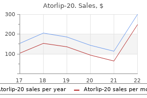
Atorlip-20

Atorlip-20
Atorlip-20 dosages: 20 mg
Atorlip-20 packs: 10 pills, 30 pills, 60 pills, 90 pills, 120 pills, 180 pills, 270 pills

In the presence of a basilar fracture in anterior cranial fossa cholesterol levels of seafood atorlip-20 20 mg generic with amex, a nasogastric tube should by no means be inserted due to the chance of getting in to the mind through the fracture cholesterol test without blood atorlip-20 20 mg discount without a prescription. Persistent leakage past 2 weeks may require operative restore with a dural patch cholesterol in food good or bad order 20 mg atorlip-20 overnight delivery. This sign can also be seen when the bloody drainage is utilized to a paper towel or filter paper. Common etiologies embody motorcar accidents, au to versus pedestrian accidents, and sports activities accidents. Blood accumulates outside the dura mater, dissecting the dura from the inner table of the cranium and compressing underlying mind because it expands beneath arterial strain. Because the bleeding is under arterial strain, it usually accumulates rapidly, causing a mass effect with shift of the ipsilateral hemisphere and, finally, transtentorial herniation. The subdural hematoma is crescent-shaped and located between the dura matter and the brain parenchyma. Depending on the dimensions of the hematoma and the degree of underlying cerebral edema, a mass effect will happen with obliteration of the ventricles and cisterns and shift of cerebral structures across the midline which will culminate in transtentorial herniation. Symptoms at this stage include headache, diminished focus and application, and variable focal neurologic deficits. Symptoms could also be very refined or may be suggestive of elevated intracranial pressure. In truth, the original head trauma may have been comparatively minor and utterly forgotten by the point the affected person presents for care. Rebleeding is seen as a hyperdense assortment of blood superimposed on an encapsulated continual hematoma. Seizure prophylaxis ought to be offered in all cases for the primary week to lower the incidence of early post-traumatic seizures. Typically it exhibits as fingerlike white projections, more obvious within the sylvian fissure. Note the effacement of the posterior horn of the left lateral ventricle and midline shift. During the acute part, intense vasospasm brought on by subarachnoid blood may worsen symptoms and outcome by causing ischemia in adjoining regular mind. Use of calcium channel blockers has proven some utility in lowering this vasospasm. Long-term sequelae embody obstructive hydrocephalus which can require creation of a ventriculoperitoneal shunt for decompression. Intraventricular Hemorrhage Hemorrhage in to the ventricular system is more frequent following hypertensive bleeds than 1. Complications embody obstructive hydrocephalus which can require placement of a ventriculoperitoneal shunt. Bleeding in these cases is often tamponaded by surrounding tissue however could turn into in depth sufficient to produce a midline shift. Coagulopathy increases with rising severity of brain damage, and is particularly common after penetrating injuries. This coagulopathy may be current on admission or may manifest as late as 3�4 days after harm, and as such serial examination of coagulation factors must be carried out. Cerebral Contusion Cerebral contusion happens because the brain parenchyma strikes fastened parts of the skull during sudden deceleration or acceleration. It is often associated with a coup�contrecoup damage and may be discovered relatively distant from the site of head influence. Certain places are significantly prone to contusion: the frontal lobe hanging the anterior cranium and the temporal lobe striking the projections of the sphenoid bone are the 2 most common areas for cerebral contusion. Frontal lobe contusions are characterized by agitation, confusion, perseveration or repetitive questioning, impaired short-term memory, and aggressiveness that always requires bodily or chemical restraint. Over a interval of hours to days, localized cerebral edema develops, causing a mass effect that probably may end up in transtentorial herniation. Management with diuresis and intracranial pressure monitoring is helpful with contusions that develop a mass effect. Eventually the edema regresses and the hematoma reabsorbs completely or liquefies, leaving a cystic fluid-filled construction. Cerebral contusions, especially frontal or temporal, are additionally characterized by a high incidence of posttraumatic seizures. Short courses of anticonvulsants have been shown to significantly lower the chance of early posttraumatic seizures; in contrast, extending remedy with anticonvulsants has not been confirmed to be helpful in prevention of delayed onset seizures or danger of posttraumatic epilepsy. Traumatic epilepsy can manifest in a delayed method with the first reported incidence occurring higher than 1 yr from damage in 50% of instances and first incidence occurring larger than 4 years from damage in as much as 20% of circumstances. These are devastating accidents that regularly end in death or profound incapacity in survivors. As a bullet enters the cranium, it produces a number of high-velocity fragments (both from shattered skull fragments and from bullet fragmentation) that cause multiple injuries. Although the cranium absorbs some kinetic vitality, the bullet retains enough vitality to trigger a strain wave once it enters mind parenchyma. The abrupt deformation of mind tissue ends in laceration and contusion of brain parenchyma, accumulation of blood in the epidural or subdural spaces, intraparenchymal bleeding, and direct laceration of mind tissue by bullet and bone fragments. Because of the excessive kinetic vitality imparted to the brain, subsequent cerebral edema is frequent. Other types of penetrating injuries involve less kinetic power and have a better prognosis. Stab wounds, impalement accidents, and low-velocity shrapnel wounds can produce the entire similar injuries but most commonly lead to open skull fracture and direct laceration of mind parenchyma. Many different objects could also be concerned, but knives and metal rods are the most typical. A cautious physical examination is indicated with stab wounds of the scalp to ensure that the knife blade has not broken off inside the skull. Surprisingly, many of those sufferers are relatively asymptomatic and often have a traditional neurologic examination in spite of a dramatic presentation. Plain anteroposterior and lateral radiographs of the skull will precisely delineate the placement and depth of penetration of radiopaque impaled objects. Impaled foreign our bodies ought to be eliminated only after an angiogram to rule out a vascular damage or within the working room, the place prompt vascular control may be obtained once the thing is removed. As with open skull fractures, bleeding from a gunshot wound of the top may be profuse, however hemorrhagic shock is uncommon with isolated head accidents, and other related injuries must be sought to clarify the shock. Foreign bodies should be removed only after an angiogram or in the operating room. Injuries that result in exposed brain matter, particularly gunshot wounds, are related to a excessive incidence of coagulopathy (massive launch of tissue thromboplastin from the injured brain), diabetes insipidus, hemodynamic instability, arrhythmias, hyperglycemia, and neurogenic pulmonary edema. The prognosis after gunshot wounds to the top is poor and the mortality is greater than 90%. However, these cases ought to be handled aggressively because they usually turn out to be organ donors. If compression of the brainstem happens, loss of vital features similar to respiration and vasomotor control result in fast demise. Uncal herniation: the most typical form of herniation results from edema or hematoma in a single cerebral hemisphere that causes a shift of that hemisphere throughout the midline, underneath the falx, and downward throughout the tentorium, leading to compression of the midbrain. Compression of the ipsilateral cerebral peduncle leads to weak spot or irregular posturing (decorticate, then decerebrate) of the contralateral limbs. Respiratory abnormalities progress from central neurogenic hyperventilation to Cheyne�Stokes respiration, to ataxic respiration, and at last to apneustic respiration. Central herniation: Compression of the brainstem by a frontal or apical mass lesion that expands downward produces pinpoint pupils, downward gaze preference, and other brainstem dysfunction described beforehand. Cingulate gyrus herniation: Pressure in one cerebral hemisphere could result in herniation of the ipsilateral medial cingulate gyrus under the falx. Cerebellar tonsillar herniation: Mass lesions or edema of the cerebellum can lead to expansion of the cerebellar tonsils in to the foramen magnum, compressing the posterior brainstem. This presents as sudden lack of consciousness and lack of brainstem operate with consequent apnea and hypotension. This situation has extremely high mortality, so cerebellar lesions must be recognized before the onset of herniation to salvage the patient. Cerebral perfusion pressure is maintained by infusion of fluids and pressors if needed.
These spiculated folds (arrows) end result from weakening of main peristalsis at the junction of the striated and clean muscle parts of the upper thoracic esophagus cholesterol lowering diet in spanish cheap atorlip-20 20 mg visa. A susceptible cholesterol levels hdl atorlip-20 20 mg purchase without a prescription, proper anterior indirect single distinction spot image shows an extrinsic indentation (arrows) on the proper posterolateral wall of the upper thoracic esophagus secondary to a distinguished right inferior supra-azygous recess in the mediastinum impinging on the esophagus cholesterol control foods eat atorlip-20 20 mg discount without a prescription. Note how the outer wall of the esophagus is clearly visible in this area because of air in the lung abutting the esophagus. The esophagus could additionally be indented by regular extrinsic impressions from the aortic arch, left major bronchus, and left facet of the guts. A clean, gently sloping indentation may be seen on the right posterolateral wall of the upper thoracic esophagus between the thoracic inlet and aortic arch in about 10% of patients because of a normal anatomic variation in which an unusually outstanding proper inferior supra-azygous recess indents the esophagus. When this anatomic variant is present, the outer wall of the esophagus may be outlined by air within the adjoining lung, so the esophageal wall is visualized as a skinny stripe paralleling the esophagus from the thoracic inlet to the aortic arch. This is a clinically helpful observation, as it signifies that the esophagus is indented by a recess of lung projecting in to the mediastinum rather than a pathologic mediastinal mass. When esophageal motility is evaluated, aged sufferers not sometimes have splitting of the peristaltic wave at or close to the level of the aortic arch, so a few of the barium bolus passes in a retrograde trend in to the extra proximal esophagus. This phenomenon is assumed to end result from focal weakening of primary peristalsis at the junction of the striated and clean muscle portion of the esophagus, also known as proximal escape. Multiple fantastic transverse striations are seen within the gastric antrum as an indication of chronic antral gastritis. In some patients, double contrast research may reveal fantastic transverse folds or striae within the gastric antrum as an indication of chronic antral gastritis. The areae gastricae are visualized with higher frequency in older sufferers, most likely due to continual H. A supine double distinction spot image of the abdomen shows enlarged areae gastricae in the gastric antrum and body as a sign of continual an infection by H. The enlarged lymphoid follicles are manifested by innumerable tiny, spherical nodules carpeting the gastric antrum. Note punctuate collections of barium in central umbilications within most of the nodules. Occasionally, the cardiac rosette could additionally be unusually prominent or nodular due to invagination of the gastroesophageal junction in to the gastric fundus. If the cardiac rosette is regular, this construction will vanish because the lower esophageal sphincter opens and barium passes in to the stomach. If a real lesion is current at the cardia, nevertheless, the abnormal findings will persist even when the sphincter is open. This easy method is subsequently extremely useful for differentiating pseudomasses from true tumors at the cardia. A proper lateral double distinction spot picture of the gastric fundus reveals irregular ulceration (white arrows) within a big mass (black arrows) in the area of the cardia. Duodenum When the duodenal bulb is sufficiently distended, the floor of the bulb is normally easy and featureless on double contrast radiographs. In some sufferers, nevertheless, double distinction studies could reveal a fantastic velvety or reticular pattern within the bulb as a standard variant. In different sufferers, the duodenal bulb may contain shallow pits that fill with barium, simulating the looks of duodenal erosions. Double contrast research generally could reveal multiple small, discrete, angular or plaque-like defects close to the bottom of the duodenal bulb because of heterotopic gastric mucosa, a finding of little or no scientific significance. When viewed en face, the gastric cardia could additionally be recognized en face on double distinction radiographs by three or four stellate folds radiating to a central level on the gastroesophageal junction, also referred to as the cardiac rosette. Is this a true cardiac mass (black arrows) containing a small central space of ulceration (white arrow) or an unusually outstanding cardiac rosette As the affected person sips, extra barium exhibits how these findings vanish as the lower esophageal sphincter opens and barium enters the abdomen. This was subsequently a pseudolesion at the cardia brought on by transient invagination of the gastroesophageal junction in to the gastric fundus. A proper lateral double contrast spot picture of the gastric fundus shows a possible tumor on the gastric cardia. As the affected person sips, further barium exhibits how this mass lesion (large arrows) persists even because the lower esophageal sphincter opens and barium enters the stomach. Also notice eccentric narrowing (small arrows) of the distal esophagus by tumor on this affected person with carcinoma of the cardia invading the distal esophagus. A double contrast spot image exhibits tiny barium collections (arrows) filling tiny mucosal pits within the duodenal bulb. This regular anatomic variant must be differentiated from true varioliform erosions in which the central barium collections are surrounded by radiolucent mounds of edema. A double distinction spot image reveals multiple discrete, angular defects within the duodenal bulb, a finding of little or no scientific significance. Philadelphia: Elsevier, 2008:311�322) 36 Chapter 2: Examination of the esophagus, abdomen, and duodenum: methods and regular anatomy. Barium trapped within a redundant fold on the internal side of the superior duodenal flexure mimics the appearance of an ulcer (white arrow) with a surrounding mound of edema (black arrows) or an ulcerated submucosal mass. However, the attribute look and location of this discovering ought to counsel the proper analysis. This affected person has a mushroomshaped defect (arrows) at the base of the duodenal bulb because of antral mucosa that has prolapsed by way of the pylorus. Still different sufferers could have a mushroom-shaped defect at the base of the duodenal bulb abutting the pylorus. Occasionally, nevertheless, benign or malignant gastric tumors prolapsing via the pylorus may be manifested by polypoid defects within the bulb that are contiguous with the pylorus. This finding ought to due to this fact increase concern about the possibility of a prolapsed antral neoplasm. In the descending duodenum, the anatomy of the papilla of Vater may be properly demonstrated. The gastric remnant has been anastomosed to the proximal jejunum via a extensively patent gastrojejunostomy (arrows). Gastric outlet obstruction When gastric outlet obstruction is suspected, preliminary fluoroscopy of the abdomen ought to be carried out before administration of barium to decide whether or not the abdomen is dilated or crammed with stable meals that would compromise the barium research. If an emergent barium research is carried out within the setting of gastric outlet obstruction, the affected person should be given high-density barium with the desk upright and then positioned in a proper aspect down lateral position because the desk is lowered to its horizontal setting so as to facilitate passage of barium in to the distal stomach and higher delineate the trigger of obstruction. Gastric resection After partial gastrectomy for ulcer illness or tumors, the double distinction study should be modified to compensate for the absence of a pylorus and fast emptying of barium from the abdomen. Because of the surgical anatomy, the esophagus must be examined at the end of the study and the abdomen first to assess the radiographic findings before excessive barium within the abdomen and small bowel limits evaluation of this region. Multiphasic examination of the esophagogastric area for strictures, rings, and hiatal hernia: analysis of the individual methods. Focal spiculation of the higher thoracic esophagus: a normal variant on doublecontrast esophagography. The right inferior supraazygous recess: a reason for upper esophageal pseudomass on double-contrast References 1. Barium sulfate as contrast medium for analysis of postoperative anastomotic leaks. Esophageal perforation: comparison of use of aqueous and barium-containing distinction media. Evaluation of three effervescent brokers for double-contrast upper gastrointestinal radiography. Spontaneous gastroesophageal reflux during double-contrast upper gastrointestinal radiography with glucagon. Comparison of film-screen combination and digital fluorography in gastrointestinal barium examinations in a medical setting. Esophageal motility: assessment with synchronous video tape fluoroscopy and manometry. Visualization of areae gastricae on double-contrast higher gastrointestinal radiography: relationship to age of patients. There is a dilated esophagus with absent main peristalsis and tapered, beak-like narrowing (arrow) on the gastroesophageal junction as a result of incomplete opening of the decrease esophageal sphincter. Achalasia Achalasia may be categorised as main when it happens as an idiopathic condition involving the myenteric plexus of the esophagus or as secondary when it results from different circumstances, most commonly malignant tumors involving the gastroesophageal junction (carcinoma of the cardia and metastases to this region). In contrast, secondary achalasia most commonly results from tumor at the gastroesophageal junction invading the ganglion cells within the distal esophagus, so that it simulates the findings of primary achalasia.
The feline esophagus is normally observed as barium refluxes from the stomach cholesterol test accuracy discount atorlip-20 20 mg on line, as in this patient cholesterol test of 8 purchase atorlip-20 20 mg without prescription. This finding should be differentiated from the mounted transverse folds attributable to longitudinal scarring from reflux esophagitis cholesterol medication starting with a 20 mg atorlip-20 discount otc. Early reflux esophagitis may be manifested on double contrast studies by a finely nodular or granular look with poorly outlined radiolucencies that fade peripherally because of edema and inflammation of the mucosa. Timing of the radiographic exposures is important for exhibiting the granular mucosa of reflux esophagitis, as this finding can easily be obscured by excess pooling of high-density barium within the distal esophagus, a frequent problem often identified as move artifact. With more superior illness, barium studies could reveal shallow ulcers and erosions within the distal esophagus. The ulcers can have a punctate, linear, or stellate configuration and are sometimes associated with surrounding mounds of edema, radiating folds, or sacculation of the adjacent wall. In such instances, nevertheless, the ulceration virtually at all times extends distally to the gastroesophageal junction. The presence of one or more ulcers which may be confined to the higher or midesophagus should subsequently recommend another explanation for esophagitis. Reflux esophagitis may also be manifested on barium research by thickened longitudinal folds as a result of edema and inflammation that stretch in to the submucosa. There are innumerable poorly defined, radiolucent nodules that fade peripherally as a result of edema and irritation of the mucosa. Note how this granularity extends proximally from the gastroesophageal junction as a steady area of disease. Multiple tiny ulcers (arrows) are seen within the distal esophagus above a small hiatal hernia. This patient has a single flat ulcer (arrow) on the posterior wall of the distal esophagus a quantity of centimeters above the gastroesophageal junction. There are thickened longitudinal folds within the esophagus because of edema and inflammation extending in to the submucosa. This affected person has a thickened fold (short arrows) extending from the gastric cardia in to the distal esophagus, where it terminates as a smooth, polypoid protuberance (long arrow). The lesion represents a heaped up space of inflammatory and granulation tissue because of persistent reflux esophagitis. There is a clean, tapered segment of concentric narrowing (arrow) within the distal esophagus above a hiatal hernia. A short, ring-like constriction (arrows) is seen in the distal esophagus above a small hiatal hernia. In advanced esophagitis, in depth ulceration, edema, and spasm may trigger the esophagus to have a grossly irregular contour with serrated or spiculated margins and variable narrowing. These strictures usually appear as concentric areas of clean, tapered narrowing above a hiatal hernia. Scarring from reflux esophagitis can typically lead to longitudinal shortening of the esophagus and the event of fastened transverse folds, producing a classic stepladder appearance because of pooling of barium between the folds. Also observe longitudinal scarring with barium trapped between fastened transverse folds (black arrows), producing a classic stepladder look. Infectious esophagitis Candida esophagitis Candida albicans is by far the most typical explanation for infectious esophagitis. There is a light stricture within the midesophagus (black arrow) with a fragile reticular pattern of the mucosa extending distally a considerable distance from the stricture ( to white arrows). Note how many of the plaques have a linear configuration and are oriented longitudinally in relation to the lengthy axis of the esophagus. Only about 50% of sufferers with Candida esophagitis have oropharyngeal candidiasis. Double distinction esophagography has a sensitivity as excessive as 90% in detecting Candida esophagitis,sixteen primarily because of the power to demonstrate mucosal plaques with this system. Candida esophagitis is typically manifested on double contrast research by discrete plaque-like lesions, seen as linear or irregular filling defects on the mucosa. These plaque-like lesions are inclined to be oriented longitudinally in relation to the long axis of the esophagus and are usually separated by segments of regular intervening mucosa. When typical findings of Candida esophagitis are seen on barium research, affected people can be handled with antifungal brokers similar to fluconazole with out need for endoscopy. Herpes esophagitis the herpes simplex virus is another frequent explanation for infectious esophagitis. Most patients with this situation are immunocompromised, but herpes esophagitis could occasionally develop as an acute, self-limited disease in otherwise healthy patients. Herpes esophagitis is typically manifested on double distinction research by small, discrete ulcers within the higher or midesophagus on a traditional background mucosa. In the suitable medical setting, the radiologic finding of multiple small, discrete esophageal ulcers with out plaques must be extremely suggestive of herpes esophagitis, since ulceration in candidiasis almost all the time happens on a background of diffuse plaque formation. One of the plaques has sloughed, producing a deep ulcer (arrow) superimposed on a background of diffuse plaque formation. There are multiple small, discrete ulcers within the midesophagus with surrounding radiolucent mounds of edema (arrows). In the appropriate medical setting, these findings ought to be extremely suggestive of herpes esophagitis, since ulceration in candidiasis solely happens on a background of diffuse plaque formation. Otherwise wholesome sufferers with herpes esophagitis sometimes present with a flu-like syndrome consisting of fevers, complications, myalgias, and higher respiratory symptoms for a period of 3 to 10 days previous to the sudden onset of extreme odynophagia. A big, ovoid ulcer (arrows) is seen en face within the midesophagus with a clean, skinny rim of edema surrounding the ulcer. This ulcer is unimaginable to differentiate from the human immunodeficiency virus ulcer illustrated in. A large, ovoid ulcer (arrows) is seen en face in the midesophagus with a thin rim of edema surrounding the ulcer. Double distinction esophagograms typically reveal a number of giant, ovoid or diamond-shaped ulcers surrounded by a thin radiolucent rim of edema, often related to small satellite tv for pc ulcers. Other inflammatory conditions Eosinophilic esophagitis Eosinophilic esophagitis (EoE) is an inflammatory situation of unknown etiology characterised by intraepithelial eosinophilia in the esophagus. During the previous decade, EoE has been recognized with greater frequency because of an growing prevalence or an growing consciousness of the disease, or maybe a mixture of both. In adults, EoE tends to have an result on younger men who present with long-standing dysphagia and occasional food impactions. These sufferers typically, but not all the time, have an atopic historical past, asthma, or peripheral eosinophilia. The diagnosis is confirmed on endoscopic biopsy specimens displaying more than 20 eosinophils per high-power subject. There is a protracted, easy stricture (arrows) with tapered proximal and distal margins in the midesophagus. A mild stricture is seen in the distal esophagus with a number of distinctive ring-like indentations (arrows) in the region of the stricture. Patients with EoE might develop higher, mid-, or distal esophageal strictures, typically seen on esophagography as lengthy segments of symmetric narrowing with a smooth contour and tapered margins. Other patients with EoE could have a variable variety of distinctive ring-like indentations (typically within the region of a stricture), producing a ringed esophagus. Affected people sometimes ingest the medicine with little or no water instantly earlier than going to bed. Prolonged contact of the esophageal mucosa with the drugs presumably causes a focal contact esophagitis. The radiographic findings in drug-induced esophagitis depend on the character of the offending medication. Tetracycline and doxycycline are associated with the development of small, discrete ulcers in the upper or midesophagus indistinguishable from these in herpes esophagitis. In contrast, different offending medicines might cause more severe esophagitis, leading to the development of large ulcers. Acute radiation esophagitis often happens 2 to four weeks after the initiation of radiation remedy. This situation could additionally be manifested by ulceration or by a granular look of the mucosa and 50 Chapter three: Esophagus.
At occasions cholesterol deep conditioner atorlip-20 20 mg cheap with mastercard, comparability views of the opposite extremity will be needed serum cholesterol chart atorlip-20 20 mg cheap without prescription, and occult fractures must be suspected in youngsters with tenderness over the physis cholesterol and testosterone atorlip-20 20 mg cheap amex. Long bone fractures should be splinted before transferring the affected person to the radiology suite. Immobilization of the fracture reduces ache and bleeding and prevents further damage to the neurovascular constructions. Fractures are first described by anatomic location, and lengthy bones are often divided in thirds describing the location of the damage. Open or compound fractures discuss with fractures with a break in the overlying pores and skin, in contrast to closed fractures which have normal pores and skin integrity. The fracture line or sample is then described and follows the following widespread convention: 1. Displacement refers to the degree of offset of the bone ends relative to each other, and utterly displaced fractures are inclined to be extra unstable injuries. Displacement is described by outlining the place of the distal bone relative to the proximal end. A bayonet deformity refers to accidents with 100% displacement and overriding of the bone ends with shortening of the affected extremity. Angulation refers to the angle between the longitudinal axes of the principle fracture segments. Fractures with important angulation generally require reduction to maintain good operate. They are outlined as fractures involved with the outside environment and thus require a break in the pores and skin overlaying the fracture website. The skin break may be massive and apparent or a small puncture wound harm, and thus determination if a fracture is open can typically be tough. The Gustilo classification is often used when describing open fractures: Type I: Puncture wound <1 cm and relatively clean. Emergency remedy includes covering the wound with sterile saline dressings, together with the suitable tetanus immunization and ache administration. Patients needing emergent operation for extreme related accidents can have exterior fixation carried out whereas steady patients are eligible for inner fixation. There is a mixture of orthopedic trauma, in depth soft tissue injury, and harm to the neurovascular buildings. The most immediate priority is to control bleeding, often by method of tourniquets within the appropriate cases. The preliminary medical evaluation should embody evaluation of the extent of skeletal and delicate tissue damage, the presence of peripheral pulse and skin perfusion, and motor�sensory function. Patients with obvious nonsalvageable limbs must be transferred to the working room for bleeding management and amputation. Detection may be straightforward on physical examination or could also be subtle, requiring 164 Musculoskeletal Injury adjunctive testing. Any deep wound in proximity to a joint must be thought of as entering the joint. Plain x-rays can show an related fracture, air, or a overseas body throughout the joint however may be normal. Careful exploration beneath sterile situations might reveal a wound track instantly penetrating the joint capsule. In questionable cases, a saline or methylene blue arthrogram may present the reply by revealing dye leakage through the wound site. Emergency therapy consists of parenteral antibiotic coverage and tetanus immunization. Injuries most frequently happen within the zone of hypertrophic cartilage cells, and the germinal cells are normally undamaged; thus, fortuitously growth is commonly not affected. The most commonly used classification of epiphyseal accidents is the Salter�Harris classification. Type I: A quite common slip through the zone of provisional calcification without fracture. No germinal layer is concerned, and the fracture often heals with out consequence. These fractures are sophisticated, and important development disturbance can occur except good anatomic discount is achieved. As with any pediatric damage consideration must be given to the potential of nonaccidental trauma. A greenstick fracture is an incomplete angulated fracture of a protracted bone recognized by a bowing look. With an extension fracture, this line intersects the anterior one-third of the capitellum or passes totally anteriorly. C D stronger than the bone, and thus hyperextension damage typically causes bone fracture while adults typically undergo a posterior dislocation of the elbow with a similar mechanism. Supracondylar fractures are of two frequent types: flexion and extension, with extension fractures the overwhelming majority. These extension accidents are often the end result of a fall on an arm with the elbow totally prolonged. On examination, the elbow will be swollen, often with a joint effusion and with important pain and tenderness. Careful neurovascular examination of the arm is important as many of those fractures are complicated by brachial artery and median, radial, or ulnar nerve harm. In addition, compartment syndrome can be seen with displaced fractures and needs to be considered. Radiographically, these fractures are sometimes detected on the lateral view of the elbow. Because many of these fractures are transverse they may not be readily seen on the anteroposterior view. In addition, up to 25% of these are fractures of the greenstick variety with the posterior cortex remaining intact. The solely abnormality seen may be a posterior fats pad sign or an abnormal anterior humeral line. Supracondylar fracture should be suspected in any child with acute elbow trauma, swelling, and pain, in spite of normal radiography. Most undisplaced fractures are handled nonoperatively with casting, and most displaced fractures undergo percutaneous pinning. In contrast, upper extremity amputations, especially involving the thumb, are often reimplanted, given the severe disability that happens with the loss of that single digit. Reimplantation is less prone to be successful if the nice and cozy ischemia (room temperature) time has been more than 6�8 hours. If the half has been correctly cooled and cared for, then this window of time could additionally be efficiently extended to 12�24 hours. In addition, clear, sharp amputations are extra likely to achieve success than crush accidents. In basic, amputated elements should be thought of as candidates for reimplantation, and even severely crushed components can be utilized for pores and skin coverage. The amputated half must be cared for properly to maximize the possibility of successful reimplantation. The half should be irrigated with regular saline and then wrapped loosely in sterile soaked gauze. No antiseptics are used, but prophylactic systemic antibiotics and tetanus immunization must be administered. Diagnosis of partial injuries is tougher, as tendon function is usually still intact. A careful examination of the laceration through the complete vary of movement is important, as the injured area of tendon may retract out of the field of view. Flexor tendon repair must be performed by an skilled hand surgeon, often in an working room setting, although extensor tendon harm over the hand and fingers can be repaired within the emergency division. Prophylactic antibiotic, tetanus immunization, and splinting are essential elements of emergency administration. Fortunately, blunt trauma hardly ever causes vascular harm besides with markedly displaced fractures and dislocations. Prompt identification and repair is necessary, given the relatively brief "golden period" of about 6 hours, after which irreversible ischemic insult will happen. Physical examination is necessary for early analysis, and most authors divide examination findings in to "onerous" and "soft" signs.
Diseases
Incisions are made over the limb on both the medial and lateral features to obtain full eschar launch cholesterol in eggs good or bad atorlip-20 20 mg buy amex. The underlying osteofascial compartments must be examined for compartment syndrome cholesterol ratio good but total high generic atorlip-20 20 mg with amex, and if suspected cholesterol food examples generic atorlip-20 20 mg without a prescription, standard fasciotomies are performed. Circumferential burns of the thorax may current with decreased chest wall compliance leading to oxygenation and ventilation compromise. Thoracic compartment syndrome must be suspected if elevated peak airway pressures, hypercarbia, and hypoxia are encountered. Properly performed escharotomies lead to quick enhancements in chest wall compliance and improved arterial blood fuel. Water at 60 degrees Celsius can cause fullthickness burns in three seconds, and governmental laws to lower sizzling water tank temperatures have led to a decrease in such injuries. Scalds on pores and skin with light clothing are inclined to be worse than on exposed skin areas, as the clothes tends to retain heat. Cooking oil and grease can attain 200 levels Celsius and is another widespread explanation for scald injury. Characteristic "immersion" patterns ought to promptly alarm the clinician of such chance. The diploma of tissue destruction is dependent on a number of elements together with the concentration, form, and pH of the offending agent, duration and volume of contact, and the extent of irrigation. On the other hand, the coagulative necrosis brought on by acids is a self-limiting process once the denatured proteins and broken tissue form a coagulum, thus limiting further acid penetration and tissue harm. Hydrofluoric acid, typically present in family rust removing and cleaning merchandise, is a particular circumstance in that the acid produces liquefactive necrosis. With intensive exposure to biologic tissues, the fluoride ion precipitates calcium and can cause extreme systemic hypocalcemia. Four mechanisms of harm exist: direct contact, electrical arc, flame, and flash burn. Current passing by way of the body from the point of entry to the grounding exit topics the skin and underlying tissues to direct electrothermal injury. Electrical burns are a unique entity in that the exterior signs of damage are deceptively benign compared to the extent of sentimental tissue injury. Electrical currents trigger totally different quantities of tissue harm based on distinct tissue resistive properties. Nerves, vessels, and muscular tissues are more vulnerable to electrothermal damages whereas bone and skin are much less so. Treatment is based on aggressive fluid resuscitation to achieve a urine output of 1 cc/kg per hour. Other unproven but generally practiced remedies embody the use of mannitol to induce an osmotic diuresis, and urine alkalinization with bicarbonate to prevent myoglobin precipitation within the renal tubules. Patients ought to be monitored for no less than 6 hours, and often as much as 24 hours, for cardiac arrhythmias and the event of extremity compartment syndrome. When suspected, decompressive fasciotomy should be promptly performed to minimize ischemic and pressure-induced myonecrosis. Long-term sequelae embody permanent debilitating peripheral neuropathy and cataracts. Most of the mortalities occur on the ends of the age spectrum with 38% as a end result of multisystem organ failure and only 4. Increased burn survival over the previous a quantity of decades raises necessary quality of life issues. Studies have proven that probably the most significant predictors of improved quality of life include the scale of fullthickness burn, age, degree of functional recovery of the hands, and perceived social help. Sixty-six % of burn patients return to work, the likelihood and timing of that are instantly associated to the scale and severity of the harm. Cosmetic outcomes range from minor skin discoloration or hypertrophic scars, to debilitating contractures in untreated wounds. There can be noticeable pores and skin discoloration in areas of superficial second-degree burn that healed without surgery. Extensive soft tissue trauma following penetrating accidents could happen after high-velocity bullet wounds, closed-range shotgun accidents, and explosions (see Chapter 10, Ballistics). Meticulous systematic and local examination ought to be performed to rule out other related injuries. Locally, the physician should consider for underlying vascular, nerve, tendon, and bone injuries Avulsion type injuries happen when a flap of tissue is separated from underlying tissue structures. The most excessive type of this damage is a degloving damage, which occurs when all of the pores and skin and subcutaneous tissues are separated from the underlying fascia. The wound needs strain irrigation and intravenous antibiotics to scale back the risk of osteomyelitis. Attempts to salvage these limbs are virtually always unsuccessful and result in serious complications and extended hospitalization. Mangled extremity accidents often contain soft tissues, neurovascular buildings, and bones. These accidents require a multidisciplinary method due to their complexity and excessive danger of great problems, including death, amputation, renal failure, and infection. The native care of enormous open wounds should be supplied in the working room, often beneath general anesthesia. Antibiotic prophylaxis ought to be administered routinely and tetanus prophylaxis must be thought of within the applicable circumstances. Initial priorities should embrace hemorrhage control, a quick neurological examination, and pho to documentation. If digital strain is unable to management bleeding a business tourniquet or inflated blood pressure cuff may provide short-term hemostasis. Documentation of neurologic perform and extent of tissue damage is important in instances the place the extremity is unsalvageable and primary amputation essential. Primary restore must be thought of only in selected circumstances with clean incising wounds. In the vast majority of instances of in depth delicate tissue trauma the wound must be debrided and left open to heal by secondary intention. Negative strain dressings may be helpful in eradicating effectively any contaminated exudates and stop the retraction of the wound edges. In extremity injuries the muscle compartments should be monitored clinically and strain measurements and timely decompressive fasciotomy should be performed within the applicable circumstances (see Section 9. Complications the next systemic issues could occur after in depth soft tissue trauma: 1. Hypovolemic shock, due to extravasation of blood and fluid externally or in to the tissues. Electrolytic abnormalities: Hyperkalemia (release of potassium from damaged cells), hypocalcemia (deposition of calcium within the injured tissues), or hyperphosphatemia. Compartment pressures ought to be considered in suspicious extremity accidents to rule out compartment syndrome. Most of those bites occur in children and younger adults and are normally secondary to unintentional provocation or perceived threatening habits exhibited by the victim. Canine bites mostly affect the extremities, adopted by the top and neck, and trunk. However, children are more likely to endure injuries to the pinnacle and neck as a result of their smaller stature. Injuries in this age group can be devastating as canines can create depressed cranium fractures, large scalp avulsions, intracranial bleeding, or major vascular injury within the neck or thoracic inlet. Similarly, massive breeds can inflict critical wounds in adults as well with chew forces exceeding 450 kilos per square inch. Dog bites might trigger a wide range of harm patterns together with punctures, avulsions, tears, abrasions, and severe crush injury. Additionally, a high diploma of suspicion for occult vascular injury must be maintained in assaults from bigger canine breeds or law enforcement animals. Tenets of wound care embody preliminary cleansing and irrigation which serves to remove particles and bacteria, and has been shown to decrease the transmission of the rabies virus. For large wounds, x-rays are important to rule out retained overseas bodies and underlying fractures. Liberal consultation of an orthopedic specialist ought to be obtained when the chew involves the hand. The hand incorporates numerous bones, nerves, and joints enclosed inside a small space and relatively superficial location. Additionally, an infection can rapidly progress alongside the fascial planes and tendon sheaths within the hand leading to everlasting disability if not rapidly handled.
During the oral or nasal and esophageal section of intubation cholesterol test blood atorlip-20 20 mg purchase free shipping, the guide wire is retracted and the delicate tip of the catheter is placed in the mouth or nostril cholesterol lowering foods for coeliacs atorlip-20 20 mg cheap without prescription. The affected person tilts his or her chin inferiorly show cholesterol chart discount 20 mg atorlip-20 amex, as the radiologist passes the catheter in to the throat. The affected person is instructed to swallow, and the radiologist advances the catheter through the pharyngoesophageal segment. If the patient is unable to speak or sounds hoarse, the catheter is probably lodged behind the epiglottis, in the larynx, or in the trachea and must be retracted. The catheter can then be superior under fluoroscopic monitoring, with the patient in a lateral position. The position of the catheter in relationship to the tracheobronchial tree is rechecked. If the catheter coils within the esophagus or at the esophagogastric junction, the catheter is pushed in to the abdomen and uncoiled by withdrawing the catheter over the guidewire. Forward progression of the catheter may be tough in sufferers with large hiatal hernias. Placing a patient within the erect place might reduce the size of the hiatal hernia, facilitating passage of the catheter. Once the catheter enters the stomach, it typically programs to the left toward the larger curvature, then heads retrogradely towards the fundus. In this affected person, no oral barium was given to assist the radiologist understand the anatomy. The location of the duodenal bulb to the left and posteriorly made it tougher to information the catheter in to the medially rotated duodenum. To manipulate the catheter toward the antrum, the affected person is turned in to a proper aspect down decubitus place (right lateral position), the catheter is withdrawn to the gastric cardia, then the catheter is torqued to the right and handed along the lesser curvature. Alternatively, the patient could be placed in a semi-erect or erect place to advance the catheter in to the gastric antrum. In some patients, the wire can be retracted, and the delicate tip of the catheter can catch on the larger curvature, allowing the catheter to move alongside the larger curvature in to the antrum. The enteroclysis catheter may be trapped within the gastric fundus in patients with a "cascade abdomen," in which the gastric fundus lies below the upper gastric physique. This problem could also be overcome by insufflating room air through the enteroclysis catheter to distend the abdomen or by turning the patient in to a right side down, erect, or erect lateral position. Patients with a transversely oriented abdomen are also difficult to intubate, as the catheter tip might catch on the higher curvature and head in a retrograde course in to the gastric fundus. Some radiologists will loop the catheter within the gastric fundus in patients with a transversely oriented abdomen;4 other radiologists will compress the stomach with a gloved hand or place the patient in an erect position. Catheter passage is made simpler by knowing the situation of the pylorus and duodenal bulb from the preliminary check swallow of barium or from air insufflated in to the abdomen. If the catheter follows the greater curvature of the abdomen simply proximal to the pylorus, the tube will not be directed toward the pylorus. The guide wire, due to this fact, should be retracted from the tip of the catheter in order that the tip heads toward the pylorus. Catheter passage through the pylorus is facilitated by turning the affected person in to either a proper facet down or left posterior indirect position. With the affected person in the left posterior indirect position, the radiologist locations his or her proper hand on the abdomen, with the thumb heading towards the pylorus and fingers along the higher curvature. The radiologist pushes the stomach to the left, straightening the relationship between the stomach and duodenal bulb. Tube passage via the flexure at the apex of the duodenal bulb/postbulbar duodenum is also facilitated by placing the affected person in a left posterior oblique place, straightening the postbulbar duodenum. If the small quantity of barium has been administered orally before the examination, the placement of the duodenum is apparent. If methylcellulose enteroclysis is carried out the catheter should be passed in to the first loop of jejunum, no much less than 10 cm past the duodenojejunal junction. In partially obstructed sufferers, the catheter should also be handed to the first loop of the jejunum. If the catheter loops in the duodenum, the loop could be a hundred and twenty Chapter 6: Examination of the small gut: techniques and normal anatomy. Peristalsis has pulled the tip of the catheter in to the second loop of the jejunum, foreshortening the jejunum. This "accordion effect" is manifested as focal angulation of the jejunum proximal to the balloon, and crowding of its folds (thin arrows). For double distinction enteroclysis utilizing Entero-H and methylcellulose, we employ 60 ml "Luer-lock" syringes to inject the barium, then infuse methylcellulose with the electric pump. Other radiologists use the electrical pump to infuse each barium and methylcellulose. Conversely, if the infusion fee is simply too quick, the proximal jejunum overdistends, leading to small bowel hypotonia. The radiologist, therefore, must consider the response of the small bowel to the speed of distinction infusion and regulate the circulate price accordingly. Types of enteroclysis Once the affected person is intubated, both the patient and radiologist breathe a sigh of relief, and the examination could begin. Each sort of enteroclysis has advantages and downsides that are discussed under. The duodenojejunal junction is commonly a site of adverse tube passage, because the conventional duodenojejunal junction usually makes an abrupt bend anteriorly and to the left as it enters the peritoneal cavity. Turning the patient to the left and towards a susceptible position (left anterior indirect with respect to the table) opens the duodenojejunal junction. Patient coughing or deep respiration can also help the catheter move the duodenojejunal junction. If the patient senses any discomfort throughout distention of the balloon, the balloon is deflated till the discomfort subsides. In some patients, the small bowel shortens across the catheter or balloon tip, leading to an appearance resembling an accordion. After passage of the enteroclysis catheter in to the proximal duodenum, between 600 and 1200 ml of low-density barium are instilled in to the duodenum at a price of about seventy five ml/minute. Single distinction research are easier to carry out, as only one contrast agent is employed. Reflux of barium alone in to the proximal duodenum and stomach hardly ever induces vomiting. The disadvantage of single distinction enteroclysis is that evaluation of the mucosal surface en face relies on compression and evaluation of fold patterns, which is clearly inferior to double distinction visualization of the mucosa. Higher magnification spot radiograph demonstrates that the tip of the catheter and balloon (B) were positioned in the fourth portion of the duodenum. The villi are seen en face as tiny barium-coated nodules on the surfaces of the valvulae conniventes (arrows) and as a barely perceptible reticular pattern (arrowhead). However, in other loops, en face element is obscured partly or utterly by dense barium pools (B). Air contrast enteroclysis Air distinction enteroclysis is a standard technique utilized in Japan and has turn into a favorite method of some radiologists. The barium is infused at a rate so peristalsis is preserved within the jejunum and uniform distention of proximal and mid small bowel is achieved. Room air or carbon dioxide injection commences when pelvic small bowel is partially crammed with barium2 or when barium reaches the terminal ileum. Not all loops are visualized in air contrast, even when the affected person is turned in varied positions and compression radiographs are obtained. It is harder to manipulate the barium pool throughout air contrast enteroclysis than throughout a double contrast barium enema or upper gastrointestinal sequence, and a few loops of small bowel are regularly crammed with barium. The radiologist should be patient and persistent to obtain air distinction in the distal ileum. Overlap of barium-filled loops with air-filled loops is one other downside, specifically when too much barium has been infused. With continuing injection of air and patience, air contrast is obtained within the distal ileal loops. Turning the affected person prone and utilizing compression will improve pelvic loop visualization. There is a lot barium within the pelvic ileum (I) that it may be tough to see all loops in air distinction. Full luminal distention straightens the valvulae conniventes, enabling superior evaluation of fold measurement and increases conspicuity of low-grade or partially obstructing lesions.
When familial (inherited as an autosomal dominant disease) cholesterol medication guidelines discount atorlip-20 20 mg free shipping, multicentricity amounts to 25% to 50% serum cholesterol ratio uk discount 20 mg atorlip-20 amex. Very rare benign lower bad cholesterol foods generic atorlip-20 20 mg, indolent epithelial middle ear tumor, arising from modified respiratory mucosa. Otoscopically, a pink soft tissue mass is seen behind an intact tympanic membrane. Intratemporal hemangiomas are rare extraneural vascular lesions arising from capillaries across the facial nerve, which might secondarily grow in to the nerve. Middle ear adenoma Well-marginated, rounded enhancing delicate tissue mass within the middle ear with minimal erosion. Facial nerve schwannomas are most commonly centered on geniculate ganglion and emanating from facial canal. Jacobson nerve schwannoma reveals erosion of the cochlear promontory, attainable enlargement of the inferior tympanic canaliculus, and absence of facial nerve involvement. Poorly marginated, intensely enhancing mass with "honeycomb" bony matrix and/or spiculated look with intratumoral bone flecks. Most common sites of involvement are the labyrinthine segment of the facial nerve and the geniculate fossa space, but any phase of the facial nerve can be affected. Hemangioma (continues on web page 224) Temporal Bone: Diseases of the Middle Ear and Mastoid 223. The tumor pedunculates in to the middle ear cavity (arrow) and produces an growth of the bony canal (arrowheads). Tegmen tympani and mastoid bones are affected in tegmen tympani meningioma; sigmoid plate and middle ear flooring are affected in jugular foramen meningioma. Middle ear element may symbolize "tip of the iceberg" for larger jugular foramen or tegmen tympani meningioma. May prolong up from jugular foramen in to center ear, extend down from dura overlying tegmen tympani, or come up within center ear. Patients are usually middle-aged and feminine with conductive listening to loss (M:F 1:three, common age at presentation forty five y). Destruction of the tegmen tympani or sinodural plate leads to intracranial involvement. Primary malignant center ear tumors in adults are uncommon and normally associated with a historical past of chronic otitis media. Squamous cell carcinoma can happen, as can various types of adenocarcinoma, notably adenoid cystic carcinoma. Although uncommon, rhabdomyosarcoma is the commonest primary center ear tumor in the pediatric age group (bimodal: in youngsters 5 y and teens 15�19 y) with male predominance. Clinical symptomatology normally includes bloody otorrhea and ear pain; 30% of sufferers have neurologic deficits and nodal metastases on the time of prognosis. Primary sites include breast, lung, kidney, prostate, head and neck squamous neoplasms, and abdomen. Middle ear rhabdomyosarcoma Large, irregular, middle ear/mastoid mass with osteolytic, damaging bone and ossicle adjustments. Possible areas of extension are the external auditory canal, inner auditory canal, center and posterior cranial fossae, nasopharyngeal carotid space, masticator house, and parotid area. Metastases involving the middle ear and mastoid normally prolong from elsewhere, significantly the petrous apex, or disseminate hematogenously, and may be osteoblastic or osteolytic. Metastases Trauma Damage to the temporal bone sometimes requires the applying of nice force and should cause fracture, hemorrhage, nerve trauma, vascular harm, or disruption of the middle or internal ear structures. Associated intracranial injuries, such as extra-axial hemorrhage, shear (or diffuse axonal injury), and brain contusion, are frequent. It begins within the pars squamosa, mastoid, or external auditory canal, extends through the posterosuperior bony external auditory canal, continues across the roof of the middle ear area anterior to the labyrinth, and ends anteromedially within the middle cranial fossa in close proximity to the foramen lacerum and ovale. The most common course of the fracture is anterior and extralabyrinthine; however, although uncommon, intralabyrinthine extension is feasible. The indirect fracture crosses the external auditory canal in a horizontal plane and then extends upward obliquely towards the middle fossa. The fracture misses the otic capsule and may prolong toward the petrous apex, where the fracture line could prolong to the foramen lacerum. Longitudinal fractures of the temporal bone are most typical (70%�90% of all temporal fractures). Longitudinal fractures usually present with classic findings of laceration of the ear canal, tympanic membrane perforation, hematotympanum, ossicular damage (most generally the incus), facial paralysis, and listening to loss. The hearing loss is predominantly conductive but could have a sensorineural part as properly. Facial paralysis, typically delayed and incomplete, occurs in 10% to 20% of longitudinal fractures. Also seen are related ossicular derangement and hemotympanum, pneumolabyrinth, and air throughout the internal auditory canal. Middle ear fluid is present, as properly as an irregular density filling of the mastoid cells. Medially situated fractures contain the vestibule, cochlea, fundus of the interior auditory canal, and crus commune. A more uncommon kind of transverse fracture occurs medial to the vestibule and bisects the internal auditory canal. Laterally positioned fractures contain the promontory, vestibule, and horizontal and posterior semicircular canals. Comments Transverse fractures of the temporal bone are less widespread (10%�30% of all temporal fractures). The fracture generally begins within the vicinity of the jugular foramen or foramen magnum and extends to the center cranial fossa. Clinical findings include persistent vertigo (due to transection of the vestibule, vestibular nerves, or vestibular aqueduct, perilymph fistula, labyrinthine concussion, or cupulolithiasis), usually with spontaneous nystagmus, and everlasting sensorineural listening to loss (due to damage to the cochlea or transection of the cochlear nerve). Facial paralysis is common (50%), usually instant and full, due to edema, intraneuronal hematoma, impingement by fracture fragments, and full transection. Trauma to the ossicular chain is a frequent complication of temporal bone damage after blows to the temporal, parietal, or occipital region, blasts, barotraumas, and lightning. Ossicular disruption can even occur following direct trauma to the ear by penetrating damage by way of the external auditory canal. Incudostapedial and incudomalleolar disarticulation and dislocation of the incus and malleoincudal advanced are common accidents, whereas stapediovestibular dislocation is rare. There is a excessive incidence of conductive listening to loss secondary to ossicular damage. Incudostapedial joint separation appears as abnormal enlargement of the darkish cleft between the top of the stapes and the long means of the incus, as a fracture of the lenticular process of the incus, or as a fracture via the stapes superstructure. Dislocation of the incus: When incudomalleolar joint separation is associated with incudostapedial joint separation or a fracture of the stapes, the incus could stay in the epitympanic recess with rotation and superiorly, posteriorly, and laterally displace, prolapse in to the lower a part of the tympanic cavity or external auditory canal, or even disappear. Dislocation of the malleoincudal advanced could additionally be associated with an incudostapedial joint separation. Fracture of the malleus occurs at the neck or manubrium and is normally related to different extreme derangements. Fractures of the incus affect the lengthy or lenticular process or the body of the incus. Fractures of the stapes could contain one crus or the arch and the footplate with or with out displacement of fragments. Affect the mastoid air cells with surrounding extensive pneumatization of the skull base. There is focal or diffuse thinning of the encircling bony constructions and loss of the bony trabeculae. Air may be current within the atlanto-occipital joints and within adjoining extracranial delicate tissues. Aggressive nonneoplastic lesion in the temporal bone, typically bilateral or with different associated osseous lesions, inflicting extensive bone destruction with related gentle tissue mass; extracalvarial and/or intracranial extradural in younger children with conductive hearing loss and otorrhea. Uncommon symptomatic acquired lesion with irregular pneumatization of the cranium base extending from the temporal bone. Persistently elevated intraluminal stress has been proposed as a mechanism of pneumocele formation that causes the mastoid cells to increase all through the skull base. Associated abnormalities: small or absent inside auditory canal, hypoplastic or absent petrous apex, flattened medial wall of center ear (because neither the promontory nor the lateral semicircular canal bulges in to the tympanic cavity), ossicle absence or fusion. Vestibule, semicircular canals, and inside auditory canal are variably affected: normal, hypoplastic, or dilated. Labyrinthine, geniculate ganglion, and anterior tympanic portions of facial nerve course occupy site where cochlea must be.
Technique In men cholesterol lowering foods in india buy atorlip-20 20 mg line, the urethral meatus and penis is cleansed and draped as described above for bladder catheterization cholesterol in eggs benedict 20 mg atorlip-20 generic overnight delivery. A 14 French Foley catheter is mostly used and the catheter could be flushed with distinction prior to cholesterol reduction purchase atorlip-20 20 mg mastercard insertion to forestall injection of huge air bubbles. The catheter is advanced in to the urethra, until the balloon is within the fossa navicularis, which is a slightly wide space in probably the most distal penile urethra, just proximal to the meatus. The balloon must be inflated with fluid quite than air, as fluid is less compressible and the catheter much less prone to slip out of the urethra during the examination. It is necessary to not over distend the catheter balloon to be able to keep away from mucosal damage within the fossa navicularis, however the balloon has to be sufficiently distended to retain the catheter throughout the urethra through the examination. The penis is then gently extended and positioned in a cephalad path to straighten the curve at the penoscrotal junction, and show the entire anterior urethra with out overlap of adjoining segments. When the penis is absolutely prolonged for the study, the syringe attached to the catheter factors in course of the shoulder of the affected person. In very overweight sufferers, the Technique for retrograde urethrogram that is the best examine to consider the male anterior urethra. The balloon of the catheter is inflated with distinction in the fossa navicularis (white arrow). There is a high-grade stricture within the proximal bulbar urethra (black arrow) via which a small amount of distinction is flowing in to the posterior urethra and urinary bladder. Notice the obliquity of the affected person and the traction on the penis which straightens out the anteroposterior curve of the urethra on the penoscrotal junction. Continued urethral distention causes intravasation in to the corpus spongiosum (white arrowheads, B and C), not seen on the early image (A). There is traumatic occlusion of the urethra on the prostatomembranous junction due to pelvic fracture (same affected person as in. After catheter placement, contrast is injected under fluoroscopic steering, till the anterior urethra is nicely distended to the level of the external sphincter. It is important to not overdistend the urethra, significantly if strictures are current, as distinction can intravasate in to the corpus spongiosum across the urethra, with venous filling. Intravasation in to the venous structures will cause considerable bleeding from the urethra when the catheter is removed, and handbook compression of the penis must be initiated till bleeding stops. Images are obtained solely in the 45-degree oblique place that the affected person has been placed in. Although the whole urethra may not be optimally evaluated with this method, the photographs usually suffice to evaluate for urethral trauma and decide if a Foley catheter could be safely advanced in to the urinary bladder. Normal anatomy Urinary bladder During early filling, the bladder wall might appear slightly irregular because of the undistended mucosal folds, and should have a spherical or oval shape within the horizontal direction. A absolutely distended bladder is oval within the craniocaudal path, and the wall of the bladder is smooth. When a catheter has been present for longer than a few days in the urinary bladder, the dome of the bladder might reveal slight irregularity due to irritation by the catheter, referred to as catheter-induced cystitis. A small amount of vesicoureteral reflux may also be seen in such patients, due to irritation of the trigone and resultant incompetence of the vesicoureteral junction. This is likely related to irritation by the suprapubic Foley drainage catheter (catheter-induced cystitis). Urethra In females, the urethra is short, of comparatively uniform caliber, and programs obliquely downward and ahead from the bladder neck to the urethral meatus. The posterior urethra is finest imaged with a voiding cystourethrogram, as it has clean muscle in its partitions (referred to because the posterior urethral sphincter complex) which is constricted within the resting state and completely relaxed solely during voiding. The posterior urethra extends from the bladder neck to the external sphincter and consists of the prostatic urethra and the membranous urethra. The prostatic urethra passes via the prostate and is recognized by an impression on its posterior side, the verumontanum; the utricle (a vestige of the mullerian ducts) opens in the heart of the verumontanum and the prostatic ducts and the ejaculatory ducts open on both facet of the verumontanum. The membranous urethra is approximately 1 cm lengthy and is situated instantly distal to the verumontanum. The bulbomembranous junction has a cone-shaped look and the urethral caliber modifications on the level of the membranous urethra (thick arrow) where the external sphincter is located. The posterior urethra is opacified as a end result of retrograde circulate of distinction but the prostatic and membranous urethra (both parts of the posterior urethra) fully distend solely when the patient voids (B). The bladder is trabeculated with several small diverticulae as a outcome of bladder outlet obstruction caused by prostate enlargement. There is retrograde filling of the prostatic ducts (arrows), giving a "prostatogram. Evaluation for leaks and fistulas Bladder leaks Evaluation of the urinary bladder for leak or extravasation could additionally be required in the following clinical conditions: blunt or penetrating abdominopelvic trauma; following surgery similar to partial cystectomy for neoplasm, nephroureterectomy with bladder cuff excision for upper monitor urothelial carcinoma, ureteral reimplantation, renal transplant with anastomosis of the donor ureter to urinary bladder (ureteroneocystostomy), 196 Chapter 10: Fluoroscopic evaluation of the bladder, urethra, and urinary diversions A B. When the bladder was filled additional till a detrusor contraction occurred, intraperitoneal extravasation is seen, with distinction outlining bowel loops within the pelvis. Intraperitoneal injuries due to blunt trauma normally require operative repair however biopsy associated leaks will often heal with Foley catheter drainage alone. Multiple sites of extravasation are seen in to the perivesical extraperitoneal space. Images must be obtained in multiple obliques for optimum analysis and confident exclusion of a leak. Bladder trauma Motor automobile crashes are the most common explanation for injuries to the bladder. The majority of sufferers with bladder accidents as a end result of blunt trauma have related pelvic fractures (60�90% of patients), while one-third of sufferers with pelvic fractures may have an related bladder harm. The bladder may also be injured by penetrating trauma due to stab wounds or gunshot wounds, or be concerned in iatrogenic injuries similar to after deep bladder biopsies. This distinction is an important issue within the administration of bladder trauma as most intraperitoneal accidents require operative restore of the laceration, while extraperitoneal accidents largely heal with bladder drainage alone. Patients who present with pelvic fractures and have gross hematuria have a excessive probability of bladder harm, ranging from 32 to 85% in different collection,8,9 and will endure imaging to consider the bladder. In the absence of pelvic fractures, microscopic or gross hematuria are thought-about to be relative indications for imaging evaluation, with the necessity for imaging decided by clinical signs such as suprapubic ache or voiding difficulties. Extraperitoneal ruptures may be either simple or complicated,6 depending on whether the distinction extravasation is confined to the pelvic extraperitoneal house alone (simple), or associated with disruption of fascial planes so that the extravasated distinction extends exterior the pelvis to contain the anterior stomach wall, perineum, or the external genitalia (complex) 6�8. One-third of bladder accidents are intraperitoneal and result from harm to the lower stomach when the urinary bladder is distended. These accidents involve the dome of the bladder, which is covered by peritoneum, and so an injury at this site causes intraperitoneal extravasation; there could additionally be no associated pelvic fractures. The presence of radiographic distinction material exterior the urinary bladder signifies bladder rupture. There is in depth extraperitoneal extravasation which is advanced and extends in to the perineum. Most urothelial neoplasms of the bladder are treated with a radical cystectomy for an extirpative cure. A Foley catheter is positioned to promote bladder healing and a cystogram may be requested to confirm full healing of the bladder prior to removal of the Foley catheter. Since the ureter inserts posteriorly on the urinary bladder, images in steep contralateral indirect projection are necessary to consider the surgical web site. Small amount of reflux is seen around the proper ureteral stent in to the proper pelvic ureter (arrow) (C). The ureter is anastomosed to the superior dome of the bladder, and the bladder has a tented appearance on the facet of the reimplant. The surgical procedures utilized for ureteral reimplantation are both a psoas hitch or a Boari flap combined with a psoas hitch, that are well described in Rassweiler et al (2007). Radical prostatectomy Radical prostatectomy is carried out for organ-confined prostate most cancers, and open or laparoscopic methods may be used. The prostate and seminal vesicles are resected away from the bladder neck and the distal urethra, and a new vesicourethral anastomosis created between the bladder neck and the urethra simply proximal to the exterior sphincter. There is an institutional variation in whether or not a routine postoperative research is carried out or not in these patients. Frontal and bilateral oblique projections are essential to consider the anastomosis completely, and if a leak is seen, elimination of the catheter is deferred till the leak is totally healed. Renal transplant Following renal transplantation, the ureter is often anastomosed to the anterosuperior aspect of the dome of the urinary bladder (referred to as the ureteroneocystostomy), and the ureterovesical anastomosis is best depicted within the ipsilateral posterior indirect position. Evaluation of the bladder for leaks may be requested in sufferers in whom surgical drains have a high output of fluid; drain fluid which is excessive in creatinine is suspicious for a urinary leak.


