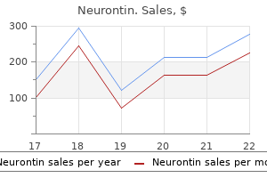
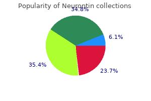
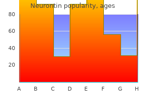
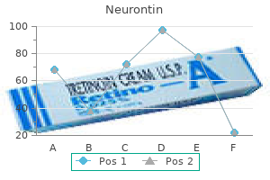
Neurontin

Neurontin
Neurontin dosages: 800 mg, 600 mg, 400 mg, 300 mg, 100 mg
Neurontin packs: 30 pills, 60 pills, 90 pills, 180 pills, 120 pills, 270 pills, 360 pills

This is a blind sac that discharges pus from its exterior opening treatment quadriceps pain 600 mg neurontin purchase mastercard, positioned at the anterior border of the sternocleidomastoid muscle treatment 0 rapid linear progression 100 mg neurontin discount mastercard. Branchial fistulae are congenital openings that discharge secretions medications ending in pril 600 mg neurontin cheap otc, with the external openings lying on the degree of the decrease third of the sternocleidomastoid. The tract passes between the inner and the exterior carotid arteries and lies superior to the hypoglossal nerve. It mostly arises from the second arch, and in such circumstances the internal opening is on the stage of the tonsillar fossa. On rare occasions, it might come up from the fourth arch, when the interior opening might be within the ipsilateral pyriform sinus. Unusual Swellings Paragangliomas Carotid physique tumours or chemodectomas are the most common paraganglioma seen in the neck. They usually present as slow-growing tumours in the fourth or fifth decade of life. Carotid body tumours could additionally be hereditary, happen bilaterally, be related to paragangliomas elsewhere (jugulare, tympanicum or phaeochromocytoma) or be secretory in nature (<5 per cent). Schwannomas/Neurilemmomas these are tumours arising from the Schwann cells surrounding the nerve. The nerve of origin is stretched by the tumour and patients could current with nerve dysfunction. The head and neck is a standard web site, lipomas having a predilection for the nape of the neck and the posterior triangle. They are often situated within the subcutaneous aircraft but can be deep-seated and are characterised by a mobile, soft to rubbery, pseudofluctuant swelling whose edge slips beneath the palpating finger. Arteries and Aneurysms the commonest pulsatile swelling in the neck is a prominence and tortuosity of the frequent carotid artery, predominantly on the right facet. The subclavian or innominate artery might sometimes be affected, which manifests as a pulsatile swelling in the lower neck or the suprasternal house of Burns. Pseudoaneurysms might occur in relation to the carotid artery following surgery and trauma. Midline Neck Swellings Dermoid cysts normally current along the line of fusion of the neck in young kids. Thyroglossal cysts, sinuses and fistulas happen alongside the course of the thyroglossal duct and are described in additional element in Chapter 27. A ranula is a cystic swelling within the floor of the mouth brought on by mucous extravasation or a retention cyst as a outcome of blockage of the sublingual or much less commonly the submandibular duct. Swellings in individuals underneath forty years of age are often benign, whereas those in individuals over 40 years of age are normally malignant. Diagnosis is helped by identifying the situation of the non-nodal swelling in relation to the specific anatomical triangles of the neck. With malignancy, the extent of the concerned lymph nodes helps to establish the location of the first. Enlarged nodes often warrant additional investigation when the dimensions of the node is: a 5 mm b 5�10 mm c 10 mm d Any size Answer b They are generally situated in the muscular triangle i. A cystic hygroma is a congenital lymphangioma that affects the paediatric age group. It is a lymphatic sequestration manifesting as a swelling in the lower neck and posterior triangle. The nodes in non-pathological lymphadenopathy hardly ever reach greater than 1 cm in diameter. Nodes larger than 1 cm constitute significant lymphadenopathy and warrant further analysis with nice needle aspiration cytology. The fan sign is also called the platysma sign; its name is derived from its resemblance to the inverted Japanese fan. It is caused by the puckering of the platysma and overlying skin due to infiltration and is pathognomonic of neoplastic nodes. Ultrasonography of his neck reveals a number of neck nodes with bilateral parotid cysts. On examination, this measures three cm � 3 cm, is fluctuant and is brilliantly transilluminant. Therefore a brilliantly transilluminant fluctuant neck swelling is likely to be a lymphatic cyst. Most of these are benign, but in scientific follow up to 5�15 per cent of them could also be malignant. The incidence of malignancy in a multinodular goitre is decrease than in a solitary thyroid nodule in endemic areas. Diffuse enlargement of the thyroid gland occurring at puberty is referred to as a physiological or simple goitre. These are normally not related to hypo- or hyperfunctioning of the gland but are a response to increased metabolic calls for. The gland may initially hyperfunction, however the course of ultimately leads to hypothyroidism. Examination of the thyroid is finest performed both from behind and in front of the patient with their neck in slight extension. Nodules occurring on the extremes of age (younger than 15 and older than forty five years) and people associated with a previous history of radiotherapy, a family historical past of thyroid cancer, a latest onset or lymphadenopathy usually have a tendency to be malignant. Long-standing multinodular goitres might cause tracheomalacia as a end result of persistent compression (a scabbard trachea). Goitres generally tend to extend to the mediastinum as a outcome of the adverse intrathoracic stress and lack of attachment of the deep layer of the cervical fascia inferiorly. Hoarseness results from stretching of the recurrent laryngeal nerve, but this is extra widespread with malignant infiltration. The fast enhance normally signifies an aggressive histology (poorly differentiated or anaplastic), lymphoma or haemorrhage right into a nodule in a multinodular gland. Papillary cancer of the thyroid gland is the most typical thyroid malignancy (80 per cent of cases). Medullary cancers, arising from parafollicular C cells of neuroendocrine origin, are seen much less commonly (5�10 per cent of cases). Thyroid enlargement could additionally be associated with the signs and symptoms of hyperthyroidism or hypothyroidism. Hypothyroidism is encountered 5�10 times more regularly in medical practice than hyperthyroidism. Hypothyroidism is often a medically treated situation for which surgery has a restricted or no position. Severe hypothyroidism in infancy is recognized as cretinism and has hallmark features of psychological and growth retardation with delayed milestones. The child suffers from failure to thrive, impairment of progress with dwarfism (the limbs being disproportionately shorter than the trunk), a delay in the onset of puberty, delayed tooth eruption, protruberance of the abdomen and dry skin, hair and nails. Unrecognized and untreated hypo- or hyperfunction of the gland may end in life-threatening conditions. Myxoedema coma is now unusual but can result from extended, untreated hypothyroidism. It is often precipitated by triggering factors similar to hypothermia or infection. Patients current with hypothermia, hypotension, hyponatraemia, hypoventilation, hypoglycaemia, bradycardia, an altered sensorium, lethargy, stupor and delirium that progresses to coma. It manifests clinically with hyperpyrexia, tachycardia and hypertension that progresses to cardiac failure. Abnormalities of descent of the thyroid or non-obliteration of the thyroglossal tract leads to the following: � Ectopic thyroid: this can be a residual thyroid that occurs anyplace in the path of the embryological descent. The lingual thyroid is the most common location; this occurs at the junction of the anterior two-thirds and posterior one-third of the tongue. It presents, normally in the first decade of life, as a swelling that strikes on protrusion of tongue. Around 70 per cent of thyroglossal cysts occur in the midline, with others lying laterally as far as the tip of the hyoid.
Syndromes
However medications you can give dogs cheap 400 mg neurontin visa, if pregnancy occurs treatment laryngomalacia infant 100 mg neurontin generic, chorionic gonadotropins from the growing placenta avert atresia of the corpus luteum in treatment 2 cheap neurontin 100 mg amex, which produces progesterone to support the gestation. The Menstrual Cycle the menstrual cycle is outlined as the time between the onset of menstrual bleeding from one interval to the subsequent. The common cycle size is 28 � three days, and the mean period of menstrual hemorrhage is 4 � 2 days. At the tip of one menstrual cycle, circulating ranges of estrogen and progesterone wane as a result of attrition of the oocyte and corpus luteum, respectively. Subsequent to the rising levels of estradiol, glandular progress of the endometrium begins. A regular ovulatory cycle is divided into a follicular and a luteal part; the corresponding cyclic adjustments within the endometrium are the proliferative and the secretory phases, respectively. During this section, the maturing follicle secretes rising quantities of estrogen that outcome in the proliferation of endometrial glands. Luteinized granulosa cells kind the corpus luteum, which produces massive amounts of progesterone and some estradiol. Progesterone stimulates endometrial glandular cells to secrete glycogen and mucus. A second peak within the serum degree of estradiol occurs through the luteal section, and rising ranges of progesterone and estradiol perpetuate endometrial development (the secretory phase). Concurrent with increasing progesterone ranges, the basal body temperature increases (by about zero. Near the tip of the luteal section, plasma estradiol and progesterone levels decrease. This decrease causes intense vasospasm of endometrial spiral arterioles; hemorrhage and desquamation of the endometrium happen due to ischemic necrosis. Currently, the common age of onset of menopause within the United States is fifty one years. After menopause, plasma gonadotropin levels begin to rise because of a decrease in estrogen production by the ovaries. Reduced manufacturing of estradiol is instantly associated to a gentle decline in the number of ova. Plasma gonadotropin ranges peak roughly 5 to 10 years after the onset of menopause and remain constant until the eighth to ninth decade, when levels begin to fall. The most typical situations accompanying menopause are vasomotor instability, atrophy of the epithelium, osteoporosis, and a decrease in breast size. Atrophy of the endometrium, urethral mucosa, vaginal mucosa, and pores and skin happens secondary to estrogen deficiency. The spectrum of anomalies includes various degrees of uterine or vaginal fusion and uterine septal resorption, which may be related to cornual atresia or hypoplasia. Class I anomalies include agenesis or hypoplasia of the vagina, uterus, or fallopian tubes, both alone or together. The ovaries have regular function, however could also be ectopic in location in these sufferers. The tissue dividing the 2 cornua is composed of myometrium and may extend to the interior cervical os (bicornuate unicollis) or to the external cervical os (bicornuate bicollis). The septate uterus is designated class V and is the most typical m�llerian anomaly. A septum could divide the endometrial cavity alone (partial septate uterus), or might extend to the external cervical os or upper vagina (complete septate uterus). There has been debate as to whether or not this represents a standard anatomic variant. Although all kinds of abnormalities of the uterus, cervix and fallopian tubes have been reported, the T-shaped configuration of the endometrial cavity is the most common. Approximately 25% of women with uterine anomalies have fertility problems, similar to issue in sustaining a standard pregnancy or spontaneous abortion. Some have obstetric complications, corresponding to premature start, irregular uterine activity during supply, irregular fetal presentation, and dystocia. Others present with stomach pain or a mass ensuing from hematocolpos, hematometra, or hematosalpinx. Other abnormalities of the urinary tract, such as horseshoe kidney, pelvic kidney, and accumulating system duplication, happen with increased incidence with full m�llerian agenesis. A, A rudimentary uterus (arrow) is seen in the best facet of the pelvis on this patient who additionally had vaginal hypoplasia. Coronal (A) and transverse (B) T2-weighted images demonstrate a cigar-shaped uterus (arrow) located to the right of midline. The signal intensity of the endometrium, junctional zone, and outer myometrium is regular. Renal anomalies, significantly unilateral renal agenesis, are generally associated with significant uterine anomalies. In particular, the septate uterus may be managed with hysteroscopic excision of the septum. However, this analysis can only be achieved if the endometrial cavity is patent and if it communicates with the cervical canal. B, the proper hemivagina is markedly distended by blood (hematocolpos) secondary to an obstructing transverse vaginal septum. D, Compensatory hypertrophy of the left kidney is noted secondary to agenesis of the best kidney. Cervical duplication, which is detected on pelvic examination, is one other feature of this anomaly. A smooth and sometimes refined indentation of the fundal endometrial contour is the only abnormality. With a 3D quantity acquisition scan of the uterus, the external contour of the uterus can be assessed along with the configuration of the uterine cavity. From a hysterosalpingogram, a ratio could be derived between the depth of the fundal indentation into the endometrial cavity and the space between the lateral edges of the endometrial cavity on the uterine horns. This distinction is important for the number of the appropriate surgical method. Another helpful distinguishing characteristic is the intercornual angle, or the angle outlined by the medial margin of the endometrial hemicavities. A, Hysterosalpingogram shows distinct uterine cornua, separated by an indeterminate intercornual angle of 85 levels. B, Oblique coronal T2-weighted image demonstrates a flat fundal contour (arrowheads), which is in maintaining with septate uterus. C, the oblique coronal and the oblique transverse photographs clearly show myometrial tissue between the cornua. This is extra typical of a bicornuate uterus, but may be seen here in a septate uterus. This is a less reliable characteristic than the exterior shape of the uterus for differentiating septate and bicornuate uteri. However, appreciable overlap in the imaging options of the dividing tissues in these two entities has been discovered. Catheter hysterosalpingogram demonstrates a small uterine cavity with focal strictures (arrows) of the uterine corpus. Axial Generalized hypoplasia Irregular quick strictures of uterine corpus T shape Vaginal and cervical carcinomas Increased miscarriage price and premature birth Not associated with urinary tract abnormalities T2-weighted picture shows extensively divergent uterine cornua with a deep fundal concavity (long arrow), in addition to two cervices (short arrow). Primary dysmenorrhea is often brought on by prostaglandins, usually occurs with menstruation, and lasts 2 to 3 days. Crampy decrease abdominal ache is widespread and normally responds to nonsteroidal anti-inflammatory medicine or oral contraception pills. By distinction, secondary dysmenorrhea attributable to issues of the reproductive tract is much less often associated to menses, might develop in older women, and may be related to different signs and symptoms, such as irregular bleeding or infertility. Uterine causes of secondary dysmenorrhea embrace leiomyomas, adenomyosis, uterine polyps, uterine anomalies, and cervical stenosis. Divergent endometrial canals are seen; nevertheless, the exterior contour of the uterine fundus is normal.
The signs vary treatment jiggers generic neurontin 600 mg overnight delivery, but pupillary inequality medicine of the prophet 800 mg neurontin order mastercard, lengthy tract indicators symptoms zyrtec overdose neurontin 100 mg generic free shipping, upgoing plantar reflexes, dysphasia and matches may result. The majority of strokes are ischaemic in origin and occur when the traditional blood circulate to the brain is blocked, usually due to an embolism or thrombosis. The signs replicate the vascular territory concerned and provides a clue to the localization of the vessel involved and the pathology. An ischaemic deficit that rapidly resolves (within 24 hours) is termed a transient ischaemic assault. Berry aneurysms happen on the junctions of the cerebral vessels within the circle of Post-concussion Syndromes these are persisting symptoms of headache, dizziness, seizures, reminiscence impairment and lack of concentration following a head injury. Seizures and haemorrhage are the widespread presenting features of arteriovenous and cavernous malformations. There may be signs of cerebral compression and raised intracranial stress with papilloedema. Vomiting and headache are frequent features, together with focal neurological deficits that depend upon the placement of the intracranial haemorrhage. The anatomical distribution of the an infection could also be diffuse (encephalitis or meningitis) or focal. Infections frequently current as medical emergencies requiring prompt recognition and management. Tuberculosis Mycobacterium tuberculosis could cause neurotuberculosis, which begins with the development of small tuberculous foci (Rich foci) in the mind, spinal cord or meninges. It normally manifests as tuberculous meningitis and less commonly as tubercular encephalitis, intracranial tuberculoma or a tuberculous brain abscess. Immunocompromised topics have a higher risk of creating not only an infection of higher severity, but additionally atypical mycobacterial infections (Mycobacterium avium or Mycobacterium intracellulare). Intracranial Conditions 329 Tubercular meningitis typically presents with the classic symptoms of fever, headache and meningismus (a stiff neck) along with focal neurological deficits, behavioural modifications and alterations in consciousness. Depending on the stage of presentation, neurological signs range from lethargy and agitation to coma. The medical manifestations of tuberculoma and tuberculous mind abscess depend largely on their location, size, quantity, stage of evolution and extent of surrounding reaction; sufferers usually present with headache, seizures, papilloedema or different signs of increased intracranial pressure. Brain abscesses may result from: � direct extension of cranial infections: osteomyelitis, mastoiditis, sinusitis and subdural empyema; � penetrating head injuries: extreme head injury, gun shots, stab wounds and neurosurgical procedures; � haematogenous unfold: bacterial endocarditis, congenital heart illness with a right-to-left shunt or intravenous drug abuse; � unknown causes. Intracerebral abscesses and multiple abscesses might result from the systemic unfold of serious an infection. Intracerebral abscesses could also be secondary to lung abscesses, bronchiectasis or different pyaemic states. The signs end result from increased intracranial strain and a mass effect associated with fever. Subdural Empyema A surgical empyema is a collection of pus between the dural and arachnoid membranes secondary to cardio, anaerobic or tubercular infections. Coexisting signs and symptoms of meningitis, paranasal sinusitis, mastoiditis, otitis, cranial osteomyelitis and trauma may give a clue to the source of an infection. Neoplasms Primary brain tumours can come up from the mind tissue, meninges, nerves, pituitary gland and varied embryonic tissues and developmental abnormalities (Table 19. In adults 80 per cent of tumours are supratentorial, whereas in children infratentorial lesions are the commonest. Symptoms of raised intracranial tension, headache, an altered sensorium, focal neurological deficits, endocrine disturbances and seizures may occur. Malignant tumours develop pretty quickly and due to this fact end in rising intracranial stress. Headache Headache is one of the most commonly encountered symptoms and has an extended record of differential diagnoses. In secondary headaches, an underlying structural, vascular, metabolic or infective trigger could be detected. The related symptoms and indicators often provide a clue to the trigger, for instance vomiting (migraine or elevated intracranial pressure), fever (infections), visual symptoms (blurring of imaginative and prescient, photophobia), an infection, acute closed-angle glaucoma, migraine, cluster complications, space-occupying lesions, idiopathic intracranial hypertension, preceding aura (migraine), focal neurological deficits (intracerebral haemorrhage, subdural haematoma, a mass lesion or infection) or seizures (epilepsy, infection or mass lesions). Epilepsy could be idiopathic/cryptogenic with no recognized aetiology, or symptomatic with a recognized structural abnormality. Syncope, breath-holding spells, pseudoseizures, panic attacks and paroxysmal rapid eye movement sleep can all mimic epilepsy. Tumours within the posterior strip of the frontal lobe � the motor strip � could trigger focal motor seizures: Jacksonian epilepsy. Here the fit begins in a localized area of the contralateral half of the body however then spreads to affect the whole half and should turn into generalized. The cerebral cortex is the organ most vulnerable to hypoxic harm, adopted by the brainstem. The myocardium is rather more resistant, so in any of the above crises the heart and other physique organs survive preferentially. The brainstem survives so spontaneous respiration happens and the guts continues to beat independently; this is termed a vegetative state. Death can subsequently be declared when brainstem demise is identified quite than when the guts stops. This has important implications for the withdrawal of ventilatory help and for organ donation. Brain death outcomes from head damage in approximately 50 per cent of circumstances and from subarachnoid haemorrhage in about 30 per cent more. It should be noted that alcohol, neuromuscular relaxants and hypothermia could trigger a quick lived absence of brainstem function so must be withdrawn or corrected before the prognosis can be made. A sagittal sequence demonstrating a sellar and suprasellar mass that confirmed contrast enhancement and was suggestive of a pituitary adenoma. Intracranial Conditions 331 � no motor response to pain; � a lack of laryngeal response to motion of the endotracheal tube; � no caloric or vestibulo-ocular response � syringing the exterior auditory meatus with ice cold water usually results in vestibular nystagmus. Once these criteria have been met, the final check is that there must be no respiratory motion after disconnection of the ventilator for a period that permits the arterial Pco2 to exceed 60 mmHg when 6 L/min of oxygen is being delivered via the endotracheal tube. Features of raised intracranial strain embody the entire following except: a Early morning headache b Vomiting c Papilloedema d Scalp swelling Answer b It is a retention cyst of a hair follicle. A sebaceous cyst is a closed sac under the pores and skin, crammed with a cheese-like, oily material, that virtually all typically arises from a swollen hair follicle. All the opposite options are examples of craniostenosis, the place the top dimension is often small in some dimension, relying on the suture concerned. The triad of raised intracranial stress comprises early morning headache, vomiting and papilloedema. The irregular respiration is due to lowered perfusion of the brainstem from swelling or attainable brainstem herniation. Brain contusion or center cerebral artery damage may produce an intracerebral haematoma. Middle meningeal arteries are vessels in the dura mater; injury to these may produce an extradural haematoma. Seizures and intracerebral haemorrhage are common modes of presentation of which one of many following: a Dermoid b Arteriovenous malformations c Venous malformation d Telangiectasia 9. The most typical malignant main brain tumour is: a Pituitary tumour b Chordoma c Acoustic neuroma d Glioma 10. Arteriovenous malformations are an irregular cluster of vessels within the brain, and are normally congenital. They might rupture spontaneously, leading to intracerebral haemorrhage or seizures. The commonest primary brain tumour � those that begin in the brain and have a tendency to keep in the brain � is a meningioma. In the absence of any brainstem function, the patient is declared mind useless, which is legally thought-about to be dead. The ultimate confirmatory take a look at for declaring brain death is the apnoeic take a look at, a test to observe for respiratory motion after rising the Pco2 to above 60 mmHg. Abnormalities of eyes embody anophthalmos (absent eye), microphthalmos (small eye) and buphthalmos (congenital glaucoma). Defects of the pinna can vary in severity from microtia (a small rudimentary pinna) to anotia (complete absence of the pinna). Midfacial, jaw and palatal abnormalities include micrognathia (mandibular hypoplasia) or macrognathia/megagnathia (large mandible), retrognathia (retracted hypoplastic mandible), cleft lip and cleft palate. The differential diagnosis of a congenital midline nasal swelling contains the next lesions: � Dermoids are stable, non-compressible and non-transilluminant.
If a rectal most cancers is palpated on digital rectal examination medications in canada neurontin 600 mg order on-line, the sphincter tone should be assessed to determine involvement of the sphincter muscular tissues medicine 319 pill purchase 300 mg neurontin fast delivery. Ruptured mucoceles of the appendix or ovaries may lead to medications not to take after gastric bypass neurontin 600 mg discount mastercard pseudomyxoma peritonei, by which in depth gelatinous fluid fills the abdomen. The illness is slow to progress, but the presentation is often similar to that of a gynaecological malignancy. These embrace: � � � � � nerve root lesions such as pre-eruption herpes zoster; diabetes; hyperparathyroidism; porphyria; tabes dorsalis, which is a form of syphilis. Gastroenteritis Fortunately, there are many benign, self-limiting circumstances that cause non-acute, generalized stomach pain. Bacterial or viral gastroenteritis presents with vague, crampy stomach ache, nausea, vomiting and diarrhoea. Key Points A thorough history and physical examination allow the healthcare supplier to formulate a differential diagnosis upon which to base additional testing and interventions if essential. When a affected person localizes their abdominal ache to a selected area, the history and bodily examination must be tailor-made to distinguish between the situations that generally affect the recognized area. A 65-year-old woman presents with complaints of obscure, poorly localized belly ache over the last 3 weeks. She has additionally noted some abdominal distension and skinny, blood-tinged stools over the last three months. A 19-year-old woman complains of progressive right decrease quadrant pain over the last 5 days. The stomach is gentle, with tenderness to deep palpation of the right lower quadrant. A 57-year-old man with hypertension and diabetes reports 4 days of proper higher quadrant and epigastric ache. The affected person has constitutional symptoms suggestive of malignancy and progressive obstructive symptoms without a prior screening colonoscopy. Gallstone ileus usually presents as a small bowel obstruction, and never with proper upper quadrant ache. For each of the following eponyms, choose the correct website of the metastatic deposit. Each possibility could additionally be used as quickly as, greater than once or under no circumstances: 1 Ovaries 2 Supraclavicular lymph node 3 Rectovesical pouch four Periumbilical lymph node a Blumer b Virchow c Sister Mary Joseph d Krukenberg Answers a 3 Rectovesical pouch. It must be remembered that nodal or peritoneal metastases from gastrointestinal malignancies could additionally be palpable on bodily examination. Each choice may be used as soon as, greater than as quickly as or by no means: 1 Right heart failure 2 Ovarian cancer 3 Liver failure 4 Colon cancer a Ascites, gynaecomastia, caput medusae b Ascites, a agency palpable left decrease quadrant mass, bloody stools c Ascites, a pulsatile liver d Ascites, and a firm adnexal mass Answers a three Liver failure. Patients with liver failure due to cirrhosis could show ascites, gynaecomastia and caput medusae. Patients with superior colon cancer may have ascites, a palpable mass in the lower left quadrant and bloody stools on rectal examination. A detailed medical and surgical history and a radical bodily examination help to slim the differential prognosis. During the interview, specific characteristics of the ache need to be evaluated (Table 36. Characteristics of the Abdominal Pain the embryology and innervation of the belly organs determine the type and site of the ache: � Visceral ache is described as boring, cramping and poorly localized. It is felt in a precise location similar to the somatic innervation of the overlying muscle group and corresponds to the organs that underlie the world anatomically. For instance, ipsilateral subscapular or shoulder pain could additionally be felt with diaphragmatic irritation, or ache in the groin or genitalia with the passage of a ureteral stone. The gastrointestinal contents leak into the peritoneal cavity, causing first chemical, then inflammatory and eventually infectious peritoneal irritation. The perforation often develops acutely and presents with a sudden onset of pain that rapidly builds up to maximal intensity. Inflammation could end result from infectious (purulent, faeculent) or chemical (bilious) irritation of the peritoneal cavity. Depending on the illness, the peritonitis could additionally be diffuse (from a perforated viscus) or focal (with cholecystitis or an intraabdominal abscess). Torsion is an acute twist of the organ (such as the bowel or ovary) round its axis, normally the vascular pedicle. Initially, the abdomen is delicate and the tenderness is localized to the affected organ. Torsion of a section of the gastrointestinal tract (volvulus) sometimes results in bowel obstruction. Whereas a rotation of lower than 180� across the axis could lead to partial obstruction, a rotation of over 360� leads to full visceral obstruction and interruption of the blood supply (one of the causes of ischaemia). Bowel obstruction is related to nausea, vomiting, constipation and distension as materials fails to move normally through the gastrointestinal tract. The belly pain is of a visceral sort and is due to intestinal distension and peristalsis. With overdistension of the bowel, ache and stomach tenderness might turn out to be severe and fixed. With ongoing distension (as in full bowel obstruction), bowel wall ischaemia may develop. With advancing ischaemia, signs of parietal peritoneal irritation and physical findings of guarding develop. Full-thickness ischaemia of the bowel wall might lead to its necrosis and perforation. During the period of observation, a change from a beforehand vague pain to a pointy, fixed, parietal peritoneum irritation pain implies a surgical emergency except confirmed in any other case. Any related signs are usually non-specific but may be helpful in determining the severity of the illness and the differential analysis. Constitutional signs, including fever, chills, malaise and anorexia, may accompany any inflammatory course of. Gastrointestinal symptoms such as nausea, vomiting and alterations in bowel behavior may accompany various surgical and medical conditions. Certain clinical traits (bilious vomiting, haematemesis, melaena) may assist to narrow the differential analysis. Always ask about genitourinary symptoms of dysuria, pyuria and haematuria, and obtain a cautious menstrual historical past in women. Therefore always assess haemodynamic and respiratory stability early and proceed to monitor it. In the early levels of an acute surgical situation, systemic indicators may be minimal and stomach signs predominate. As the pathological process evolves, the systemic inflammatory response and sepsis develop. Patients with a delayed presentation have obvious indicators of peritoneal irritation and indicators of progressing shock. Classically, patients with advanced peritonitis or bowel ischaemia, no matter its aetiology, are tachycardic and have a thready pulse and labile blood pressure. They are tachypnoeic and will have altered psychological standing and poor tissue perfusion, manifested by a low urine output and funky and cyanotic pores and skin. Tachycardia is a very important signal of an early physiological response to the acute sickness. In morbidly obese sufferers, it may be the earliest sign of an intra-abdominal catastrophe. Always do not neglect that sufferers receiving beta-blockers could not manifest modifications in coronary heart rate. As the intra-abdominal process progresses, the stomach turns into more distended secondary to paralytic ileus. During analysis, take notice of the place of the patient, their facial features and their overall consolation degree. Patients with intra-abdominal sepsis look unwell and lie still in order to shield a tender stomach. Note the respiratory actions, and whether or not the patient can draw in or blow out the abdominal wall without discomfort. A diploma of abdominal distension could also be arbitrarily assigned relative to the level of the costal margin. In a non-obese patient within the supine place, the abdomen is described as scaphoid, mildly distended (distension at the degree of the costal margin) or considerably distended (the distension protrudes above the costal margin).
It is usually a quickly progressive condition that begins on the ideas of the interdental papillae in treatment neurontin 800 mg best, spreads alongside the gingival margins and destroys the periodontia symptoms zithromax neurontin 800 mg discount without a prescription, which sometimes leads to medicine in motion buy 600 mg neurontin with mastercard cancrum oris. In acute leukaemia of childhood, gingivitis could also be a presenting signal because of the abnormal white cell perform. The area requires a defence from an infection but the only response the cells can mount is to infiltrate the gums in giant numbers. Dental Cysts and Gingival Tumours A cyst is a fluid-filled cavity lined with epithelium, and dental cysts are very common in the oral cavity. They happen both because of an contaminated tooth or are associated with an impacted or missing tooth. Rarely, cysts may additionally be developmental in origin, growing from epithelial remnants of the dental follicle. In this, a pocket varieties between the enamel and gums during which plaque and food debris accumulate. There is a progressive lack of alveolar bone, and the tooth steadily turns into cell and is eventually lost. Juvenile and Rapidly Progressing Periodontitis Aggressive periodontitis can occur even earlier than puberty or in early grownup life. Malignant Tumours Malignant tumours arising from dental tissue are rare, however oral squamous cell carcinoma is sort of common and arises from the oral mucosa and gingiva. They are the most typical benign odontogenic tumours of epithelial and mesenchymal origin. They could be broadly categorised into two classes: compound odontomes, which resemble a tooth structure, and complex odontomes, which encompass a haphazard conglomerate of enamel, dentine and pulp. Herpes is transmitted by mucosal contact, often kissing or the sharing of objects corresponding to toothbrushes and utensils. These Ameloblastomas Ameloblastomas (previously known as an adamantinomas) are benign however locally aggressive tumours arising from the mandible, or much less generally from the maxilla. They are slow-growing and have a tendency to present in the second to fifth many years of life, with no gender predilection. Vesicles or ulcers develop at the again of the throat and palate, and last for about 10�14 days. Lesions usually happen within the cutaneous distribution of a quantity of branches of the trigeminal nerve. There is a prodromal phase of itching and tingling adopted after hours or days by the eruption of a cluster of small blisters full of clear fluids and involving just one side of the face alongside the distribution of the department of the trigeminal nerve. The lesions heal in about 2 weeks, but they may get secondarily infected or, in uncommon circumstances, lead to a neuralgic kind of ache in the distribution of the concerned nerve. They are characterized by a spherical or oval ulcer involving any part of the oral mucosa. There are three forms of apthous ulcer: � Minor apthous ulcers, which are smaller than 10 mm, often final for 7�10 days and heal with out scarring. Oral Candidiasis Oral candidias, also called thrush, is regularly attributable to Candida albicans or less commonly by Candida glabrata or Candida tropicalis. Syphilis Syphilis is a sexually transmitted an infection attributable to the spirochaete Treponema pallidum. Oral manifestations may be seen in all of the three stages of this disease: � In the first stage, a painless sore, open, wet ulcer referred to as a chancre affects the lip and tongue. Although typically arising idiopathically, pemphigoid could also be drug-induced, notably by penicillamine. Lichen Planus Lichen planus is a common idiopathic lesion that impacts the skin and oral mucosa. The erosive or atrophic forms of lichen planus could current as irregular erosions on the tongue or palate; this is doubtlessly malignant in lower than 3 per cent of circumstances. Pemphigus and Pemphigoid Pemphigus is the time period given to a bunch of potentially deadly problems that are all characterised by autoantibodies directed against intercellular substances or stratified squamous cell epithelium. It is a subepithelial immune illness by which the autoantibodies act in opposition to zones of the Leukoplakia Leukoplakia is a potential premalignant situation that appears as a white patch involving the oral mucosa. Approximately three per cent of leukoplakia lesions endure malignant transformation over time. The two common varieties are homogenous, which has a daily appearance and a flat or barely raised surface, and nonhomogenous, which has an irregular, raised, thick or erythematous look. Sublingual keratosis is a white patch seen on the ground of the mouth and has the next incidence of malignant transformation. It is mostly associated with the utilization of tobacco and has a excessive potential to endure malignant transformation. The risks associated with this syndrome embody a powerful tendency to develop most cancers in multiple websites. While the hamartomatous polyps themselves have solely a small malignant potential (less than 3 per cent), sufferers with the syndrome have an increased threat of creating carcinomas of the pancreas, liver, lungs, breast, ovaries, uterus, testes and different organs. There is also a severe burning sensation within the oral cavity, with intolerance to spicy food. This is a premalignant situation with a 5 per cent incidence of malignant transformation to oral squamous cell carcinoma over a 15 yr interval. Multiple endocrine neoplasia syndromes happen in three patterns � types 1, 2A and 2B � though the kinds sometimes overlap. Leukoedema Leukoedema is a benign abnormality of the buccal mucosa characterized by a filmy, opalescent-to-whitish gray look as a outcome of folding of the mucosa. White Spongy Naevus White spongy naevus is an autosomal dominant lesion that appears more commonly in childhood or early adult life. Fordyce Spots Multiple, pinhead-sized, whitish to yellowish papules on the buccal mucosa or lips caused by ectopically positioned sebaceous glands. Genetic factors, smoking, alcohol consumption and chronic irritation from the sharp cusp of a tooth are the opposite aetiological factors. Features of the local invasion of an oral squamous cell carcinoma are outlined in Table 23. Most basal cell carcinomas are thought to be caused by longterm publicity to ultraviolet radiation from sunlight. The situation is continual and often exacerbated by eating certain foods or during instances of stress, sickness or hormonal surges (particularly in girls before menstruation). There could also be speech abnormalities, and sufferers maintain their mouth open, causing drooling of saliva. At instances, benign tumours or malformations, such as haemangiomas, lymphangiomas or neurofibromas, can lead to enlargement of the tongue. It varies in severity from gentle circumstances characterized by mucous membrane bands to complete ankyloglossia during which the tongue is tethered to the ground of the mouth. Median Rhomboid Glossitis Median rhomboid glossitis is the time period used to describe a smooth, pink, flat or raised nodular space within the midline in relation to the circumvallate papillae. Hairy Tongue Hairy tongue is a condition brought on by elongation of the filliform papaillae on the dorsal floor of the tongue. The explanation for this situation is an opportunistic an infection by the Epstein�Barr virus. It is a white patch on the side of the tongue with a corrugated or bushy look. Pernicious Anaemia Pernicious anaemia is characterised by the triad of paraesthesia, sore tongue and weakness. Pellagra Pellagra is a vitamin deficiency disease generally brought on by a persistent lack of niacin (vitamin B3) within the diet. It commonly arises from the sublingual salivary gland and sometimes from the submandibular gland. Agranulocytosis Agranulocytosis is an acute situation involving extreme and dangerous neutropenia. The ulcers are necrotic, with a whitish or greyish floor, usually with out indicators of irritation. Lingual Torus A mandibular torus is a compact bony lesion that occurs alongside the lingual side of the mandible, often on each side of the midline. Wasting and Deviation of the Tongue Unilateral losing of tongue is associated with long-standing hypoglossal nerve palsy, with the tongue deviating to the affected site. This can lead to an obstruction of the circulate of saliva from the gland, leading to recurrent swellings of the submandibular gland. It has a delicate, doughy consistency, is properly encapsulated and has no associated cervical lymphadenopathy. It may characterize the one functioning thyroid tissue, and its removing might render the patient hypothyroid.
Dogwood Jamaica (Jamaican Dogwood). Neurontin.
Source: http://www.rxlist.com/script/main/art.asp?articlekey=96531
Some segments of the mesonephric duct medicine for yeast infection 300 mg neurontin with mastercard, also called the wolffian duct medicine hunter buy generic neurontin 300 mg on-line, persist and become segments of the genital system treatment zoster ophthalmicus buy discount neurontin 300 mg. In men, the wolffian duct forms the efferent ductules of the testes, the epididymis, and the vas deferens. During the fifth week of gestation, the metanephros begins to develop into the definitive kidney. The metanephros develops from two separate cell strains, each with totally different potentials. The ureteric bud develops as an outgrowth of the mesonephric duct, proximal to the cloacal entry. The ureteric bud finally gives rise to the ureter, renal pelvis, calyces, and collecting tubules of the renal medulla. The metanephric blastema develops from the caudal portion of the nephrogenic wire and gives rise to the excretory a half of the kidney. Development of the metanephric blastema into the excretory system must be induced by contact with the ureteric bud. This ampullary section later provides rise to the renal pelvis, whereas the rest of the ureteric bud will become the ureter. After the ampullary portion of the ureteric bud contacts the metanephric blastema, a number of divisions of this portion of the ureteric bud commence. Division of the ureteric bud is dichotomous but asynchronous, and this dividing process continues for 12 to 14 generations. Each generation of the ureteric bud invaginates deeper into the metanephric tissue. First- and secondgeneration divisions give rise to the main calyces, and the minor calyces arise from the third by way of fifth generations. All subsequent generations present the basis for development of the amassing tubules. These tubules are radially arrayed round minor calyces, forming the renal pyramids. Divisions of the ureteric bud in the polar areas of the kidney temporarily lag behind development in the interpolar segment of the kidney. This divisional delay usually ends in the event of fewer and fewer fully divided compound calyces within the polar regions of each kidney. Metanephric blastema tissue forms a cap overlying the terminal ampullary segments of the ureteric bud. The metanephric blastema tissue is carried with the dividing and rising ureteric bud. Differentiation of the metanephric blastema eventually results in the event of Bowman capsule, the proximal and distal convoluted tubules, the loop of Henle, and supporting tissue of the renal parenchyma. The creating glomerulus is supplied by branches of the renal artery and is related to the developing convoluted tubules and the loop of Henle. The tubules come to talk with the ampullary section of the ureteric bud, allowing for excretion of urine into the accumulating tubules. The metanephric blastema surrounding this creating excretory system differentiates into the interstitial supporting tissue of the renal parenchyma. Because the ureteric bud growth is dominant in the embryology of the kidney, its branching sample and induction of metanephric blastema define the renal lobe. A single renal lobe consists of a calyx, amassing ducts, and its overlying renal cortex. Coronal reconstructed computed tomography picture show delicate indentations along the margin of the left kidney, typical of persistent fetal lobation. Note also the fat that extends from the higher renal sinus on the proper to the perirenal fat (arrow); that is associated to the junctional parenchymal defect, an anomaly as a end result of partial fusion of embryonic parenchymal plenty often identified as renunculi. In utero, renal lobar anatomy is evident as early because the fourth month of gestation. During maturation, cellular multiplication in the renal cortex continues and results in loss of definition of gross lobar anatomy in most individuals. Hypertrophy leads not to an elevated number of glomeruli, however somewhat to enlargement and increased capacity of the existing glomeruli. This enlargement can outcome in international or focal enlargement of the kidney, as is often seen in patients after loss of substantial volumes of functioning renal tissue. The remaining kidney enlarges and increases its excretory capacity to compensate for the lost renal parenchyma. In older people, however, the capability for renal hypertrophy is lower than in children. When focal, this may find yourself in masslike areas of prominent, however regular renal parenchyma, generally known as a pseudotumor. At start, the kidneys lie within the higher lumbar region because of the differential migration of fetal tissues during gestation. This obvious ascent of the kidney is actually because of the fast longitudinal growth of the embryo within the lumbar and sacral areas caudal to the growing kidney. This cephalic migration to the adult position happens from the fourth to the eighth week of gestation. A and B, Axial unenhanced computed tomography pictures through the upper and lower aspects of the kidney and (C) a coronally reconstructed image show bulging areas of parenchyma within the left higher pole and the best lower pole. The affected person had an extended historical past of urinary tract infections starting as a younger woman. During ascent from the true pelvis, vascular supply to the kidneys comes from progressively larger branches off the aorta, and the inferior branches regress. The renal artery then arises laterally from the aorta at roughly the level of the second lumbar vertebra. Although progressive ascent often results in regression of the inferior blood vessels, anomalous vessels are generally seen supplying the kidney. In addition, failure of complete ascent resulting in anomalous renal position, as seen with pelvic and horseshoe kidneys, is almost always associated with coexistent anomalous blood supply to the affected kidney, which displays persistence of these inferior branches. Obviously, kidney improvement includes a fancy sequence of developmental processes during gestation. It is attention-grabbing to observe that successive development and maturation of the primitive excretory organs- the pronephros, mesonephros, and metanephros- recapitulate the complicated evolution of excretory organs in species of varying levels of sophistication. A, An unenhanced computed tomography image obtained for left flank pain reveals a stone in the left kidney, and the kidney is enlarged (compensatory hypertrophy). B, Axial image through the pelvis reveals absence of the proper seminal vesicle (arrow). Abnormalities of Number Renal Agenesis Renal agenesis results from failure of the ureteric bud to attain the metanephric blastema as a result of the ureteric bud fails to form or degenerates prematurely. These embrace absence of the ipsilateral ureter and its related hemitrigone, or presence of a blind-ending ureteral stump, a remnant of the incompletely developed ureteral bud. Axial computed tomography reveals the normal wishbone form of the best adrenal gland, and the flattened, single limb of the left adrenal gland (arrow) within the absence of the left kidney from the renal fossa. A, An enhanced computed tomography image through the upper abdomen reveals right renal agenesis and left renal compensatory hypertrophy. B, An image via the anatomic pelvis reveals two obvious uterine horns on this affected person with a bicornuate uterus. These advanced m�llerian duct anomalies are thought-about part of the Mayer-Rokitansky-K�ster-Hauser syndrome. Absence of the ipsilateral adrenal gland is related to renal agenesis in 10% of patients. With absence of one kidney, bowel (duodenum or colon on the proper and colon on the left) might fall into the empty renal fossa. With absence of the left kidney, the descending colon may course medially with respect to the distal transverse colon leading to a looped configuration of the splenic flexure (Box 2-4). Bilateral involvement is rare, occurring approximately as quickly as in every 3000 reside births. With bilateral renal agenesis, intrauterine growth does happen as a end result of the placenta serves because the excretory organ for the fetus.
The absence of those two findings has a negative predictive worth of 93% for the absence of a ureteral stone medications ending in pam order 800 mg neurontin with visa. Because many stones are detected acutely medications and breastfeeding generic neurontin 600 mg with visa, detection of hydronephrosis and hydroureter could also be subtle medications lexapro neurontin 300 mg generic on line. However, comparability with the unaffected aspect often demonstrates important asymmetry with dilatation and a column of urine in a mildly dilated ureter on the affected facet cephalad to the stone. Periureteric and perinephric stranding is prone to be because of lymphatic engorgement and edema around the affected kidney and ureter. Stranding seems as linear soft-tissue bands in the perinephric area, subcapsular area, and adjacent to the ureter. As described earlier, these findings at the facet of hydroureter are extremely suggestive of acute obstruction. Nephromegaly is often seen because of parenchymal edema from highgrade ureteral obstruction, and its presence might alert one to the presence of a ureteral stone and different secondary signs of obstruction. The presence of kidney stones, although not as important an indicator of acute ureteral obstruction, does point out that the patient has a propensity to develop ureterolithiasis and will have a ureteral stone causing symptoms. The location of the stones in the ureter, the size of the stones in the ureter, and patient signs are the major determinants of treatment for patients with ureteral calculi. Larger stones (>4 mm) within the upper ureter are less prone to pass spontaneously and will require intervention corresponding to ureteral stenting. The potential dangers of imaging for analysis of urinary tract stones are opposed distinction reactions and radiation publicity. Radiation publicity for the patient is a concern to be thought of when selecting the most effective test for detection of urinary tract calculi. Stranding and heterogeneity of one or each the kidneys with out hydronephrosis could indicate the presence of pyelonephritis, renal infarction, or acute renal vein thrombosis. As with all radiologic research, the radiologist should be alert to the potential for detecting surprising abnormalities such as renal cell carcinoma, other renal neoplasms, congenital urinary tract anomalies, and extrarenal pathology. In some conditions, similar to uncontrollable patient motion, morbid weight problems, or improper scanning strategies, some stones will be undetectable. There are also a couple of reported cases of stones, which have formed from condensation of chemicals, that are insoluble in urine beneath sure circumstances, and are excreted in excessive concentrations within the urine. The finest recognized of these is composed of indinavir sulfate, a protease inhibitor used within the therapy of human immunodeficiency virus�positive patients. In up to 5% of patients taking this drug chronically, concretions of pure excreted indinavir sulfate or indinavir sulfate complexed with calcium oxalate form within the urine and obstruct the ureter. The single most necessary step is analysis of a plain stomach radiograph with attention to the world of ureteral pathology. Urolithiasis is the commonest reason for ureteral obstruction, and tons of urinary tract stones are radiopaque or visible on a highquality abdominal film. It is especially useful to distinguish uric acid stones from calcium-containing stones. Pure calcium phosphate stones and calcium monohydrate stones are the densest per volume of stone (Table 5-1). This plain abdominal radiograph demonstrates a densely calcified left renal stone. Magnesium ammonium phosphate (struvite) stones have low radiopacity, or could additionally be fully radiolucent. However, these stones are usually complicated with calcium phosphate, which will increase their radiopacity. These stones type partially because of urinary an infection with micro organism that harbor a urea-splitting enzyme. This radiograph demon- strates prominent laminations within the oval stone in the proper renal pelvis. These laminations are typical of an infection-based stone composed largely of struvite. Incidental notice is made of multiple gallstones overlying the lateral portion of the best kidney. This plain abdomi- nal radiograph demonstrates large bilateral infection-based struvite calculi. Fragmentation and extraction of these stones via a percutaneous nephrostomy tract is the popular treatment in most patients. Cystine stones comprise no calcium and are rendered radiopaque by their sulfur content material. Their look is typically described as being opalescent or just like that of floor glass. [newline]Although most urothelial carcinomas are transitional cell carcinomas, combined histology may be present, with some being predominately composed of squamous cell carcinoma or adenocarcinoma. Because remedy and prognosis are largely dependent on cell grade and tumor stage somewhat than the subtype of carcinoma, many pathologists now classify all of these tumors as urothelial carcinomas. Urothelial neoplasms are mucosal lesions arising from the inner lining of the pelvocalyces, ureter, bladder, or upper urethra. Two thirds of urothelial carcinomas grow into polypoid lesions, prolong into the lumen of the urinary tract, and cause radiolucent filling defects in the excretory phase of contrast imaging. The remaining urothelial carcinomas infiltrate the urinary tract wall and lead to stricturing and wall thickening, as described earlier in this chapter. Computed tomogra- phy urography considered with bone home windows and ranges reveals a polypoid filling defect (arrow) in an antidependent location, attached to the renal pelvic mucosa. These stones are probably to have comparable density to excreted contrast and could be very troublesome to determine following intravenous injection of distinction materials. Typically, these tumors avidly improve and are readily detected in the course of the nephrogram phase. When viewing excretory-phase photographs, they want to be evaluated with commonplace soft-tissue settings, and likewise with extensive bone-window-and-level settings to detect intraluminal filling defects. Searching for lots and urothelial thickening are the objectives of this evaluation. Focal areas of avid enhancement are suspicious for urothelial carcinoma and require additional investigation, usually by an urologist. If the mass is bigger than 5 mm and urine cytology is optimistic or suspicious, the positive predictive value of most cancers is 92%. Masses smaller than 5 mm are normally not malignant and follow-up imaging or ureteroscopy is advisable for further evaluation. If thickening is current with constructive or suspicious cytology, then the optimistic predictive worth for malignancy will increase to larger than 90%. Other Ureteral Lesions Other causes of filling defects in the ureter bear further remark. Blood clots may result in transient ureteral obstruction and symptoms typical of ureteral colic. However, the presence of urokinase in normal urine leads to clot lysis, normally inside a matter of hours. Therefore the looks of a blood clot adjustments with repeated examinations, and complete resolution often happens inside days of presentation. Papillary necrosis can even trigger focal cavities communicating with calyces the place necrotic material has been sloughed. On contrast studies, this causes contrast-filled outpouchings extending from calyces, described as a ball-on-tee appearance. Bilateral retrograde pyelograms show multifocal papillary necrosis (arrows). A triangular radiolucent filling defect (arrowhead) is noted in the proper renal pelvis. A, Intravenous urography shows an elongated lucent filling defect (arrow) in the left ureter because of a blood clot on this patient with gross hematuria from a bleeding renal pseudoaneurysm. B, Noncontrast computed tomography in a special patient who has hemophilia and gross hematuria exhibits high-attenuation blood clot (arrow) within the lower right ureter. Fungal debris or fungus balls can lead to filling defects in the calyces or ureter. The filling defects brought on by blood clots or fungal debris within calyces have often been described as the hand-in-glove appearance. Blood and fungal debris are most likely to conform to the form of their surroundings, and a thin rim of contrast material in the calyces will outline this radiolucent materials. Infectious particles or frank pus has an identical look, mimicking blood or fungal debris. Clinical symptoms and urine cultures are essential for establishing the proper analysis.
Focal tenderness is the cardinal sign of pus medications like zoloft 300 mg neurontin order free shipping, and demonstration of this tenderness is a vital diagnostic tool treatment 12th rib syndrome 400 mg neurontin discount, significantly when trying to find deep an infection corresponding to in a tendon sheath medications peripheral neuropathy generic neurontin 600 mg line. Streptococci (50 per cent) and staphylococci are the commonest infecting organisms, but wounds may turn into infected with coliforms (19 per cent) and Bacteroides and different anaerobes. Viral infections, for example herpes simplex, can occur, and opportunistic organisms must be considered in immunosuppressed people. The infection arises beneath torn nails or broken cuticles, with subeponychial suppuration spreading around the nail fold, usually to the collateral side. The space is full of compact fat, partly partitioned by septa and separated from the remainder of the finger by a definite transverse septum on the degree of the epiphyseal line of the terminal phalanx. In the previous (collar stud abscess), the deeper component may solely be recognized after deroofing the subcuticular abscess. Infection over the Middle and Proximal Phalanges these infections have an identical aetiology and medical course to terminal pulp area infections, however the oedema is more marked because of higher skin laxity. Web area infections produce gross oedema of the online house and lengthen over the dorsum of the hand. The differential prognosis of tendon sheath infection is difficult until pus varieties locally, when the redness turns into extra focal. Thenar Space Infection the thenar compartment encloses the short thenar muscles and lengthy tendons to the thumb. Infection of the Tendon Sheaths Infection of the tendon sheaths often follows penetration by a pointy pointed object such as a needle or thorn, notably over the digital flexor creases the place the sheath is close to the surface. The entire sheath is rapidly involved and within a couple of hours of the harm throbbing pain is felt in the affected digit, with an accompanying pyrexia. Although the finger could be moved backwards and forwards by lumbrical action, energetic finger flexion is absent. There is marked tenderness alongside the tendon sheath, this being excessive on the proximal and distal limits of the sheath the place it extrudes outside the fibrous containing bands. The little finger communicates with the ulnar bursa, and the thenar flexor sheath with the radial bursa. Hand Infections 91 of tenderness with bursal involvement extends proximal to the flexor retinaculum. Associated palmar and dorsal swelling can make the differential prognosis from different palmar infections difficult, but the absence of extension and the intense pain on movement are often diagnostic. These are serious accidents, and the harm could be underestimated in the early stages because of small entry wounds, lack of bleeding, numbness masking the pain and the world being chilly on palpation quite than demonstrating the classical signs of irritation. Debridement can thus be delayed, with subsequent intensive subcutaneous necrosis that will involve the tendons and tendon sheaths. Infection of the Dorsal Space that is uncommon however might accompany boils and carbuncles, and extensions of these lesions can involve the extensor tendon sheaths. Focal tenderness over the contaminated space differentiates it from dorsal swelling related to palmar lesions. Animal and human bites may cause a variety of superficial and deep infections from a range of pathogenic organisms. The healing of penetrating accidents may be followed by implantation dermoid cysts, most commonly seen within the pulps of the fingers. In addition to the infections described above, the hand is topic to a number of different attribute lesions. The superficial component proved to be a collar stud abscess involving the mid-palmar area. The vesicular macular lesions break down and are usually painless till secondary an infection occurs. The lesions are present in these coming into direct contact with contaminated animals or animal products. The lesions consist of a black, firmly adherent scab surrounded by oedematous, purplish vesicles. Systemic infection may comply with, typically affecting the lungs, gut and more hardly ever the meninges. The digital lesions are painful blisters, initially containing clear fluid however later exuding pus and particles. Penetrating injuries could allow subcutaneous infection to produce collar stud abscesses, and web area infection to spread through the tissue planes and track into the plantar or dorsal elements of the foot. Mycetoma (Madura Foot) this fungal infection is endemic in tropical Africa, India and South and Central America. It is almost completely confined to individuals who stroll barefoot and are liable to the contamination of delicate abrasions with road mud. The first manifestation is a agency, painless, rather pale nodule, usually on the foot. In the early phases, especially if the sole is involved, a malignant melanoma may be suspected. In approximately a week, vesicles seem on the floor of the nodules, and these soon burst to reveal the mouth of a sinus, which discharges purulent, mucoid fluid. The black selection spreads mainly subcutaneously, however the pink and yellow varieties unfold early to the muscular tissues and underlying bone. Tight, rigid footwear can produce blisters over the heel and outstanding joints, and strain on the good toenail can precipitate the problem of an ingrowing toenail. Neuropathic feet are vulnerable to unnoticed harm, with secondary infection; this is notably common in diabetic sufferers (see p. The dense fascial layers of the foot and their numerous septa tend to localize any an infection, due to spread from one compartment to one other being slow. Pus inside a compartment produces extreme local tenderness and throbbing pain that may interfere with sleep. As within the hand, related oedema might appear over the dorsum or lateral features of the foot somewhat than over the only. If the an infection is within the heel area, the affected person dare not put the foot to the ground. Infection of the deep fascial areas Differential Diagnosis of Multiple Sinuses in the Foot A large variety of tropical infections may cause foot ulceration and sinus formation. The diagnosis is dependent upon a information of the local endemic infections and on isolating the causative organisms. It could additionally be precipitated by tight sneakers or by chopping the corner off the nail; the sharp residual edge then penetrates the nail fold as it grows forwards rather than growing clear of the pores and skin. The lesion is very tender and the patient wears capacious sneakers or sandals to prevent a painful limp. In its extra aggressive form, it produces purplish-red, raised pores and skin around the internet, which can blister. It also can infect the nail fold and nail, producing chronic paronychia and a ridged and brown pigmented nail. There are a variety of totally different classifications of wounds associated to their position, their depth and the amount of tissue injury, for instance incision, laceration and contusion. A common classification of surgical wounds is by their potential for an infection: � Clean wounds are these related to elective operations carried out underneath sterile conditions with entry through non-infected or non-contaminated skin. There may be gross spillage from the gastrointestinal tract, or entry into the biliary or genitourinary tract within the presence of infected bile or urine. Open fractures, penetrating trauma lower than 4 hours old and chunk injuries are contaminated wounds. The risk of wound an infection is considerably elevated in these cases, which should be closely monitored for and promptly handled if it occurs. Although such classifications are of sensible worth, every wound should be assessed independently for associated issues since even a small insect bite could end in a lethal anaphylactic response or be followed by marked cellulitis. The body responds to all injuries with an inflammatory response, this being capillary dilatation and the manufacturing of an inflammatory exudate incorporating both mobile and humoral responses to the damaged tissue. In the abdomen, where there may be rigidity � coughing, straining and distension � stitches are left in for 10�14 days. Delayed main repair may be desired if a wound is initially contaminated; in this the wound is cleaned and left unapproximated before surgical closure 3 or 4 days later. In non-apposed wounds, therapeutic is by secondary intention, the inflammatory response producing a slough of damaged and dead tissue, international materials and organisms. The capillary growth and fibroblast activity occur towards the surface and progressively exchange the green slough with pink granulating tissue, with epithelial ingrowth from the encompassing edge. Drains may be placed in wounds to take away current or anticipated blood, pus or body fluids, or to drain useless areas where such collections may happen. Complications Postoperative healing is promoted by the fragile dealing with of the tissues, minimizing tissue harm and bleeding, removing international or useless materials, obliterating any useless space and ensuring the meticulous alignment of the pores and skin edges, apposing the tissues with out rigidity.
The sulcus signal demonstrates the inferior instability medicine search neurontin 100 mg purchase on-line, whereas the load shift test reveals anterior and/or posterior instability treatment kidney infection neurontin 400 mg buy low price. Humeral shaft fractures are related to radial nerve palsy in as a lot as symptoms you need a root canal generic neurontin 300 mg 18 per cent of cases. The nerve is especially in danger at the junction of middle and distal thirds of the humeral shaft the place is emerges from the spiral groove. Injury to the nerve results in weak point of the wrist extensor muscles, resulting in wrist drop. For every of the next situations, choose the more than likely affiliation from the list under. Each possibility could additionally be used as quickly as, more than as soon as, or by no means: 1 Radial nerve palsy 2 Axillary nerve palsy 3 Suprascapular nerve palsy 4 Long thoracic nerve palsy 5 Ulnar nerve palsy a Humeral surgical neck fracture b Mid-clavicular fracture c Humerus shaft fracture d Atrophy within the supraspinatus fossa e Medial winging of the scapula Answers a 2 Axillary nerve palsy. The nerve winds around the surgical neck of the humerus roughly 7 cm distal to the tip of the acromion. Loss of sensation over the shoulder area and/or or loss of arm abduction is a possible complication resulting from denervation of the deltoid muscle. On uncommon events, medial cord damage and ulnar nerve palsy, for instance, can occur. Fractures of the humeral shaft, particularly at the junction of the center and distal thirds of the humerus, endanger the radial nerve because it emerges from its spiral groove. Atrophy within the supraspinatus fossa may be appreciated by seeing and palpating a despair over the fossa of the scapula when analyzing the higher back. It may end up from overtension of the nerve secondary to a torn and retracted rotator cuff tendon. Medial winging of the scapula presents with shoulder and scapular pain, weakness when lifting objects and discomfort when sitting on a chair. It is more commonly seen in young athletes due to lengthy thoracic nerve damage from repetitive stretch or chest compression injuries throughout sports. The serratus anterior muscle is then denervated and the scapula elevates off the chest wall and migrates medially. The tip of the olecranon and the medial and lateral epicondyles type an equilateral triangle when the elbow is flexed. This relationship is preserved in supracondylar fractures but misplaced in dislocations. Effusions are seen as a bulge emphasizing the concavity between the olecranon and the lateral epicondyle. The triangular sulcus is a landmark for entry to the elbow joint because it lies between these two constructions and the radial head. An elevated angulation is called cubitus valgus, and decreased angulation as cubitus varus, a common deformity in the malunion of supracondylar fractures. Flexion and extension happen on the elbow joint, the vary of motion being 0�150� degrees. Supination and pronation happen as the radial head rotates, the range of movement being 80�. The radial head, and its rotatory motion, is palpable l cm distal to the lateral epicondyle. There is ache alongside the lateral border of the elbow, which radiates proximally and distally. Grasp the decrease forearm in the left hand and, while the patient continues to try to maintain the Tennis Elbow (Lateral Epicondylitis) this could be a common overuse situation, typically provoked by heavy repetitive bodily activity involving rotation at the elbow. Radial Tunnel Syndrome this condition causes lateral elbow and radial forearm pain. The downside is produced by radial nerve compression as it passes via the radial tunnel. The maximal level of tenderness is 2�3 cm anterior and distal to the lateral epicondyle. The ache can be provoked by extension of the center finger towards resistance and by supination of the forearm against resistance. There is pain, aggravated by throwing, and tenderness situated on the medial epicondyle on the widespread flexor origin. In the vicinity of this landmark are different potential compression websites including the medial intermuscular septum proximally and the flexor carpi ulnaris aponeurosis distally. Cubitus valgus or varus deformities, burns and occupational or athletic overuse additionally predispose to growing this condition. There could additionally be losing of the muscle tissue of the hypothenar eminence, in addition to of the intrinsic muscles of the hand. Rupture of the long head of biceps occurs within the elderly and is as a outcome of of attrition of the tendon as it passes through the bicipital groove. Biceps and Triceps Tendinitis Bicipital tendinitis causes pain, either proximally alongside the bicipital groove within the upper arm or distally at the insertion of the tendon over the antecubital fossa. Tricipital tendinitis causes posterior elbow pain and tenderness at the triceps insertion. Arthritis of the Elbow Joint Osteoarthritis Osteoarthritis is manifest by ache, swelling and a loss of vary of movement. Occasionally, there are severe degenerative adjustments together with osteophytes, subchondral cysts and intra-articular loose our bodies that lead to the signs of intermittent locking. The syndrome has numerous parts: � Ulnar collateral ligament accidents: Valgus stresses may cause a partial or complete rupture There is medial elbow ache and tenderness, simply distal to the medial epicondyle. The muscle losing related to tuberculous infection and joint swelling produces a fusiform configuration across the elbow joint. Rupture of the Biceps the biceps is subject to rupture of its muscle stomach at both the proximal and distal attachment. Rupture of the belly is often the Elbow Joint 219 � Medial epicondylar apophysitis: In younger folks with open epiphyses, there may be pain throughout sporting activities with tenderness over the medial epicondyle. This repetitive microtrauma results in osteochondritis dissecans and even the looks of osteochondral loose bodies, resulting in locking and crepitus. This extension overload could lead to the development of osteophytes, chondromalacia and loose our bodies. There is posterior ache, tenderness over the olecranon, loss of extension, and ache when the examiner passively extends the elbow with a valgus pressure. The distal fragment of the humerus is often displaced posteriorly and tilted dorsally (an extension-type injury). Pain on passive stretch of the fingers is an important purple flag for compartment syndrome. Less commonly, the median, radial or ulnar nerve is affected, and these usually bear a gradual restoration. Malunion is common and should current with an elbow in a valgus, varus, extension or flexion deformity. Intercondylar T-shaped Fractures these fractures constitute grownup elbow fractures which may be sustained by way of a fall on the elbow. Such fractures involve the articular surface, and, as in kids, the neurovascular structures are also in danger. Fractures of the Epicondyles Fractures of the lateral epicondyle in adults and the epiphysis in children are produced by lateral angulatory forces applied to the fully prolonged elbow. The lateral epicondyle is topic to delayed or non-union, and healing of the medial epicondylar epiphysis is often by a fibrous union. Cubitus valgus or varus, degenerative joint illness and ulnar nerve harm are potential complications of those injuries. Fractures of the Olecranon Fractures of the olecranon can range from simple avulsion to a comminuted fracture-dislocation. In a big fracture, the ability to extend the elbow against gravity, as a end result of the motion of triceps, is lost. An effusion is present, and the elbow is painful in flexion, extension, and principally supination and pronation. There may be an related impaction of the capitellum and its overlying cartilage. A pulled elbow is a subluxation of the radial head by way of its annular ligament in a toddler, resulting from an ascending pressure on a stretched arm.
Peritonsillar Abscess (Quinsy) A quinsy is an abscess mendacity between the capsule of the tonsil and the lateral pharyngeal wall treatment restless leg syndrome neurontin 800 mg buy discount on line. It causes a excessive pyrexia symptoms 5 days post embryo transfer cheap neurontin 600 mg online, progressive ache within the throat treatment 1st metatarsal fracture neurontin 300 mg order line, dysphagia to solids followed by liquids, otalgia, drooling of saliva, a plummy voice and recent-onset trismus. It Chronic Pharyngitis Chronic pharyngitis is brought on by continual irritation (smoking, dusty working environments, acid reflex, allergic postnasal drip and publish tonsillectomy). Primary pharyngeal tuberculosis is rare and could additionally be seen in kids; it affects the tonsils, adenoids and cervical lymph nodes. It presents as multiple, shallow ulcers or widespread miliary tuberculosis of the pharynx. They may cause nasal obstruction leading to breathing via the mouth, crowding of the front tooth, a toneless voice and nostrils with a pinched appearance. Eustachian tube obstruction causes serous otitis media resulting in conductive deafness. Malignant Tumours Squamous cell carcinoma is most the common tumour arising in the pharynx. Carcinoma of the Nasopharynx that is associated with the Epstein�Barr virus, being of Chinese race and sure environmental factors, similar to occupational exposure and diets containing salty fish (nitrosamines). It presents with cervical lymphadenopathy, epistaxis, obstruction and a postnasal drip. Parapharyngeal Abscess A parapharyngeal abscess lies on both facet between the pharynx and the parotid gland, and extends from the skull base to the higher cornu of the hyoid bone. The most common causes are tonsillitis, quinsy and dental an infection due to gram-negative cardio an infection. Intraoral swellings behind the tonsil, fluctuant abscess within the neck and torticollis are salient features. Retropharyngeal Abscess An abscess lies within the potential area between the buccopharyngeal and prevertebral fascia. Acutely, the situation could additionally be brought on by suppuration of the retropharyngeal lymph nodes or be secondary to penetration by a international physique, the positioning then turning into contaminated. This normally occurs within the toddler and is manifested as fever, neck stiffness, respiratory and suckling difficulties. Tuberculosis of the retropharyngeal nodes or unfold from tuberculous involvement of the cervical vertebrae could give rise to a continual retropharyngeal abscess. A plain lateral radiograph reveals a lack of the normal curvature of the cervical spine with a delicate tissue bulge in front of the spine. On imaging, the parapharyngeal house is displaced from posteromedially to anterolaterally. Carcinoma of the Oropharynx A sore throat, dysphagia, blood in the saliva, enlarged cervical nodes, referred otalgia and altered speech and swallowing may be the presenting signs, and are classically seen in elderly men who smoke and present alcohol habit. If the affected person is a younger man and not using a historical past of addiction, human papillomavirus is the doubtless aetiology. Bilateral cervical lymphadenopathy, a poorly differentiated tumour, thyroid gland invasion and distant metastases are the peculiarities of hypopharyngeal most cancers. Plummer�Vinson (Paterson�Kelly) syndrome predisposes to the event of post-cricoid carcinoma. There is a relationship with iron deficiency, angular stomatitis, glossitis and koilonychia. Pyridoxine deficiency, abnormal tryptophan metabolism and gastrectomy are additionally related. Dysphagia for solids and later liquids develops on account of a persistent, atrophic inflammation of the cricopharyngeal region and concentric stenosis along with a web. Neoplasms of the deep lobe of the parotid gland are the most typical prestyloid tumours, whereas neurogenic tumours adopted by paragangliomas are the most common poststyloid tumours. Malignant tumours are mainly direct extensions from the nasopharynx, tonsil, parotid and oral cavity. Large parapharyngeal tumours may current with neck swelling, dysphagia, otalgia, trismus as a result of pterygoid muscle involvement and obstructive sleep apnoea. Laryngeal cysts, subglottic haemangiomas and laryngomalacia trigger reduction of the laryngeal inlet. Unilateral vocal cord palsy presents with hoarseness and aspiration, however bilateral paralysis presents mainly with stridor. Laryngeal clefts, tracheo-oesophageal fistulas and vascular anomalies give rise to dysphagia. Congenital subglottic stenosis and post-intubation stenosis also lead to laryngeal obstruction. Infants with cri-du-chat syndrome (trisomy 5) develop a high-pitched cat-like cry due to developmental abnormalities within the larynx. An inside laryngocoele is confined to the framework of the larynx, whereas an exterior laryngocoele passes by way of the thyrohyoid membrane, in all probability through the factors of entry of the superior laryngeal nerve and artery; the 2 sorts are generally combined. Laryngitis Acute Laryngitis Acute laryngitis causes hoarseness, a sore throat, cervical lymphadenitis, breathlessness and a dry cough. This may happen as a outcome of an anatomical weakness, spasm of the cricopharyngeus muscle or incoordination of the pharyngeal muscular tissues. It could additionally be associated with gastro-oesophageal reflux or a lower oesophageal carcinoma. Patients are usually elderly and malnourished, and complain of a long-standing dysphagia and regurgitation of undigested food. Patients could current with pulmonary issues because of aspiration, together with pneumonia, lung abscess, bronchiectasis or lung collapse. In youngsters, upper respiratory tract infections produce a more pronounced obstruction of the subglottic space due to the greater quantity of surrounding lymphoid tissue. This often occurs in the 1�2 years age group and is the results of an infection with parainfluenza virus, respiratory syncytial virus or rhinovirus. A frontal X-ray reveals narrowing of the subglottic airway (the church steeple sign). This is attributable to Haemophilus influenzae and tends to affect 2�3-year-old old boys. The children are unwell with a high fever, tachycardia, dysphagia, odynophagia, cervical lymphadenopathy, drooling of saliva, a young larynx and an acute onset of stridor. There is a danger of life-threatening higher airway obstruction; this could be precipitated by inspecting the throat and may warrant rapid intubation. A lateral radiograph exhibits the swollen epiglottis (the thumb sign) and absence of the deep vallecula (the vallecula sign). A youngster with a congenital internet may current with cyanosis, stridor, feeding issues, breathlessness and hoarseness. Diphtheria Diphtheria presents with a sore throat, fever, nasal discharge and cervical lymphadenopathy. On examination, a grey-white inflammatory membrane is seen to contain the vocal cords. This is particularly harmful in younger youngsters because it may possibly result in respiratory misery. Chronic Laryngitis Chronic laryngitis produces persisting hoarseness and cough after chronic publicity to irritants. It is graded according to the presence of erythema, diffuse oedema, a granular and ulcerated mucosa and granuloma formation. Radiation-induced persistent laryngitis is related to the dose of and response to the radiotherapy. Syphilis may affect the larynx � in the secondary stage as erythematous papules, and within the tertiary stage as painless nodular infiltrates. The most common laryngeal discovering is a gumma of the epiglottis, but diffuse infiltration could happen. Leprosy might end in cartilaginous destruction, scarring and stenosis with stridor due to vocal weakness. Referred Otalgia Unexplained ear ache must raise the suspicion of laryngeal or oropharyngeal conditions (neoplastic or inflammatory). The laryngeal mucosa is innervated by the interior department of the superior laryngeal nerve.


