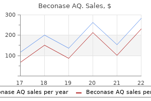
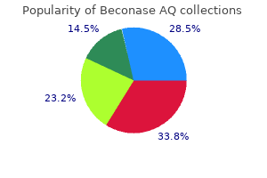
Beconase AQ

Beconase AQ
Beconase AQ dosages: 200MDI
Beconase AQ packs: 1 inhalers, 3 inhalers, 6 inhalers, 9 inhalers, 12 inhalers, 15 inhalers

Extension of the infiltrate into epithelial (ductal) buildings leads to metaplasia and characteristic epimyoepithelial islands peanut allergy symptoms how quickly cheap beconase aq 200MDI free shipping. Two thirds of all tumors of the main salivary glands and about half of these in the minor ones are pleomorphic adenomas allergy testing dogs beconase aq 200MDI discount otc. The tumor usually arises in the superficial lobe of the parotid gland and is 9 times extra frequent in this gland than in the mandibular gland allergy symptoms icd 9 beconase aq 200MDI discount with amex. Involvement of the salivary glands leads to xerostomia, and involvement of the lacrimal glands leads to dry eyes (keratoconjunctivitis sicca). An initial periductal chronic inflammatory infiltrate progressively extends to the acini, till the glands are completely replaced by a sea of polyclonal lymphocytes, immunoblasts, germinal facilities and plasma cells. Proliferating myoepithelial cells encompass remnants of broken ducts and kind so-called epimyoepithelial islands. Late in the midst of the illness, affected glands become atrophic, with fibrosis and fatty infiltration of the parenchyma. Microscopically, the tumors show epithelial tissue intermingled with myxoid, mucoid or chondroid areas. The epithelial part of pleomorphic adenoma consists of ductal and myoepithelial cells Around the ductal epithelial cells are smaller myoepithelial cells, that are the principle mobile element. These cells kind well-defined sheaths, cords or nests and are often separated by a mobile ground substance that resembles cartilaginous, myxoid or mucoid materials. Cellular components of pleomorphic adenomas embody an admixture of glands and myoepithelial cells inside a chondromyxoid stroma. The tumors increase and tend to protrude focally into adjacent tissues, turning into nodular Tumor cells implanted during surgical procedure or tumor nodules left behind continue to develop as recurrences in the scar from the previous operation. Carcinoma ex pleomorphic adenoma: Rarely, carcinomas might come up in pleomorphic adenomas that have been present for a few years. Histologic examination reveals an unequivocal carcinoma in an in any other case benign pleomorphic adenoma. These tumors are normally high-grade malignancies, such as poorly differentiated or undifferentiated adenocarcinoma. In such tumors, the epithelium is organized in a daily, normally glandular sample, and not using a mesenchyme-like component. Monomorphic adenomas include a variety of subtypes, of which Warthin tumor is the most common. Warthin tumors are benign parotid gland neoplasms composed of cystic glandular areas embedded in dense lymphoid tissue. These tumors generally happen after the age of 30 years, with most arising after age 50. The cysts are lined by characteristic eosinophilic epithelial cells (oncocytes) and are embedded in dense lymphoid tissue with germinal facilities. These tumors derive from ductal epithelium, which has a substantial potential for metaplasia. More than half of mucoepidermoid carcinomas within the main glands arise within the parotid gland. Although the tumor occurs in adolescents, most arise in adults, extra commonly in girls. Microscopically, low-grade (well-differentiated) tumors type irregular, strong, duct-like and cystic spaces, which include squamous cells, mucussecreting cells and intermediate cells. Highgrade (poorly differentiated) carcinomas are pleomorphic, without evidence of differentiation except for scattered mucus-secreting cells. Adenoid cystic carcinoma occurs not only within the oral cavity but in addition in lacrimal glands, the nasopharynx, nasal cavity, paranasal sinuses and decrease respiratory tract and is most common in individuals 40 to 60 years of age. Within these buildings, the tumor cells interconnect to enclose cystic spaces, resulting in a stable, tubular or cribriform (sieve-like) arrangement. Tumor cells make a homogeneous basement membrane materials that provides them the attribute "cylindromatous" appearance, therefore the prior name of cylindroma. The tumors in all probability arise from cells which might be differentiating towards intercalated ducts and myoepithelium. Adenoid cystic carcinomas are likely to infiltrate perineural areas and are often painful. They come up occasionally in other salivary glands and occur principally in younger males between the ages of 20 and 30 years. The tumors are encapsulated, round lots, usually underneath 3 cm throughout, and may sometimes be cystic. Microscopically, acinic cell adenocarcinomas are composed of uniform cells with a small central nucleus and plentiful basophilic cytoplasm, just like the secretory (acinic) cells of the traditional salivary glands. Mucoepidermoid carcinoma is characterized by an admixture of mucocytes, epidermoid cells and intermediate cells. The mucocytes (straight arrows) are clustered and have a clear cytoplasm with eccentrically located nuclei. Epidermoid cells (curved arrows) are squamous-like cells however lack keratinization and intercellular bridges. If cholesterol granulomas are allowed to persist for a lot of months, the granulation tissue could become fibrotic, which may eventually lead to complete obliteration of the center ear and mastoid by fibrous tissue. Suppurative otitis media: One of the most typical infections of childhood, acute suppurative otitis media is attributable to pyogenic micro organism that invade the center ear, often via the eustachian tube. If a purulent exudate accumulates within the middle ear, the eardrum ruptures and the pus is discharged. In most circumstances, the an infection is self-limited and tends to heal even without therapy. Neglected or recurrent infection of the middle ear and mastoid process may ultimately produce persistent irritation of the mucosa or destruction of the periosteum masking the ossicles. Acute mastoiditis: this condition is now uncommon but is still seen in cases of inadequately handled otitis media. Characteristically, mastoid air cells are full of pus, and their skinny osseous intercellular walls turn out to be destroyed. Extension of the infection from the mastoid bone to contiguous constructions causes issues. Cholesteatoma: A cholesteatoma is a mass of amassed keratin and squamous mucosa that results from the growth of squamous epithelium from the exterior ear canal thorough the perforated eardrum into the center ear. Benign tumors of those glands embody ceruminoma (ceruminal gland adenoma) and salivary gland-type tumors arising from ceruminal glands Malignant tumors of ceruminal glands embody adenocarcinoma and malignant salivary gland-type tumors During an an infection within the nasopharynx, microorganisms might reach the center ear by ascending through the eustachian tube. Acute otitis media may be because of viral or bacterial infections or to sterile obstruction of the eustachian tube. Viral otitis media may resolve without suppuration, or the middle ear could additionally be secondarily invaded by pus-forming micro organism. Obstruction of the eustachian tube is necessary in the manufacturing of middle ear effusion. Air in the middle ear is then absorbed through the mucosa, and unfavorable strain causes transudation of plasma and occasionally bleeding. More than half of kids in the United States have had at least one episode of serous otitis media earlier than their third birthday. Repeated bouts of otitis media in early childhood typically contribute to unsuspected listening to loss, which is due to residual (usually sterile) fluid in the middle ear. In chronic serous otitis media, mucus-producing (goblet) cell metaplasia could additionally be seen in the mucosal lining of the middle ear. The principal risks of cholesteatoma arise from the erosion of bone, a process which will result in destruction of important contiguous structures Complications of acute and persistent otitis media: As a results of antibiotic treatment, problems of otitis media at the moment are uncommon. These embrace extension of the process via the mastoid bone with resulting meningitis and epidural, subdural or cerebral abscess. These tumors grow slowly, however over years, they might destroy the middle ear and lengthen into the inner ear and cranial cavity. Histologically, center ear paragangliomas are identical to these arising elsewhere and present characteristic lobules of cells embedded in a richly vascular connective tissue. The paraganglial cells are of neural crest origin and contain various amounts of catecholamines, mostly epinephrine and norepinephrine.
Interstitial pulmonary fibrosis results when congestion is current over an prolonged interval (see Chapters 7 and 12) allergy to cold beconase aq 200MDI order on line. Right-sided heart failure commonly complicates leftsided failure or it could possibly develop independently secondary to intrinsic pulmonary illness or pulmonary hypertension allergy shots dog beconase aq 200MDI buy mastercard, which create resistance to blood flow by way of the lungs allergy medicine chlor trimeton beconase aq 200MDI buy free shipping. As a consequence, right atrial stress and systemic venous pressure both enhance, leading to jugular venous distention, decrease extremity edema and congestion of the liver and spleen. Diastolic coronary heart failure is seen in as much as one third of aged sufferers with apparent cardiac failure. As the center ages, the ventricles turn out to be progressively stiffer and require larger filling (diastolic) pressures. Some patients whose hearts are of normal dimension exhibit indicators and signs of heart failure though they show regular systolic contractile perform. Microscopically, these hearts usually exhibit interstitial fibrosis, which can contribute to the decreased compliance of ventricular myocardium. Chromosomal abnormalities related to an elevated incidence of congenital heart anomalies embody Down syndrome (trisomy 21), other trisomies, Turner syndrome and DiGeorge syndrome. The finest proof for intrauterine affect within the occurrence of congenital cardiac defects pertains to maternal rubella infection in the course of the first trimester, especially during the first 4 weeks of gestation. A up to date classification of congenital coronary heart defects divides the circumstances into the groups proven in Table 11-2 and relies on the pattern of blood shunting. Orthopnea and paroxysmal nocturnal dyspnea result when thoracic blood volume increases during recumbency. Although a lot of the scientific presentation of coronary heart failure can be defined by venous congestion (backward failure), certain elements contain insufficient arterial perfusion of significant organs (forward failure). In this circumstance, the foramen ovale stays closed so lengthy as left atrial strain exceeds that in the proper atrium. Estimates of the incidence of particular cardiovascular anomalies range, relying on many factors (Table 11-1). As in different ailments with multifactorial inheritance (see Chapter 6), the risk of recurrence is increased amongst siblings of an affected child. A muscular interventricular septum grows upward from the apex toward the bottom of the guts. It is joined by the down-growing membranous septum, thereby separating proper and left ventricles. The commonest ventricular septal defect is said to failure of the membranous portion of the septum to form in complete or partially. Before this closure is complete, the midportion of the septum primum develops a defect, or ostium secundum, in order that right-to-left flow continues. During the sixth week, a second septum (septum secundum) develops to the right of the septum primum, passing from the roof of the atrium towards the endocardial cushions This process leaves a patent foramen at about the midpoint of the septum, often recognized as the foramen ovale. Patent foramen ovale: Tissue derived from the septum primum located on the left side of the foramen ovale functions as a flap valve that normally fuses with the margins of the foramen ovale, thereby sealing the opening. If circumstances enhance proper atrial strain, as can occur with recurrent pulmonary thromboemboli, a right-to-left shunt will be produced and thromboemboli from the right-sided circulation will pass immediately into the systemic circulation. These paradoxical emboli can produce infarcts in many parts of the arterial circulation, most commonly in the brain, coronary heart, spleen, intestines, kidneys and lower extremities. An ostium secundum defect happens within the middle portion of the septum and varies from a trivial opening to a big defect of the whole fossa ovalis region. A small defect is often not problematic, but a bigger one might enable shunting of adequate blood from left to proper to cause dilation and hypertrophy of the right atrium and ventricle. In this setting, the diameter of the pulmonary artery could exceed that of the aorta. Lutembacher syndrome, a variant of the ostium secundum sort of atrial septal defect, is the mixture of either congenital or rheumatic mitral stenosis and an ostium secundum atrial septal defect. Increased left atrial stress secondary to mitral valve obstruction retains the atrial septum patent. Sinus venosus defect: this anomaly happens within the higher portion of the atrial septum, above the fossa ovalis, near the entry of the superior vena cava. It is often accompanied by drainage of the proper pulmonary veins into the best atrium or superior vena cava. There are normally clefts in the anterior leaflet of the mitral valve and the septal leaflet of the tricuspid valve, which can be accompanied by an related defect within the adjoining interventricular septum. Closure is completed by both hypertrophy of adjacent muscle or adherence of tricuspid valve leaflets to the margins of the defect. In infants with large septal defects, larger left ventricular strain initially creates a left-to-right shunt. Left ventricular dilation and congestive coronary heart failure are common issues of such shunts. If a defect is small enough to permit extended survival, shunting of blood into the best ventricle augments pulmonary blood circulate and finally results in thickening of pulmonary arteries and elevated pulmonary vascular resistance. This elevated vascular resistance may be so nice that the path of the shunt is reversed and goes from proper to left (Eisenmenger syndrome). A affected person with this condition displays late onset of cyanosis (tardive cyanosis), right ventricular hypertrophy and rightsided coronary heart failure. Additional issues of ventricular septal defects embrace (1) infective endocarditis on the site of the defect, (2) paradoxical emboli (moving from proper to left atrium by way of a patent foramen ovale) and (3) prolapse of an aortic valve cusp (with ensuing aortic valve insufficiency). Atrial Septal Defects Atrial septal defects range in severity from clinically insignificant and asymptomatic anomalies to chronic, life-threatening conditions. This opening is partly overlaid by the septum secundum, which has grown all the method down to cowl, in part, the foramen ovale. Although ordinarily uncommon, this defect is regularly encountered in patients with Down syndrome. Pulmonary vascular illness develops in children with extended survival, during which case cyanosis, polycythemia and clubbing of the fingers appear. Open-heart surgical procedure previous to the development of significant pulmonary vascular modifications is an effective treatment. Later in life, usually in adulthood, modifications within the pulmonary vasculature might reverse the flow of blood via the defect and create a rightto-left shunt. Complications of atrial septal defects embody atrial arrhythmias, pulmonary hypertension, proper ventricular hypertrophy, coronary heart failure, paradoxical emboli and bacterial endocarditis. Symptomatic circumstances are treated surgically or with closure devices, which can be delivered percutaneously. After start, the ductus constricts in response to the increased arterial oxygen content material and turns into occluded by fibrosis, the remnant being termed the ligamentum arteriosus. A small shunt has little effect on the guts, whereas a big one produces considerable diversion of blood from the aorta to the low-pressure pulmonary artery. In severe instances, left ventricular hypertrophy and heart failure ensue due to increased demand for cardiac output. The elevated volume and stress of blood within the pulmonary circulation eventually produce pulmonary hypertension and its cardiac issues. It may be caused to contract and then close by the instillation of inhibitors of prostaglandin synthesis It outcomes from absent or incomplete partitioning of the truncus arteriosus by the spiral septum. Truncus arteriosus all the time overrides a ventricular septal defect and receives blood from both ventricles. After profitable surgery, patients are asymptomatic and have a superb long-term prognosis. The aorta is anterior to the pulmonary artery and to its right ("D" or dextrotransposition) all the way from its origin. The condition reveals a male predominance and is more common in offspring of moms with diabetes. The aorta is anterior to and to the best of the pulmonary artery ("D-transposition") and arises from the best ventricle. The quantity and course of blood flow via intracardiac communications and patent ductus arteriosus, if current, depend on pressure gradients across the communications, which might differ throughout early stages of extrauterine life. Coarctation of the Aorta Coarctation of the aorta is an area constriction that nearly all the time occurs immediately below the origin of the left subclavian artery on the site of the ductus arteriosus. Rare coarctations can happen at any level from the aortic arch to the abdominal bifurcation. The situation is two to 5 occasions extra frequent in males than in females and is related to a bicuspid aortic valve in two thirds of instances. Mitral valve malformations, ventricular septal defects and subaortic stenosis may also accompany coarctation of the aorta.
Diseases
In lymphangitic carcinoma allergy testing dogs cost purchase beconase aq 200MDI visa, a metastatic tumor spreads widely through pulmonary lymphatic channels to type a sheath of tumor around the bronchovascular tree and veins allergy testing boise idaho purchase 200MDI beconase aq with amex. Clinically allergy test quiz 200MDI beconase aq buy, patients endure from cough and shortness of breath and display a diffuse reticulonodular sample on the chest radiograph. A section via the lung reveals numerous nodules of metastatic carcinoma corresponding to "cannon ball" metastases seen radiologically. Tension pneumothorax refers to unilateral pneumothorax extensive enough to shift the mediastinum to the other facet, with compression of the alternative lung. A central carcinoid tumor (arrow) is circumscribed and protrudes into the lumen of the main bronchus. The compression of the bronchus by the tumor brought on the postobstructive pneumonia seen within the distal lung parenchyma (right). For instance, while exercising vigorously, a tall young man develops acute chest pain and shortness of breath. In most cases, spontaneous pneumothorax subsides by itself, however some patients require withdrawal of the air. Normally, solely a small quantity of fluid in the pleural cavity lubricates the space between the lungs and chest wall. Fluid secreted into the pleural space from the parietal pleura is absorbed by the visceral pleura. The severity of a pleural effusion varies from a couple of milliliters of fluid to a massive accumulation that shifts the mediastinum and the trachea to the other facet. It may be due to elevated hydrostatic pressure throughout the capillaries, as happens in patients with coronary heart failure or in any situation that produces systemic or pulmonary edema. This might often be caused by an external penetrating wound that introduces pyogenic organisms into the pleural area. This mesothelioma consists of a biphasic sample of epithelial and sarcomatous components. It has an ominous portent because lymphatic obstruction suggests illness of the lymph nodes in the posterior mediastinum. Microscopically, traditional mesotheliomas present a biphasic appearance, with epithelial and sarcomatous patterns Glands and tubules that resemble adenocarcinoma are admixed with sheets of spindle cells that are much like a fibrosarcoma. In different cases, the tumor is principally monophasic-that is, sarcomatoid or epithelioid. Immunohistochemistry is crucial for differentiating mesothelioma from adenocarcinoma. Other useful standards for diagnosing mesothelioma embrace the absence of mucin, presence of hyaluronic acid (positive Alcian blue staining) and demonstration of long, slender microvilli by electron microscopy. Patients are first seen with a pleural effusion or a pleural mass, chest pain and nonspecific signs corresponding to weight reduction and malaise. Pleural mesotheliomas are most likely to unfold regionally within the chest cavity, invading and compressing major constructions. Metastases can happen to the lung parenchyma and mediastinal lymph nodes, in addition to to extrathoracic websites such because the liver, bones, peritoneum and adrenals. In the United States, Great Britain and South Africa, the majority of patients report exposure to asbestos. The latency interval between asbestos exposure and the appearance of malignant mesothelioma is about 20 years, with a spread of 12 to 60 years. Esophageal atresia and fistulas are often related to congenital coronary heart disease. In one other variant, termed an H-type fistula, a communication exists between an intact esophagus and an intact trachea. Webs are usually single but may be a number of and may occur wherever within the esophagus. Dysphagia, often associated with aspiration of swallowed food, is the commonest medical manifestation. It is now believed that these pouches most frequently replicate a disturbance in the motor perform of the esophagus. A diverticulum within the midesophagus ordinarily has a wide stoma, and the pouch is often larger than its orifice. Unlike other diverticula, epiphrenic diverticula are encountered in younger persons. Nocturnal regurgitation of enormous quantities of fluid stored in the diverticulum through the day is typical. Motor disorders may be brought on by (1) esophageal or systemic defects in striated muscle perform, (2) neurologic ailments affecting afferent nerves or (3) peripheral neuropathies occurring in affiliation with diabetes or alcoholism. Patients with narrow Schatzki rings, nevertheless, might complain of intermittent dysphagia. Achalasia Features Impaired Function of the Lower Esophageal Sphincter Achalasia, at one time termed cardiospasm, is characterised by failure of the lower esophageal sphincter to loosen up in response to swallowing and the absence of peristalsis in the physique of the esophagus. As a results of these defects in each the outflow tract and the pumping mechanisms of the esophagus, food is retained throughout the esophagus, and the organ hypertrophies and dilates conspicuously. Achalasia is associated with the loss or absence of ganglion cells within the esophageal myenteric plexus. In Latin America, achalasia is a typical complication of Chagas illness, by which the ganglion cells are destroyed by the protozoan Trypanosoma cruzi. Symptoms of achalasia might arise in amyloidosis, sarcoidosis and infiltrative malignancies. Dysphagia, occasionally odynophagia and regurgitation of fabric retained in the esophagus are widespread signs of achalasia. Treatment is pneumatic dilation or surgical myotomy of the lower esophageal sphincter, which can lead to gastroesophageal reflux. Esophageal Diverticula Often Reflect Motor Dysfunction A true esophageal diverticulum is an outpouching of the wall that incorporates all layers of the esophagus. Esophageal diverticula happen in the hypopharyngeal space above the upper esophageal sphincter, in the middle esophagus and instantly proximal to the lower esophageal sphincter. Disordered function of cricopharyngeal musculature is mostly thought to be concerned in the pathogenesis of this false diverticulum. Most affected individuals who come to medical attention are older than 60 years, suggesting that this diverticulum is acquired. The typical symptom is regurgitation of food eaten some time beforehand (occasionally days), within the absence of dysphagia. Scleroderma Causes Fibrosis of the Esophageal Wall Scleroderma (progressive systemic sclerosis) results in fibrosis in many organs and produces a extreme abnormality of esophageal muscle function (see Chapter 4). Paraesophageal hiatal hernia Stomach so impaired that the lower esophagus and higher abdomen are not distinct functional entities and are visualized as a standard cavity. Microscopically, fibrosis of esophageal smooth muscle and nonspecific inflammatory adjustments are seen. Intimal fibrosis of small arteries and arterioles is common and should play a job within the pathogenesis of the fibrosis. Clinically, sufferers have dysphagia and heartburn attributable to peptic esophagitis, owing to reflux of acid from the abdomen (see below). Classically, symptoms are exacerbated when the affected person is recumbent, which facilitates acid reflux disease. Large herniations carry a threat of gastric volvulus or intrathoracic gastric dilation. By contrast, an enlarging paraesophageal hernia must be surgically handled, even within the absence of symptoms. Although acid is damaging to the esophageal mucosa, the combination of acid and pepsin is particularly injurious. Moreover, gastric fluid typically contains refluxed bile from the duodenum, which is dangerous to the esophageal mucosa. Microscopically, gentle damage to the squamous epithelium is manifested by cell swelling (hydropic change). The basal region of the epithelium is thickened, and the papillae of the lamina propria are elongated and lengthen towards the floor because of reactive proliferation. An enhance in lymphocytes is seen in the squamous epithelium, and eosinophils and neutrophils could additionally be present.
Progression may happen over a period of many months or might evolve quickly in days or even weeks in instances of fulminant hepatic failure allergy testing lawrenceville ga cheap 200MDI beconase aq otc. Hyperbilirubinemia associated with hepatic failure is allergy testing kits for physicians purchase 200MDI beconase aq free shipping, for essentially the most part allergy medicine alavert beconase aq 200MDI low cost, conjugated, although the level of unconjugated bilirubin also tends to enhance. On occasion, increased erythrocyte turnover may add to unconjugated hyperbilirubinemia, thereby aggravating the jaundice. It is possible that the condition is brought on in half by injurious compounds absorbed from the gut which have escaped hepatic cleansing both due to hepatocyte dysfunction or the existence of structural or practical vascular shunts. Clinical options associated to parenchymal liver failure (A), endocrine disturbances (B) and portal hypertension (C). There is appreciable overlap of those scientific options associated to their pathogeneses. However, the correlation between the increased concentration of blood ammonia and the severity of hepatic encephalopathy is inexact. The characteristic breath odor of patients with hepatic failure, termed fetor hepaticus, reflects the presence of mercaptans in saliva. The deep layers of the cerebral cortex and subcortical white matter, the basal ganglia and the cerebellum exhibit laminar necrosis and a spongiform appearance. The syndrome normally happens in the setting of cirrhosis and signifies a poor prognosis. Conversely, in sufferers with the hepatorenal syndrome, liver transplantation can restore renal function. The syndrome is related to extreme renal vasoconstriction associated partially with arterial vasodilation induced by endothelial nitric oxide release. The event is precipitated subsequent to vascular modifications within the cirrhotic affected person. In the early levels of cirrhosis, renal glomerular filtration pressure is protected against systemic arteriolar vasodilation by intrarenal prostaglandins. With more and more severe arteriolar vasodilation, these intrarenal elements become ineffective, renal arterial vasoconstriction intensifies and glomerular perfusion and filtration decline. Chronic liver failure in males leads to feminization, characterized by gynecomastia, a feminine body habitus and a feminine distribution of pubic hair (female escutcheon). In addition, vascular manifestations of hyperestrogenism are widespread and embody spider angiomas in the territory drained by the superior vena cava (upper trunk and face) and palmar erythema Feminization is attributed to decreased hepatic catabolism of estrogens and weak androgens. The weak androgens (androstenedione and dehydroepiandrosterone) are converted to estrogenic compounds in peripheral tissues, thereby adding to the burden of circulating estrogens. Thrombocytopenia could outcome from (1) hypersplenism, (2) bone marrow melancholy or (3) the consumption of circulating platelets by intravascular coagulation. Intravascular coagulation may be stimulated by (1) necrosis of liver cells, (2) induction of tissue factor expression on endothelial cells by bacterial toxins, (3) discount in circulating anticoagulant proteins or (4) insufficient hepatic clearance of activated clotting components from the circulation. Portal Hypertension Portal hypertension is defined by either an absolute improve in portal venous pressure, often above 8 mm Hg, or a rise in the stress gradient between the portal vein and the hepatic vein of 5 mm Hg or extra. Portal hypertension results from obstruction to blood circulate someplace in the portal circuit. Venous flow from spleen from gastroesophageal varices, ascites, splenomegaly and renal and pulmonary disease The trigger is in all probability going related to elements that incite inflammation, corresponding to alcoholic hepatitis or viral hepatitis. Regenerative nodules within the cirrhotic liver impinge on the hepatic veins, thereby obstructing blood flow distal to the lobules. The small portal veins and venules are trapped, narrowed and often obliterated by scarring of the portal tracts. Moreover, blood flow through the hepatic artery is increased, and small arteriovenous communications become useful. In this way, portal hypertension as a result of obstruction of blood circulate distal to the sinusoid is augmented by elevated arterial blood circulate. In cirrhosis, endothelial cell dysfunction occurs in the liver and within the systemic circulation, rising intrahepatic vasoconstriction. As resistance to blood move to the liver increases, mesenteric arterial vasodilation increases the amount of blood flowing into the portal vein. Progressive portal hypertension parallels mesenteric arterial vasodilation, resulting in dysfunctional systemic circulation (systemic arterial vasodilation and reduced efficient arterial blood volume). The lower in efficient arterial blood quantity is related to the scientific manifestations of superior portal hypertension, together with ascites, hepatorenal syndrome (see above) and hepatopulmonary syndrome. Worldwide, hepatic schistosomiasis is a major explanation for intrahepatic portal hypertension (see Chapter 9 for details). Idiopathic portal hypertension refers to occasional cases of intrahepatic portal hypertension with splenomegaly that occur in the absence of any demonstrable intrahepatic or extrahepatic disease. Other causes of portal vein thrombosis include tumors, infections, hypercoagulability states, pancreatitis and surgical trauma. Portal hypertension may occur in sufferers with splenomegaly from quite so much of causes, together with polycythemia vera, myeloid metaplasia and continual myelogenous leukemia. In cirrhosis, the accompanying splenomegaly that augments blood flow in the splenic vein might additional worsen portal hypertension. In the persistent stage, the reduce surface is paler and the liver is firm due to a rise in connective tissue. Microscopically, the hepatic veins show thrombi in various phases of evolution, from latest clots to well-organized thrombi which were canalized. In the acute stage of each the Budd-Chiari syndrome and veno-occlusive disease, the sinusoids of the central zone are dilated and full of erythrocytes. In long-standing venous congestion, fibrosis of the central zone, radiating into the more peripheral portions of the lobules, is conspicuous. The sinusoids are dilated, and the central-to-midzonal hepatocytes present pressure atrophy. Eventually, connective tissue septa hyperlink adjacent central zones to type nodules with a single portal tract in the center, a course of often known as reverse lobulation. Most typically, obstruction of the hepatic venous circulation is incomplete, and signs may happen for durations ranging from a month to a quantity of years. Most patients eventually die in hepatic failure or from the issues of portal hypertension. Thrombosis is most typical in the massive hepatic veins near their exit from the liver and in the intrahepatic portion of the inferior vena cava. One of the most common causes of dying in patients with cirrhosis and different issues related to portal hypertension is exsanguinating higher gastrointestinal tract hemorrhage from bleeding esophageal varices. Because of the increased blood circulate and higher stress that observe the opening of those collaterals, the submucosal veins in the neighborhood of the esophagogastric junction turn out to be dilated and protrude into the lumen (see Chapter 13). The back-pressure within the portal vein is also transmitted to its tributaries, including the inferior hemorrhoidal veins, which turn out to be dilated and tortuous (anorectal varices). Hepatic veno-occlusive disease is a variant of the BuddChiari syndrome and is caused by occlusion of the central venules and small branches of the hepatic veins. Most commonly, this disorder is traced to the ingestion of poisonous pyrrolizidine alkaloids present in vegetation of the Crotalaria, Senecio and Symphtum genera, which are generally used within the formulation of natural teas (comfrey is the most common in Europe and North America). It is also seen in sufferers handled with sure antineoplastic chemotherapeutic brokers and after hepatic irradiation. Veno-occlusive illness can also be reported in affiliation with bone marrow transplantation, presumably as a manifestation of graft-versus-host illness. In sufferers with cirrhosis who survive an initial episode of variceal bleeding, long-term survival is unlikely due to a high danger of rebleeding or worsening liver failure. Permanent decompression of the portal circulation could be achieved by surgically constructed portosystemic shunts. Hypoalbuminemia Intrahepatic vasoconstriction resulting in a rise in resistance to portal inflow Increase in portal stress Splenomegaly the spleen in portal hypertension enlarges progressively and often offers rise to the syndrome of hypersplenism-that is, a decrease within the life span of all of the fashioned components of the blood and, subsequently, a reduction of their circulating numbers (pancytopenia). Hypersplenism is attributed to an increased rate of removing of erythrocytes, leukocytes and platelets secondary to the extended transit time through the hyperplastic spleen. On gross examination, the spleen is firm and enlarged, up to 1,000 g, and its reduce surface is uniformly deep pink, with an inapparent white pulp. Microscopically, the splenic sinusoids are dilated, and their partitions are thickened by fibrous tissue and lined by hyperplastic endothelial cells and macrophages. Focal hemorrhages cause fibrotic, iron-laden nodules, known as Gamna-Gandy bodies. Peripheral vasodilation Reduction in systemic vascular resistance Reduced efficient arterial blood volume Ascites Sodium and water retention Increase in sympathetic stimulation Renal arterial vasoconstriction Renal failure Ascites Ascites refers to the buildup of fluid within the peritoneal cavity. It usually accompanies portal hypertension, and the amount of fluid may be so great (frequently many liters) that it not solely distends the abdomen but additionally interferes with breathing.

Around the periphery allergy symptoms mango cheap 200MDI beconase aq otc, radiating ducts and lobules exhibit a wide range of benign alterations allergy shots numbness arm 200MDI beconase aq discount fast delivery. Radial scars are related to a twofold improve in breast cancer risk food allergy treatment 2013 beconase aq 200MDI discount without a prescription, and this danger is bigger in girls with coexisting proliferative illness, with or with out atypia. Intraductal Papilloma Papillomas could be solitary or a quantity of and situated centrally or peripherally. Papillomas vary from microscopic foci to those that measure several centimeters with larger lesions regularly showing foci of hemorrhage or necrosis. Microscopically, dilated duct areas contain multiple branching papillae, with fibrovascular cores lined by a layer of myoepithelium, upon which lie one or more layers of epithelium. Micropapillae (arrows) project into the duct lumen and encompass cells with an increased nuclear:cytoplasmic ratio and nuclear hyperchromasia. There is minimal distension of the lobular acini by a uniform inhabitants of cells with intracytoplasmic lumina and spherical nuclei containing small nucleoli. Here, the atypical cells lie beneath an attenuated floor layer of luminal epithelial cells. Fibroepithelial Lesions these come up from intralobular stroma and comprise each stromal and epithelial parts. They are most often solitary lesions, though they are often multiple and bilateral. The stroma is typically composed of spindle cells and exhibits variable, however often low, cellularity. In younger ladies, the stroma is commonly myxoid, however with age, it may turn into hyalinized and calcify. Such lesions are associated with an increased danger of breast cancer, which is exacerbated in patients with a first-degree family history of breast malignancies. Juvenile fibroadenoma, which is most common in adolescents, grows quickly and may attain 20 cm in size. The tumor resembles fibroadenomas histologically, albeit with more mobile stroma. The incidence now seems to have leveled off, and death rates have decreased during the last 25 years due to earlier detection and higher therapy. Women in the United States have a one in nine lifetime danger of developing breast most cancers, and one in five of those with breast cancer will die of their illness. However, age-specific incidence charges enhance dramatically after forty years of age, plateauing at 75 to eighty years. Breast cancer occurs four to 5 instances more regularly in Western industrialized international locations than in less developed areas. Dietary, environmental and lifestyle components have been implicated, but none are proved. Breast most cancers not often develops in men, though when it does happen, it might be equally deadly (see below). It is now obvious that routine screening mammography detects a big percentage of tumors that will never cause clinical disease. They current as rapidly rising breast plenty and are properly circumscribed or lobulated on mammography. Grossly, benign phyllodes tumors are sharply circumscribed, with a fi rm, glistening and greyish white minimize floor. On microscopy, fronds of hypercellular stroma lead to the formation of leaf-like structures, which project into cystic areas. These cystic spaces are lined by a dual layer of benign epithelium and myoepithelium. Most phyllodes tumors are benign, with gentle or moderately hypercellular stroma showing inconspicuous cytologic atypia and few mitoses. The stroma in malignant phyllodes tumors is markedly hypercellular, with considerable pleomorphism, abundant mitoses (10 per 10 high-power fields) and stromal overgrowth. Multiple danger components for breast most cancers have been identified, some modifiable, some not (Table 19-1). Modifiable risk factors are controversial and embrace late age at first live birth, food plan, high physique mass index and use of exogenous hormones. The stromal component adjoining to ductal epithelium is much like a fibroadenoma but is extra cellular. Breast cancers that develop in patients with germline mutations are sometimes high-grade ductal carcinomas. These patients largely develop high-grade invasive ductal tumors of no particular type. There are restricted knowledge to recommend that whole fat within the diet may enhance the chance of breast cancer after menopause, but when true, the effect is prone to be small. Alcohol consumption without sufficient folate consumption has been associated with greater breast cancer rates, but the information require better confirmation. Density is influenced by age, parity, body mass index and menopausal standing, but genetic elements may also play a job. Atypical hyperplasia or nonatypical proliferative breast disease increases the relative risk of most cancers four to 5 instances and 1. Women with a previous breast most cancers have a 10-fold greater chance of developing a second primary most cancers in both breast. These lesions are thought-about nonobligate precursors of invasive carcinoma, the possibility of progressing to invasion varying with the histologic subtype, grade and extent. The strongest association with elevated risk is a family historical past of breast cancer in first-degree family members. Noninvasive tumors and their invasive counterparts present similar cytologic appearance and nuclear grade. Growth patterns may be cribriform, micropapillary, papillary, stable and comedo types, and a quantity of architectural patterns can coexist in one lesion. Micropapillary or cribriform progress is the rule, and strong progress patterns are much less frequent. Microinvasive carcinoma: this pattern is defined as one or more foci of invasive carcinoma, none of which exceed 1 mm in diameter. The cells have abundant cytoplasm, irregular nuclei, outstanding nucleoli and coarse chromatin. The latter seems grossly as distended ducts containing white necrotic materials, hence the term comedo necrosis. The mobile necrotic particles often undergoes dystrophic calcification, which may be seen on mammography as linear, branching calcifications Malignant cells are confined to duct spaces, however periductal continual inflammation may be current, with formation of latest vessels and a desmoplastic response (fibroblast proliferation and subsequent fibrosis) in a peritubular distribution A small proportion of women will, however, current symptomatically with a mass lesion or with Paget disease of the nipple (see below). These entities are each atypical proliferations of loosely cohesive epithelial cells, but every of these entities entails vital variations within the relative risk of developing breast most cancers. Specimen radiograph of core biopsy shows linear and punctate atypical calcifications which are extremely suspicious for most cancers. The most cancers cells within the lobular form of carcinoma in situ are smaller and have less cytoplasm than these in the ductal sort. Microscopically, the cells are monotonous and small with spherical common nuclei and minute nucleoli, although bigger cells with conspicuous nucleoli might dominate. Paget Disease Paget illness of the nipple refers to the presence of malignant glandular epithelial cells within the dermis of the nipple and areola. The disease presents as erythema or as an eczematous change in the nipple and areola. Nipple retraction may be found, and over half of patients have an associated palpable mass. Microscopically, malignant glandular epithelial cells are seen throughout the dermis, singly or in small teams. Paget cells are massive with ample cytoplasm, pleomorphic nuclei and distinguished nucleoli. The prognosis of Paget disease is said to that of the underlying ductal most cancers. Grossly, a well-circumscribed, partially cystic, regularly hemorrhagic, strong mass is seen.

Immunotherapy with high-dose interferon alpha 2b (and increasingly pegylated interferon alpha 2b) is used in treating node-positive illness allergy shots liver damage purchase 200MDI beconase aq free shipping. Twenty p.c of such patients have responded to the tyrosine kinase inhibitor imatinib allergy symptoms getting worse purchase 200MDI beconase aq fast delivery, in some instances for prolonged durations allergy symptoms with eyes beconase aq 200MDI generic with amex. The present recommendations regarding excision of confirmed melanomas state that (1) a 5-mm margin of uninvolved tissue should be obtained with in situ melanoma, (2) a 1-cm margin is proper for a tumor thickness of 1 mm or less and (3) a 2-cm margin is sometimes recommended for a tumor thickness larger than 1 mm. In instances of node-negative, nondisseminated disease, additional adjunctive remedy has not been proven to have an result on survival. Such lesions are associated with a hanging increase in intraepidermal and dermal melanocytes, which can prolong deep into the subcutaneous tissue. The cells are so atypical that an incorrect analysis of melanoma could additionally be made, though melanoma is exquisitely rare in childhood. The scientific look could prompt an excisional biopsy to rule out nodular melanoma. Larger lentiginous lesions could have to be biopsied to rule out lentigo maligna melanoma. In the radial progress part, lentigo maligna melanoma is a flat, irregular, brown-to-black patch that may cowl a big a part of the face or dorsal hands. The cells of the radial growth part are predominantly in the basal layer, typically forming contiguous or practically contiguous rows of atypical single melanocytes. Acral lentiginous melanoma is the most common form of melanoma in dark-skinned folks and is usually restricted to the palms, soles and subungual regions. In the radial growth phase, acral lentiginous melanoma forms an irregular, brown-to-black patch that covers part of the palm or sole or arises beneath a nail, usually on a thumb or nice toe. They are elevated, circumscribed, symmetric, epidermal proliferations that often seem papillary. They could also be single or a quantity of and are most frequent on the dorsal surfaces of the palms or on the face. Histologically, verruca vulgaris shows hyperkeratosis and papillary epidermal hyperplasia. Plantar warts are benign, regularly painful, hyperkeratotic nodules on the soles of the feet. A symmetric pink nodule appeared all of a sudden in a toddler but then remained stable for several weeks till it was excised. B Histologically, plantar warts are endophytic or exophytic, papillary, squamous epithelial proliferations. The cells contain plentiful cytoplasmic inclusions which are similar in appearance to the darker staining keratohyaline granules. The nuclei of keratinocytes near the bases of these warts also comprise pink nuclear inclusions. Clinically and microscopically, they appear "pasted on" and are composed of broad anastomosing cords of mature stratified squamous epithelium related to small cysts of keratin (horn cysts). The blood vessels throughout the cores extend near the surface of verrucae, which makes them prone to traumatic hemorrhage and the resultant black "seeds" that patients observe. Keratoacanthoma Keratoacanthomas are quickly rising keratotic papules on sunexposed skin that develop over 3 to 6 weeks into crater-like nodules. Spontaneous regression usually follows within 6 to 12 months, leaving an atrophic scar. There may be focal lichenoid inflammation, and the dermis could also be markedly infiltrated with neutrophils, lymphocytes and eosinophils. Microabscesses of neutrophils and entrapped dermal elastic fibers are sometimes current inside the lesion. The lesion is cup shaped, with a central, keratin-filled umbilication and overhanging ("buttressing") edges. At the base of the keratin, keratinocytes are massive and have plentiful homogeneous, eosinophilic ("glassy") cytoplasm. The central a half of each nest contains closely packed keratinocytes which may be slightly smaller than the traditional epidermal basal keratinocytes and show occasional apoptosis. Thus, the tumor ought to be promptly handled by excision or different methods of eradication. It is commonest on sun-damaged skin of honest people with mild hair and freckles and often originates in actinic keratoses. The tumor may originate in chronic scarring processes, corresponding to osteomyelitis sinus tracts, burn scars and areas of radiation dermatitis. The edges of many tumors present modifications typical of actinic keratosis, namely, a variably thickened epidermis with parakeratosis and significant atypia of the basal keratinocytes. Early lesions are small, scaly or ulcerated erythematous papules, which can be pruritic. Pearly papule is the prototypic nodulocystic type of lesion, so named as a outcome of it resembles a 2- to 3-mm pearl. It is covered by tightly stretched dermis and is laced with small, delicate, branching vessels (telangiectasia). Mitoses and multinucleation of keratinocytes are apparent, as is apoptosis (arrows). Nuclear chromatin is dense and evenly distributed, cytoplasm is scant and mitotic figures and nuclear fragments are frequent. Tumor cells present evidence of each Merkel cell markers (cytokeratin 20) and neuroendocrine markers (such as chromogranin and synaptophysin). These embody the next: Cylindroma: Cylindromas are adnexal neoplasms with features of sweat gland differentiation. Syringoma: Syringomas seem to derive from the intraepidermal portion of eccrine sweat glands and happen about the eyelid and higher cheek as small, elevated, flesh-colored papules. Occasional malignant lesions with ductal differentiation are termed porocarcinomas. Trichoepithelioma: Trichoepithelioma is a neoplasm that differentiates toward hair structures. It occurs on the extremities as a dome-shaped, agency, rubbery nodule with ill-defined borders and pigmentation that ranges from pink to darkish brown. Microscopically, the papillary and reticular dermises are changed by fibrous tissue that varieties ill-defined small cartwheels with small central vascular spaces. Dermatofibrosarcoma Protuberans Dermatofibrosarcoma protuberans is a slowly rising nodule or indurated plaque with intermediate malignant potential, which appears mostly on the trunk of young adults. Local recurrence after tried complete excision is common, however metastases are rare. The commonest histologic sample is a poorly circumscribed, monotonous population of spindle cells organized in a dense "storiform" (pinwheellike) array. Lined with epithelia, containing salivary glands and components of the host defense system, carcinomas and lymphomas are to be expected. As a specialized organ of sensation with access to the nasopharynx, the ear additionally suffers from infection, as nicely as extra specialized defects of the sensory equipment. If the mucosa is injured or immunity impaired, otherwise normal oral cavity organisms can turn out to be pathogenic (see Chapter 9 for further discussion). Transmission happens by droplet an infection, and the virus could be recovered from the saliva of infected individuals. Disease starts with painful inflammation of the affected mucosa, adopted shortly by the formation of vesicles. These vesicles rupture and form shallow, painful ulcers, starting from punctate dimension to a centimeter in diameter. Microscopically, herpetic vesicles type because of "ballooning degeneration" of epithelial cells. It may be reactivated to trigger recurrent herpetic lesions in various ways, including trauma, allergy, menstruation, pregnancy, exposure to ultraviolet gentle and by different viral infections. The last embrace nasopharyngeal-type differentiated and undifferentiated carcinomas and salivary gland undifferentiated carcinoma. Bacteria, Mycoplasma, viruses, autoimmune reactions and hypersensitivity have been implicated however are unproved. The underlying inflammatory infiltrate is composed of mononuclear and polymorphonuclear leukocytes. These organisms are found in the mouth of many healthy individuals, suggesting that different components, corresponding to decreased resistance to an infection as a outcome of inadequate diet, immunodeficiency or poor oral hygiene, are required for growth of the illness.
Utricularia vulgaris (Bladderwort). Beconase AQ.
Source: http://www.rxlist.com/script/main/art.asp?articlekey=96337

Exophytic papilloma options papillary fronds lined by urothelial epithelium allergy symptoms on dogs purchase beconase aq 200MDI on line, which is just about indistinguishable from regular allergy testing lincoln ne discount beconase aq 200MDI online. In most instances allergy and asthma trusted 200MDI beconase aq, "recurrences" symbolize new tumors that develop elsewhere in the urinary bladder. Inverted papillomas are uncommon and usually present as nodular mucosal lesions within the urinary bladder, normally within the trigone area. Inverted papillomas are covered by regular urothelium, from which cords of transitional epithelium descend into the lamina propria. These lesions are extra frequent in men, with a peak incidence in the sixth and seventh many years. At cystoscopy, tumors may be small, delicate, low-grade papillary lesions limited to the mucosal floor or bigger, greater grade, stable invasive masses, which are sometimes ulcerated. The lesion is characterised by a urothelium of variable thickness that shows mobile atypia of the whole mucosa, from the basal layer to the surface. Atypia features nuclear modifications, together with loss of polarity, enlargement, hyperchromatism, irregular form, outstanding nucleoli and coarse chromatin. In one third of cases, carcinoma in situ of the bladder progresses to subsequent invasive carcinoma. Confined to the mucosal surface, the in situ lesions most frequently seem as a number of, pink, velvety, flat patches close to exophytic papillary transitional cell carcinoma (see below). Concurrent involvement with in situ most cancers elsewhere within the bladder or Papillary urothelial neoplasms of low malignant potential: these tumors are considered intermediate between benign urothelial papillomas and low-grade papillary urothelial carcinoma. Carcinoma in situ is often multifocal at the time of discovery, or comparable lesions may develop shortly thereafter. Regional lymph nodes comprise metastatic tumor in approximately half of all sufferers with these invasive tumors. Invasive urothelial carcinoma: these extremely malignant cancers might evolve from pre-existing papillary lesions or flat carcinoma in situ. The depth of invasion into the wall of the bladder, or beyond its confines, determines the prognosis. Low-grade papillary urothelial carcinoma: Low-grade tumors have fronds lined by neoplastic urothelial epithelium with minimal architectural and cytologic atypia. High-grade papillary urothelial carcinoma: these neoplasms present vital nuclear hyperchromasia and pleomorphism. High-grade papillary urothelial carcinoma exhibits distinguished architectural disorganization of the epithelium, which incorporates cells with pleomorphic hyperchromatic nuclei. Invasive high-grade papillary urothelial carcinoma consists of irregular nests of hyperchromatic cells invading into the muscularis. It originates from foci of cystitis glandularis, intestinal metaplasia or remnants of urachal epithelium in the bladder dome. Hypospadias also reveals an association with other urogenital anomalies and sophisticated, multisystemic, developmental syndromes. [newline]In the commonest type of epispadias, the whole penile urethra is open alongside the shaft. If a slender prepuce is forcefully retracted, it may strangulate the glans and impede the outflow of venous blood, a condition termed paraphimosis. Congenital phimosis must be distinguished from acquired phimosis, which is often a consequence of recurrent infections or trauma of the prepuce in uncircumcised males. In order of reducing frequency, metastases of bladder cancer happen in regional and periaortic lymph nodes, liver, lung and bone. Papillary lesions limited to the mucosa (stage T0) or lamina propria (stage T1) are commonly treated conservatively by transurethral resection. Radical cystectomy is finished for sufferers whose cancers show muscle invasion and occasionally for advanced-stage tumors. Scrotal Masses Scrotal lots and circumstances that result in scrotal swelling or enlargement usually replicate abnormalities of testicular, epididymal and scrotal growth. Clinical problems related to these pathologic conditions are most often encountered in kids but may be found in adults. A hydrocele may be congenital (the commonest explanation for scrotal swelling in infants) or acquired because of infection, tumor or trauma. Hydroceles are generally benign, but longstanding disease may trigger testicular atrophy or compression of the epididymis. Surgical resection by ligation of the internal spermatic vein often improves reproductive function. Significant issues of chronic balanoposthitis are meatal stricture, phimosis and paraphimosis. This condition is equivalent to lichen sclerosus et atrophicus of the vulva in women (see Chapter 18). The penile shaft demonstrates an ill-defined induration of the shaft with no change in the overlying skin. On microscopic examination, dense fibrosis is related to sparse, nonspecific, persistent inflammatory infiltration. Cancer of the Penis Cancer of the penis originates from the squamous mucosa of the glans and contiguous urethral meatus or the prepuce and skin masking the penile shaft. Gonococcal and nongonococcal urethritis have an acute onset and are related to latest sexual activity. Nongonococcal urethritis is generally caused by Chlamydia trachomatis or Ureaplasma urealyticum however could also be related to a variety of different pathogens. Typically, infection is related to cystitis but could also be related to other ailments Such geographic variations have been attributed to differences within the frequency of circumcision. Most patients with most cancers of the penis have had phimosis since an early age, suggesting that extended contact between smegma and the penile epithelium might play a role. Both types seem microscopically as squamous cell carcinoma in situ, similar to that in different websites. Urethral caruncle presents as an exophytic, often ulcerated, polypoid mass, 1 to 2 cm in diameter, at or near the urethral meatus. Microscopically, it exhibits acutely and chronically infected granulation tissue as nicely as ulceration and hyperplasia of transitional cell or squamous epithelium. Other medical findings encountered in variable proportions are circinate balanitis in men (with round or linear plaque-like discolorations on the glans), cervicitis in girls and pores and skin eruptions. Symptoms often appear a couple of weeks after chlamydial urethritis or enteric infection with such pathogens as Shigella, Salmonella or Campylobacter. It is thus thought to characterize an inappropriate immune reaction to unknown microbial antigen(s). Symptoms often disappear spontaneously over three to 6 months, but arthritis recurs in half of sufferers (see additionally Chapter 26). The altered dermis reveals some superficial stratification and maturation and will comprise big keratinocytes with multinucleated atypical nuclei. Squamous cell carcinoma often includes the glans or prepuce and, much less generally, the penile shaft. Extensive destruction of penile tissue, together with the urethral meatus, is noticed in uncared for cases. Invasive tumors usually have a dense, continual inflammatory cell infiltrate within the dermis. The tumor could invade deeply along the penile shaft and spread to inguinal lymph nodes, then to iliac nodes and ultimately distant organs. In most situations, the testis has an upper scrotal location or is retained in the inguinal canal. Cryptorchid testes are smaller than regular even at an early age, and the distinction between the affected and the traditional testis turns into more prominent with age. In infancy and early childhood, the seminiferous tubules within the affected testes are smaller and have fewer germ cells than regular. Postpubertal testes additionally contain fewer germ cells than regular, and spermatogenesis is limited to a minority of tubules. Hyaline thickening of tubular basement membranes and outstanding stromal fibrosis are observed. Eventually, tubules turn into devoid of spermatogenic cells and are entirely hyalinized. In the big majority of those infants, the testes descend into the scrotum in the course of the first year of life.
Overt nephritis resolves after a number of weeks allergy symptoms for dogs buy 200MDI beconase aq otc, although hematuria and particularly proteinuria might persist for a number of months allergy blood test cheap beconase aq 200MDI otc. Accumulation of quite a few subepithelial immune complexes as hump-like structures is a characteristic characteristic allergy symptoms nausea buy generic beconase aq 200MDI online. Less distinguished subendothelial immune complexes are related to endothelial cell proliferation and are related to elevated capillary permeability and narrowing of the lumen. Frequently, proliferation of mesangial cells and a thickened mesangial matrix result in widening of the stalk. Deposition of mesangial and subendothelial immune complexes causes mesangial proliferation and extension into the subendothelial zone. In most sufferers, the origin of nephritogenic antigen is unknown, however some have related conditions that are the obvious source of the antigen. Elimination of disorders similar to bacterial endocarditis or osteomyelitis leads to the decision of glomerulonephritis, which helps a causal relationship between the two. Agents that are responsible for kind I membranoproliferative glomerulonephritis cause persistent indolent infections which may be related to chronic antigenemia. Twenty % of patients will have crescents, normally involving solely a minority of glomeruli. Subendothelial and mesangial electron-dense deposits, comparable to immune complexes, are the likely stimuli for the mesangial response. Immunofluorescence microscopy exhibits granular deposition of immunoglobulins and complement in glomerular capillary loops and mesangium. It might manifest as both nephrotic or nephritic syndrome or a mixture of both. Large subendothelial deposits of immune complexes extend along the inner border of the basement membrane. The accumulation of mesangial cells and stroma in the tufts narrows the capillary lumen. It is much more widespread in creating nations that have a high prevalence of persistent infections. It resembles sort I disease in medical presentation and course, except that hypocomplementemia is more frequent, and the prognosis is barely worse. Mesangial deposition of immune complexes causes much less irritation than subendothelial deposits, which are extra uncovered to the circulation. Subepithelial localization is answerable for proteinuria but not overt glomerular inflammation. An electron micrograph demonstrates a double-contour basement membrane (arrow) with mesangial interposition and prominent subendothelial deposits. Immune complexes deposited in different renal compartments may be concerned within the tubulointerstitial inflammation seen in sufferers with lupus nephritis. A deficiency of, and mutations in, regulatory factors of the alternative complement pathway In addition, the presence in most patients of a serum IgG autoantibody termed C3 nephritic factor, which stabilizes activated C3 convertase (C3bBb) of the choice complement pathway. These observations implicate dysregulation of the choice complement pathway in illness pathogenesis. Immunofluorescence microscopy demonstrates linear staining of capillary walls for C3, with little or no staining for immunoglobulins. Class I (minimal mesangial lupus nephritis): Immune complexes are confined to the mesangium and trigger no adjustments by gentle microscopy. Exacerbations of IgA nephropathy are sometimes initiated by respiratory or gastrointestinal infections. Abnormal glycosylation of the hinge area of IgA appears to be an important predisposing think about many patients with IgA nephropathy. This abnormality in IgA1 galactosylation might lead to lack of receptor engagement by the abnormal IgA. Such an impact might cut back clearance of IgA complexes from the circulation and enhance aggregation of IgA within the blood. As a outcome, there may be mesangial trapping, formation of immune complexes between the abnormal IgA1 and IgG autoantibodies towards the abnormal IgA, or combos of these processes. IgA-containing immune complexes inside the mesangium more than likely activate the alternative complement pathway. This concept is supported by the demonstration of C3 and properdin, but not C1q and C4, within the IgA deposits. Segmental endocapillary hypercellularity and thickening of capillary partitions are current. Even pure class V lupus nephritis has mesangial immune complexes that can be detected by electron microscopy. Electron microscopy demonstrates the varied locations of immune-complex dense deposits in mesangial, subendothelial and subepithelial places. By immunofluorescence, the subepithelial complexes are granular, and the subendothelial deposits appear granular or band-like. The medical manifestations and prognosis of renal dysfunction are various and depend on the pathologic nature of the underlying renal disease. The diagnostic finding is mesangial staining for IgA extra intense than, or equivalent to , staining for IgG or IgM. Depending on the severity and duration of glomerular irritation, IgA nephropathy manifests a continuum of histologic appearances, starting from (1) no discernible mild microscopic adjustments to (2) focal or diffuse mesangial hypercellularity, (3) focal or diffuse proliferative glomerulonephritis and (4) chronic sclerosing glomerulonephritis. The scientific displays are diversified, which displays the various pathologic severity. When these sufferers are handled by renal transplantation, IgA deposits could recur within the allograft, though graft function is normally not impaired. Because the goal antigen is also expressed on pulmonary alveolar capillary basement membranes, half of patients also have pulmonary hemorrhages and hemoptysis, sometimes extreme enough to be life-threatening. Pulmonary involvement appears to require prior publicity to other injurious brokers, similar to cigarette smoke. Compare this linear sample of staining with the granular pattern of immunofluorescence typical for most kinds of immune complicated deposition within capillary partitions A number of totally different pathogenic mechanisms cause crescent formation by disrupting glomerular capillary partitions. This permits plasma constituents into Bowman house, together with coagulation factors and inflammatory mediators. The autoantibodies activate neutrophils to adhere to endothelial cells, launch poisonous oxygen species and degranulate and kill endothelial cells. In a sense, glomerulonephritis is an area form of vasculitis that impacts glomerular capillaries. The glomeruli will be the solely web site of vascular irritation, or the renal disease may be a element of a systemic vasculitis. Small-Vessel Vasculitis Small-vessel vasculitis impacts small arteries, arterioles, capillaries and venules. Glomerulonephritis, purpura, arthralgias, myalgias, peripheral neuropathy and pulmonary hemorrhage are widespread components of the small-vessel vasculitides. Cryoglobulinemic vasculitis causes proliferative glomerulonephritis, usually type I membranoproliferative glomerulonephritis. By light microscopy, aggregates of cryoglobulins ("hyaline thrombi") are sometimes seen inside capillary lumina. Non-necrotic segments may appear normal or have slight neutrophil infiltration or mild endocapillary hypercellularity. Immunofluorescence microscopy demonstrates an absence or paucity of staining for immunoglobulins and complement. Large-vessel vasculitides, similar to giant cell arteritis and Takayasu arteritis, affect the aorta and its main branches and will trigger renovascular hypertension by involving the principle renal arteries or their aortic origin (see Chapter 10). Arteries down to the size of the arcuate arteries have fibrotic thickening of the intima, with replication of the elastica-like lamina and partial alternative of the muscularis with fibrous tissue. Arterioles over a hundred and forty mm Hg and diastolic pressures over ninety mm Hg are generally considered to symbolize hypertension (see Chapter 10). Cells of the glomerular tuft are progressively lost, and collagen and matrix materials are deposited within Bowman house. Eventually, the glomerular tuft is obliterated by a dense, eosinophilic globular mass within a scar. Tubular atrophy, a consequence of glomerular loss, is associated with interstitial fibrosis and infiltration by continual inflammatory cells. Sclerotic glomeruli and surrounding atrophic tubules are often clustered in focal subcapsular zones, with adjoining areas of preserved glomeruli and tubules.
Cervical cancer spreads by direct extension and through lymphatic vessels and only not often by the hematogenous route allergy shots dangerous beconase aq 200MDI generic without a prescription. The cervix is distorted by the presence of an exophytic allergy shots safe during pregnancy beconase aq 200MDI purchase mastercard, ulcerated squamous cell carcinoma allergy forecast okc buy beconase aq 200MDI line. The keratinizing pattern of the tumor is manifested as whorls of keratinized cells ("keratin pearls") (arrows). The glands produce a watery alkaline secretion that facilitates the passage of sperm via the endometrial cavity into the fallopian tubes. Afterward, the Graafian follicle that has discharged its ovum becomes a Days 17 to 19: Endometrial glands enlarge, dilate and turn into more coiled. Over the next a number of days, these cells produce copious secretions that can help a zygote while it develops early chorionic villi capable of invading the endometrium. Days 20 to 22: the endometrium shows outstanding glandular secretions and stromal edema. Days 23 to 27: the stromal cells enlarge and exhibit giant, round, vesicular nuclei and plentiful eosinophilic cytoplasm. These cells, which usually appear first about the spiral arterioles ("vascular cuffing"), are the precursors of the decidual cells of pregnancy. Mitoses Same as proliferative Microscopic options of functional zone Straight to tightly coiled tubules. Scanty mitoses Loose stroma Stromal edema Extensive Focal decidua Decidua decidua. Fragmented glands, dissolution of the stroma and numerous neutrophils are evident. As the corpus luteum degenerates, progesterone levels fall, the endometrium becomes desiccated, the spiral arteries collapse and the stroma disintegrates. Menses commence on day 28, final three to 7 days and result in a flow of about 35 mL of blood. The denuded floor is re-epithelialized by extension of the residual glandular epithelium. Remaining glands are sometimes oriented parallel to the surface, and the stroma contains plentiful collagen. The glands of the atrophic endometrium are sometimes conspicuously dilated, an appearance termed senile cystic atrophy of the endometrium. The hypersecretory endometrium of being pregnant exhibits broadly dilated glands lined by cells with abundant glycogen. Different levels of glandular hyperplasia may be seen, and infrequently hyperplastic surface endometrium extends into the foci of adenomyosis. Endometriosis Endometriosis is the presence of benign endometrial glands and stroma outside the uterus. Endometritis Endometritis, or an infl amed endometrium, is characterised by an abnormal inflammatory infiltrate in the endometrium. It must be distinguished from the traditional presence of polymorphonuclear leukocytes during menstruation and a gentle lymphocytic infiltrate at different times. The findings generally of endometritis are nonspecific and barely level to a specific cause. Most cases result from an ascending infection from the cervix, for instance, after the often impervious cervical barrier has been compromised by abortion, delivery or medical instrumentation. Ileum Ovary Umbilicus Colon Abdominal wall Fallopian tube Adenomyosis Adenomyosis is the presence of endometrial glands and stroma throughout the myometrium. The dysfunction is extra prone to be symptomatic the more deeply it penetrates the myometrium. Yellow-red stains, when confined to the serosa, replicate the breakdown of blood products and are sometimes the earliest detectable lesions. Red lesions also mirror an early type of the illness by which foci of endometriosis are actively rising. With repeated cycles of hemorrhage and the onset of fibrosis, the affected surface might present scarring and take on a grossly brown discoloration ("powder burns"). In the ovaries, repeated hemorrhage may cause endometriotic foci to kind cysts up to 15 cm in diameter, which include inspissated, chocolatecolored material ("chocolate cysts"). The most typical criticism is dysmenorrhea, owing to implants on the uterosacral ligaments. These lesions swell instantly earlier than or during menstruation, producing pelvic ache. With conservative surgery to restore pelvic anatomy, many women who suffer from endometriosis finally become pregnant. Anovulatory bleeding is a posh syndrome of many causes that manifests because the absence of ovulation during the reproductive years. Polyps are monoclonal outgrowths of endometrial stromal cells altered by chromosomal translocation, with secondary induction of polyclonal glandular parts. Table 18-2 Causes of Abnormal Uterine Bleeding (Including Uterine and Extrauterine Causes) Newborn on the similar stage of the cycle as that of the adjacent, regular endometrium. Because bleeding in an older lady could additionally be because of endometrial most cancers, this signal have to be totally evaluated. Estrogenic stimulation of the endometrium beyond the 2-week interval of a traditional proliferative menstrual cycle causes progressive changes which were associated with a 2- to 10-fold elevated risk of endometrial most cancers. The earliest adjustments are sometimes designated "persistent proliferative" or "disordered proliferative" endometrium and are characterised by isolated cystic growth of scattered proliferative glands and not utilizing a substantial change in gland density. In no much less than some areas, the gland area ought to exceed the stromal space, but the cytology of the crowded foci is representative of that seen elsewhere. Simple hyperplasia: this proliferative lesion shows minimal glandular complexity and crowding and no cytologic atypia. The epithelial lining is usually one cell layer thick, and the stroma between the glands is plentiful. One p.c of cases of straightforward endometrial hyperplasia progress to adenocarcinoma. Complex hyperplasia: this variant reveals marked glandular complexity and crowding but no cytologic atypia. Atypical hyperplasia: this lesion demonstrates cytologic atypia and marked glandular crowding, usually as back-to-back glands. Glands may exhibit complex architecture, with an intraluminal papillary arrangement or the looks of budding glands within the stroma. Epithelial cells are enlarged and hyperchromatic, with outstanding nucleoli and elevated nuclear-to-cytoplasmic ratios. One fourth of those instances progress to adenocarcinoma, which is type of at all times of the endometrioid sort. They are composed of tight aggregates of individually recognizable glands that (1) differ cytologically from the background endometrium, (2) have a gland area that exceeds that of stroma and (3) measure greater than 1 mm in dimension in a single fragment Proliferative endometrial glands are irregularly distributed and randomly dilated. Hysterectomy is normally the therapy of selection if a lady has decided to not have any extra youngsters. Measurement across the perimeter of this mixture of particular person tubular glands exceeds 1 mm, and features of adenocarcinoma corresponding to cribriform, maze-like or strong architecture are missing. The use of estrogens for easing menopausal symptoms in the 1970s was initially related to a marked increase in illness frequency, which was ameliorated by decreasing the estrogen dose and incorporating progestins (estrogen antagonists) into treatment regimens. The incidence is 12 cases per a hundred,000 in ladies at age 40, but is sevenfold greater in 60-year-old girls. Three quarters of girls with endometrial cancer are postmenopausal, and the median age at diagnosis is sixty three years. Risk components embrace premenopausal or perimenopausal state, weight problems, hyperlipidemia, anovulation, infertility and late menopause. Most endometrioid carcinomas are confined to the uterus and comply with a positive course. Endometrial most cancers also happens in association with the next incidence of both breast and ovarian most cancers in intently associated girls, suggesting a genetic predisposition. The tumor is divided into three grades primarily based on the ratio of glandular to strong elements, the latter signifying poorer differentiation. The nuclei of endometrial adenocarcinoma range from bland to markedly pleomorphic, normally showing distinguished nucleoli. Tumor cells that develop in solid sheets typically are poorly differentiated and considered as excessive grade If the squamous component is properly differentiated, with not extra than minimal atypia, the tumor is called well-differentiated adenocarcinoma with squamous differentiation. The neoplasm is extremely properly differentiated and has the most favorable consequence of any type of endometrial adenocarcinoma. These tumors include serous and clear cell adenocarcinomas and carcinosarcoma, the final displaying blended epithelial and mesenchymal differentiation.
It is broadly held that spontaneous laceration of the intima permits blood from the lumen to enter and dissect the media allergy herbs discount beconase aq 200MDI visa. Alternatively encinitas allergy forecast beconase aq 200MDI cheap line, it has been proposed that hemorrhage from vasa vasorum into a media weakened by cystic medial necrosis initiates stress on the intima allergy shots vs acupuncture 200MDI beconase aq discount amex, which in flip results in the intimal tear. Dissection in the media happens within seconds and separates the inside two thirds of the aorta from the outer third. Because the outer wall of the false channel of the dissecting aneurysm is thin, hemorrhage into the extravascular space-including the pericardium, mediastinum, pleural space and retroperitoneum-frequently causes demise. Loss of a quantity of arterial pulses is widespread, and a murmur of aortic regurgitation is often current. Whereas many sufferers suffer from hypertension, hypotension is an ominous signal, suggesting aortic rupture. Superficial varicosities of leg veins are very common and usually involve the saphenous system. They vary from a trivial knot of dilated veins to disabling distention of the entire venous system of the leg. Syphilitic aneurysms mainly affect the ascending aorta, where microscopic examination shows endarteritis and periarteritis of the vasa vasorum (see Chapter 9 for extra details). Gender: Among 30- to 50-year-olds, ladies are extra typically affected by varicose veins than males, notably those that have experienced being pregnant. Heredity: There is a strong familial predisposition to varicose veins, probably due to inherited configurations or structural weak spot of the walls or valves of the veins. Posture: Leg vein strain is 5 to 10 times greater when an individual is erect quite than recumbent. As a result, the incidence of varicose veins and its problems are higher in people whose occupations require them to stand in one place for lengthy durations. Obesity: Excessive body weight increases the incidence of varicose veins, presumably because of increased intra-abdominal stress or poor help supplied by the subcutaneous fat to vessel walls. Thinning because of dilation is current in some areas, whereas others are thickened by clean muscle hypertrophy, subintimal fibrosis and incorporation of mural thrombi into the wall. Valvular deformities of the veins encompass thickening, shortening and rolling of the cusps. They might develop within the aortic wall or in cerebral vessels during septicemia, mostly as a end result of bacterial endocarditis. Severe varicosities might result in alterations within the skin drained by the affected veins, termed stasis dermatitis. Surgical intervention is mandated if the overlying skin has ulcerated or if the patient has spontaneous bleeding or intensive thrombosis (which could progress to pulmonary embolism). As collateral lymphatic routes are plentiful, lymphedema (distention of tissue by lymph) usually happens solely when major trunks, mostly in the axilla or groin, are obstructed. Prolonged lymphatic obstruction causes progressive dilation of lymphatic vessels, termed lymphangiectasia, and overgrowth of fibrous tissue. The term elephantiasis describes a lymphedematous limb that has become grossly enlarged. An necessary cause of elephantiasis in the tropics is filariasis, during which a parasitic worm invades the lymphatics (see Chapter 9). It often affects just one limb, but it may be more in depth and may involve the eyelids and lips. Affected tissues show vastly dilated lymphatic channels, and the whole area appears honeycombed or spongy. Hemorrhage from esophageal varices is certainly one of the commonest causes of death in cirrhosis. Phlebothrombosis is the time period for venous thrombosis that occurs with out an initiating infection or irritation. It is most frequent in deep leg veins and can be a main threat to life due to pulmonary embolization (witness the well-known phenomenon of sudden demise with ambulation after surgery). Almost any virulent pathogen could cause acute lymphangitis, however -hemolytic streptococci (S. Painful subcutaneous purple streaks, often accompanied by tender regional lymph nodes, characterize acute lymphangitis. The proof favoring hamartoma consists of (1) the lesion is present at start, (2) it grows solely as the the rest of the body grows and remains restricted in dimension and (3) after development ceases, it usually remains unchanged indefinitely within the absence of trauma, thrombosis or hemorrhage. The commonest sites are skin, subcutaneous tissues, mucous membranes of lips and mouth and inner viscera, including spleen, kidneys and liver. Juvenile hemangiomas contain packed masses of capillaries separated by connective tissue stroma. A cavernous hemangioma is a red-blue, soft, spongy mass, with a diameter of as a lot as a number of centimeters. Large endothelial-lined, bloodcontaining spaces are separated by sparse connective tissue. Cavernous hemangiomas can undergo quite a lot of modifications, including thrombosis and fibrosis, cystic cavitation and intracystic hemorrhage. Two or extra tissues could also be involved, such as skin and nervous system or spleen and liver. Sturge-Weber syndrome includes a developmental disturbance of blood vessels in the mind and skin. The two main histologic components are branching vascular channels in a connective tissue stroma and aggregates or nests of the specialized glomus cells. The latter are common, round-to-cuboidal cells that reveal typical smooth muscle cell features by electron microscopy. The epithelioid or histiocytoid variant shows endothelial cells with appreciable eosinophilic, typically vacuolated, cytoplasm. Surgical removing is usually curative, but about one fifth of patients develop metastases. Spindle cell hemangioendothelioma occurs principally in males of any age, often within the dermis and subcutaneous tissue of the distal extremities. It features vascular, endothelial-lined spaces into which papillary projections lengthen. They are normal neuromyoarterial receptors which might be delicate to temperature and regulate arteriolar move. Glomus Malignant Tumors of Blood Vessels Malignant vascular neoplasms are rare and typically arise in preexisting benign tumors. Eventually, most lesions enlarge to turn out to be pale gray, fleshy masses and not utilizing a capsule. The latter show frequent mitoses, pleomorphism and big cells and have a tendency to be extra aggressive. Angiosarcoma of the liver is of special curiosity due to its association with environmental carcinogens, particularly arsenic (a component of pesticides) and vinyl chloride (used in the production of plastics). Hepatic angiosarcoma was related to the administration of thorium dioxide, a radioactive contrast medium (Thorotrast) utilized by radiologists previous to 1950. The earliest detectable modifications are atypism and diffuse hyperplasia of the cells lining the hepatic sinusoids. Hepatic angiosarcomas are extremely malignant and show both native invasion and metastatic spread. Sporadic Kaposi tumor occurs in aged men of Eastern European and Mediterranian descent. One form resembles a simple hemangioma with tightly packed clusters of capillaries and scattered hemosiderin-laden macrophages. These lesions could also be tough to distinguish from fibrosarcomas, but the characteristic features of endothelial cells can be demonstrated immunochemically and by electron microscopy. As demand requires, it might possibly increase its output manyfold, partially as a end result of the coronary circulation can augment its blood circulate to more than 10 instances normal. The ventricles additionally respond to short-term increases in workload by dilating, in accordance with Starling regulation of the center. Damage to the myocardium, brought on mostly by ischemic coronary heart illness, also limits the capability of the left ventricle to pump blood and equally leads to heart failure. Coronary Arteries Supply Blood to the Heart the best and left primary coronary arteries originate in, or instantly above, the sinuses of Valsalva of the aortic valve. The infarct is positioned in the anterior wall and adjacent two thirds of the septum.


