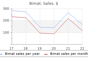
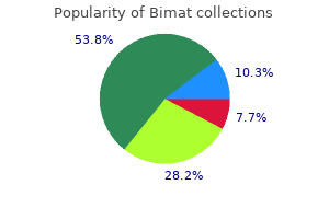
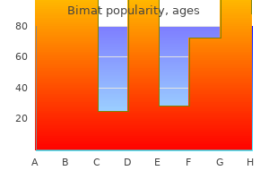
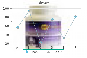
Bimat

Bimat
Bimat dosages: 3 ml
Bimat packs: 1 bottles, 2 bottles, 3 bottles, 4 bottles, 5 bottles, 6 bottles, 7 bottles, 8 bottles, 9 bottles, 10 bottles

This endobronchial papilloma demonstrates traditional architectural options: arborizing fibrovascular cores coated by a proliferative squamous epithelium medicine effects bimat 3 ml cheap line. Cavitation and surrounding lung fibrosis are additionally not uncommon; the latter may even recommend tumor invasion treatment centers for depression bimat 3 ml purchase on line. The mass lesions can grow to giant sizes and essentially exchange complete pulmonary segments medications 2 times a day discount bimat 3 ml online. Each fragment is remarkably comparable in measurement and with regard to amount of epithelial proliferation. Exophytic lesions have orderly epithelial maturation extending from the basal layer to the superficial flattened and often keratinized surface cells. Acanthosis and nonkeratinized surfaces are sometimes seen, along with intraepithelial neutrophils. Not in distinction to urinary bladder inverted papillomas, endobronchial lesions are exophytic with invaginated nests of bland squamous mucosa. In uncommon instances atypia reaching the extent of carcinoma in situ may be current and in such cases a prognosis of carcinoma arising in a squamous papilloma should be rendered. The quantities of stroma and stromal lymphoplasmacytic infiltrates vary from very scant to prominent, whereas the adjoining respiratory mucosa could also be normal, inflamed, hyperplastic or metaplastic. These rare lesions are non-keratinizing and show orderly maturation from the basal layer upward. Well-circumscribed, stable, intra-alveolar nests of cytologically bland, non-keratinizing squamous cells may fill alveolar spaces. Distinguishing this course of from invasive squamous cell carcinoma may be very troublesome. Bronchial and alveolar parenchymal involvement with laryngotracheal papillomatosis is morphologically much like the isolated lesions; however, virtually all lesions function viral cytopathic effect. Multinucleation may be seen and nuclei differ from small and pyknotic to large with evenly distributed coarse chromatin. Smaller basal cells with scant basophilic cytoplasm and round regular nuclei are also famous along with background neutrophils. Scattered single cells are also 854 Chapter 22: Benign epithelial neoplasms and tumor-like proliferations of the lung described. However, positivity appears to correlate with the morphological presence of koilocytosis. Recognizing even focal carcinoma within an otherwise benign squamous papilloma warrants a analysis of carcinoma. The illness can undergo spontaneous remission, persist as stable illness, or progress. Extralaryngeal unfold is noted in approximately 30% of children and fewer than 20% of adults. The most frequent sites of unfold in lowering order of frequency are the oral cavity, trachea, bronchi, and esophagus. As mentioned above, inflammatory polyps lack a true papillary structure, stromal cores and proliferative epithelium. Unlike papillomas, well-differentiated squamous cell carcinomas lack orderly epithelial maturation and infrequently function keratinization. Difficulties arise when one misinterprets entrapped seromucinous glands for invasion into the polyp stalk. Glandular and combined squamous cell and glandular papillomas As not extra than a dozen glandular papillomas are described within the thoracic literature, comments are very restricted. Tumors may carefully resemble Schneiderian papillomas of the higher respiratory tract. The glandular epithelium could also be ciliated, cuboidal or columnar, and interspersed mucin-rich cells are sometimes identified. A basal cell layer is commonly apparent and may be highlighted with either p63, K903 or cytokeratin17 immunohistochemical stains. Mucus gland adenomas function mucusfilled cysts and tubules, while papillary adenomas are true pulmonary parenchymal, somewhat than endobronchial lesions. All but one reported case was handled surgically and the only recurrence and death was within the affected person unable to tolerate anything more than bronchoscopic treatments. Uniform columnar cells with eosinophilic cytoplasm, round common nuclei and interspersed mucin-rich cells are surrounded with inflammatory cell-rich mucin whereas stromal cores comprise many plasma cells. Inflamed arborizing papillary cores are lined with discrete foci of glandular and squamous epithelium. Glandular atypia is considered reactive, while squamous atypia is believed to be neoplastic. Minute welldemarcated lesions owe their nodularity to thickened alveolar septa and intraalveolar macrophages. Lesional epithelial cells grow along alveolar walls and feature eosinophlic cytoplasm and spherical nuclei with vesicular chromatin. Rare multinucleated cells are famous however marked atypia, mitoses or necrosis are absent. Alveolar macrophages also contribute to the nodular character of the lesion by filling airspaces lined by the epithelial cells. Macroscopic pathology Most reported lesions lack macroscopic descriptions however several case reviews describe 0. Crowded epithelial cells within the 858 Chapter 22: Benign epithelial neoplasms and tumor-like proliferations of the lung proliferations, very tough. Multifocal micronodular pneumocyte hyperplasia additionally options extra pronounced alveolar septal thickening and intra-alveolar macrophages. Prognosis and pure historical past Multifocal micronodular pneumocyte hyperplasia is kind of always an incidental radiographic or microscopic discovering with no clinical significance. Electron microscopy Ultrastructural research verify the epithelial nature of the proliferation and describe cuboidal cells resting on a basal lamina with desmosomes and hemidesmosomes. One research additionally recognized electron-dense, membrane-limited, secretory granules with a granular matrix and abundant rough endoplasmic reticulum, suggestive of Clara cell differentiation. Papillary adenoma Introduction Classification, cell of origin and etiology this benign peripheral lung tumor is believed to arise from a multipotential stem cell or immature bronchioloalveolar cell. The etiology is unknown however comparable morphological lesions are genetically and/or chemically induced in mice. Special scientific features these adenomas have been reported in each sexes in patients ranging from 7 to 60 years of age (mean 32 years). This well-circumscribed parenchymal lesion has an obvious papillary configuration. Computed tomography reveals a clean circumscribed parenchymal nodule, with out pleural indentations or spiculations, which can bulge into a small bronchus. Well-circumscribed and sometimes encapsulated tumors have tan, gray, delicate to firm, granular and spongy minimize surfaces. Elastic fibers are absent from the stromal cores and scattered mast cells may be seen. Pulmonary and metastatic papillary adenocarcinomas might not always be infiltrative however are at all times proliferative lesions with mobile crowding and cytological atypia. Of observe, metastatic papillary thyroid carcinoma options nuclear grooves and thyroglobulin positivity. Papillary carcinoid tumor could also be architecturally identical to papillary adenoma, however granular cytoplasm and round regular nuclei with granular chromatin allow distinction. Neuroendocrine immunohistochemical stains (chromogranin and synaptophysin) could additionally be required in uncommon cases (see Chapter 31). Sclerosing hemangioma is less of a sensible consideration given its typical heterogeneous architectural patterns and dual cell population (see below). Papillary cystadenoma, a lately described entity arising in the lung of sufferers with von Hippel-Lindau illness, enters the differential analysis. In this lesion, papillae are lined with bland multilayered epithelial cells with focally clear cytoplasm and nuclear enlargement. Unlike papillary adenoma, this cystadenoma is associated with many microcysts accompanied by fibrous stroma and thin-walled vascular channels.
The prevalence of osteoporosis in sufferers with chronic obstructive pulmonary illness: a cross sectional study 5 medications bimat 3 ml generic with mastercard. Leptin medicine rock order bimat 3 ml on-line, visfatin medications pictures buy bimat 3 ml online, insulin resistance, and physique composition change in continual obstructive pulmonary illness. Genome-wide affiliation evaluation of physique mass in continual obstructive pulmonary illness. Systemic irritation and skeletal muscle dysfunction in chronic obstructive pulmonary disease: cutting-edge and novel insights in regulation of muscle plasticity. Inflammation, oxidative stress and systemic effects in mild persistent obstructive pulmonary disease. The effects of hypoxia on markers of coagulation and systemic irritation 655 Chapter 17: Chronic obstructive pulmonary disease and ailments of the airways in sufferers with continual obstructive pulmonary disease. Effects of acute hypoxia on left and proper ventricular contractility in chronic obstructive pulmonary illness. Dysmorphic lungs in a case of leprechaumism: case report and evaluate of literature. Morphogenesis of abnormal elastic fibers in lungs of sufferers with panacinar and centriacinar emphysema. Risk of renal and colonic neoplasms and spontaneous pneumothorax in the Birt-Hogg-Dube syndrome. Nonsense mutations in folliculin presenting as isolated familial spontaneous pneumothorax in adults. Mutations of the Birt Hogg Dube gene in patients with a number of lung cysts and recurrent pneumothorax. Lung cysts, spontaneous pneumothorax, and genetic associations in 89 households with BirtHogg-Dube syndrome. Lung cysts in Birt-Hogg-Dube syndrome: histopathological traits and aberrant sequence repeats. Swyer-James (MacLeod) syndrome with placental transmogrification of the lung: a case report and evaluate of the literature. Placental transmogrification of the lung, a histologic variant of large bullous emphysema. Suppurative diseases of the lung and pleura: a continuing challenge in 656 Chapter 17: Chronic obstructive pulmonary disease and illnesses of the airways developing countries. The cartilage of the intrapulmonary bronchi in normal lungs in bronchiectasis and in massive collapse. The aetiology of bronchiectasis (with particular reference to pulmonary atelectasis). Factors associated with lung function decline in adult patients with stable non-cystic fibrosis bronchiectasis. Resection of the right center lobe and lingula in youngsters for middle lobe/lingula syndrome. Rigid bronchoscopy and surgical resection for broncholithiasis and calcified mediastinal lymph nodes. Diffuse tracheo-bronchial amyloidosis: a uncommon 657 Chapter 17: Chronic obstructive pulmonary disease and diseases of the airways variant of a protean disease. Bronchiolitis obliterans organising pneumonia in patients taking acebutolol or amiodarone. Unilateral hyperlucent lung (Swyer-James syndrome) after extreme Mycoplasma pneumoniae an infection. Proliferative exercise in fibrosing lung illnesses: a comparative study of Ki-67 immunoreactivity in diffuse alveolar harm, bronchiolitis, obliteransorganizing pneumonia, and traditional interstitial pneumonia. Diffuse idiopathic pulmonary neuroendocrine cell hyperplasia: an under-recognised spectrum of illness. Brief report: idiopathic diffuse hyperplasia of pulmonary neuroendocrine cells and airways illness. Neuroendocrine cell hyperplasia and obliterative bronchiolitis in patients with peripheral carcinoid tumors. Persistent tachypnea of infancy is associated with neuroendocrine cell hyperplasia. Outbreak of bronchiolitis obliterans related to consumption of Sauropus androgynus in Taiwan. Outbreak of obstructive ventilatory impairment related to consumption of Sauropus androgynus vegetable. Association of Sauropus androgynus and bronchiolitis obliterans syndrome: a hospital-based casecontrol research. Dose-response relationship and irreversible obstructive ventilatory defect in patients with consumption of Sauropus androgynus. Segmental necrosis of small bronchi after extended intakes of Sauropus androgynus in Taiwan. Histopathological research of Sauropus androgynus-associated constrictive bronchiolitis obliterans: a new reason for constrictive bronchiolitis obliterans. Sauropus androgynus-constrictive obliterative bronchitis/bronchiolitis: histopathological research of pneumonectomy and biopsy specimens with emphasis on the inflammatory course of and disease progression. The effect of large-dose prednisolone on patients with obstructive lung disease associated with consuming sauropus androgynus. Diffuse panbronchiolitis: prognosis and distinction from numerous pulmonary ailments with centrilobular interstitial foam cell accumulations. Neutrophil survival-enhancing activity in sputum from patients with diffuse panbronchiolitis. Comparative clinicopathology of obliterative bronchiolitis and diffuse panbronchiolitis. Increased numbers of dendritic cells in the bronchiolar tissues of diffuse panbronchiolitis. Clinical similarities and variations between human T-cell lymphotropic virus sort 1-associated bronchiolitis and diffuse panbronchiolitis. Cellular distribution of bronchus-associated lymphoid tissue in rheumatoid arthritis. Chronic bronchiolitis with associated eosinophilic lung disease (eosinophilic bronchiolitis). One hundred consecutive granulomas in a pulmonary pathology consultation follow. Peribronchiolar metaplasia: a typical histologic lesion in diffuse lung disease and a uncommon reason for interstitial lung disease. Does the single-breath N2 take a look at identify the smoker who will develop chronic airflow limitation. Pulmonary operate in youngsters and adolescents with postinfectious bronchiolitis obliterans. Outbreak of bronchiolitis obliterans associated with consumption of Sauropus androgynus in Japan: alert of food-associated pulmonary issues from Japan. Mooi Pulmonary hypertension is defined as a sustained imply pulmonary arterial strain at relaxation of! The strain is measured by proper coronary heart catheterization or can be estimated by echocardiography. Pulmonary hypertension is a consequence of right ventricular adaptation to increased vascular resistance, elevated pulmonary blood flow, or a mixture of each. Initially, the symptoms of pulmonary hypertension are nonspecific and normally limited to dyspnea, significantly on exertion, or patients may be asymptomatic. As the pulmonary hypertension progresses, indicators and symptoms of right-sided heart failure develop; specifically peripheral edema, fatigue, abdominal fullness, angina pectoris and syncope. Depending on its trigger, severity and potential treatment options, pulmonary hypertension may result in demise. Pulmonary hypertension is a function of a heterogeneous group of issues which differ in threat issue profile, initiating factors, response to remedy and prognosis (Table 1). Table 1 Risk components and associated conditions for pulmonary arterial hypertension A. Definite Aminorex (Menocil) Fenfluramine (Ponderal) Dexfenfluramine (Adifax, Redux) Fenfluramine-phentermine Toxic rapeseed oil 2.
Syndromes
The system relies on three principal parts that describe the anatomic extent of illness symptoms umbilical hernia bimat 3 ml purchase without prescription. The T category describes the extent of the first tumor medications ending in zole discount bimat 3 ml otc, the N category describes the absence/ presence and extent of regional lymph node metastasis medicine reminder app buy 3 ml bimat free shipping, and the M class describes the absence/presence of distant metastases. The T, N and M categories are additional classified by numerals, which indicate progressively superior illness. Therefore for the longer term, emphasis is placed on assortment of the first measurements and anatomical descriptions. The data supplied in this chapter serves as an summary and to spotlight some necessary changes; nonetheless, readers are strongly suggested to discuss with the entire staging handbook and manual for extra detailed data. The pathological evaluation of the primary tumor (pT) entails resection of the first tumor or a biopsy adequate to consider the best pT category. Removal of nodes sufficient to validate the absence of regional lymph node metastasis is required for pN0. The pathological evaluation of distant metastasis (pM) entails microscopic examination. A pathological stage could additionally be assigned if the anatomic extent of disease has been confirmed, whether or not the primary lesion has been fully eliminated. When info is obtained for staging after induction remedy (in lung cancer, this normally implies preliminary remedy with chemotherapy or radiotherapy prior to surgery) the prefix y is used. For the staging of recurrent tumour after a disease free interval, the prefix "r" is used. T classification the T descriptors are assigned based on tumor size, anatomic relation/invasion or the state of the lung distal to the first tumor. In the seventh version, new T size minimize factors (at 2 cm, 5 cm and seven cm) had been launched, in addition to the established minimize level of three cm that historically separated T1 and T2 tumors. T1 tumors are actually sub-divided into T1a (2 cm) and T1b (> 2 cm but 3 cm), T2 tumors have been subdivided into T2a (> 3cm but 5 cm) and T2b (> 5 cm but 7 cm), and tumors > 7 cm at the second are categorised as T3. This recommendation is problematic for semisolid and groundglass opacities, since prognosis may be associated to the dimensions of the stable part only. In addition, formalin fixation-induced tumor shrinkage brought on stage shift from stage Ib to stage Ia in 10% of the research cohort. In the seventh edition, for the first time, an agreed definition of visceral pleural invasion has been given, i. However, in a considerable share of cases the elastic layer is imperceptible on hematoxylin and eosin-stained tissue sections. This layer could also be near the lung parenchyma, close to the pleural surface or in the middle. These processes end in alteration of the elastic layers, often with reduplication. First, the complicated term Table 2 T descriptors for lung most cancers "satellite nodules" has been replaced with the extra affordable descriptor "further tumor nodules". Grossly identified "extra tumor nodules" in the major tumor-bearing lobe require a T3 project. Great vessels are defined as aorta, superior vena cava, inferior vena cava, major pulmonary artery, the intrapericardial segments of the trunks of pulmonary arteries or veins. Discerning intrapulmonary metastases from synchronous primary carcinomas may be unimaginable (see below) (see Chapter 27). N classification the N descriptors are based mostly on the absence/presence and extent of metastasis in regional lymph nodes. International Association for the Study of Lung Cancer Nodal Chart with stations and zones. The extra peripheral lymph nodes at stations 12:14 are often evaluated by the pathologist in lobectomy or pneumonectomy specimens however may be individually eliminated when sublobar resections. Three of those nodes/stations must be mediastinal, including the sub-carinal nodes (station 7), and three from N1 nodes/stations. If resection has been carried out, and in any other case fulfills the necessities for full resection, it must be classified as R0. Prognosis is influenced by the presence or absence of nodal disease, but also by the extent, sample and bulk of any nodal illness. In the presence of distant disease, it may be very important document the number and sites of metastases. These embody the cLy category for the classification of lymphangitis carcinomatosis, determined on radiological studies. Unlike other tumor websites, in lung cancer the "V" classification refers not only to venous invasion but also to arteriolar invasion, a not unusual feature in lung most cancers. Direct invasion of a tumor into an adjoining lobe, across the fissure or by direct extension at some extent the place the fissure is poor ought to be classified as T2a until other standards assign a higher T category. Determining whether or not a number of carcinomas symbolize synchronous primaries or intrapulmonary metastases is a troublesome topic. Because the resected lung tissue is collapsed, pathologists might have extra issue than radiologists or surgeons figuring out small tumors. This is particularly the case with adenocarcinomas with a predominant lepidic element. Multiple tumors of comparable histological appearance ought to only be thought-about to be synchronous primary tumors if in the opinion of the pathologist, based mostly on features corresponding to differences in morphology, immunohistochemistry and/or molecular research, or, in the case of squamous cancers, being associated with carcinoma in situ, they symbolize differing subtypes of the same histopathological cell type. These circumstances are most commonly encountered when coping with lepidic-pattern adenocarcinomas (see Chapter 27). The highest T class and stage of illness should be assigned and the multiplicity or the variety of tumors must be indicated in parenthesis. Careful palpation throughout surgical procedure and by the pathologist of the resected specimen is therefore required to display screen for the presence of additional tumor nodules. Only nodules found for the primary time at pathological examination are classified as further nodules. However, many issues stay unresolved together with the validity of this method for typical versus atypical carcinoid tumors, the validity of staging multicentric carcinoid tumors, the validity of measurement cut-offs within the dedication of T worth, and the prognostic significance of thoracic lymph node metastases. Effect of formalin fixation on tumor measurement dedication in stage I non-small cell lung most cancers. Effect of number of lymph nodes sampled on end result in sufferers with stage I non-small-cell lung most cancers. Which is the better prognostic factor for resected non-small cell lung cancer: the variety of metastatic lymph nodes or the currently used nodal stage classification The impression of stage and cell type on the prognosis of pulmonary neuroendocrine tumors. International Association for the Study of Lung Cancer Staging Handbook in Thoracic Oncology. Impact of positive pleural lavage cytology on survival in sufferers having lung resection for non-small-cell lung cancer: a global particular person patient information meta-analysis. It could also be useful as a supplement to morphology in classifying major lung tumours. This subclassification is increasingly essential, as rising therapeutic options demand increasing diagnostic exactitude. The technique is also invaluable in deciding whether or not a tumor, significantly an adenocarcinoma, is a pulmonary major or arises from an extra-pulmonary site. If metastatic, immunohistochemical stains can even usually decide the primary site. Deciding whether a tumor shows positive staining also has a component of subjectivity. Some antibodies, when utilized to some tumors, have given rise to hundreds of reported instances with constant outcomes. Published results with different antibodies and tumors have given various outcomes for causes that may or will not be obvious. Particularly for much less frequent pulmonary or extra-pulmonary tumors, the printed information may be sparse within the extreme. The corpus of published data is immense and ever growing, particularly as tissue microarrays have the potential to study hundreds of tumors throughout the scope of one examine. However, when a quantity of attainable main sites exist, the relative chances of tumors metastisizing from every attainable major website are unknown. Even with the large numbers of tumors that may be examined using tissue microarrays, many tumors that will enter a differential prognosis have never been tested in sufficient numbers for any of the antibodies in use. All one can do is concentrate on the publications which present that all antibodies are lower than completely specific. The protein product is a 38 kilodalton (kDa) homeodomain-containing tissue-specific nuclear transcription protein of the Nkx2 gene family. Staining is observed in each ciliated and non-ciliated cells of the bronchial and bronchiolar epithelia and in cells lining the distal airspaces.
Peripheral tumors are often asymptomatic medications used for depression 3 ml bimat generic with visa, but could trigger ache because of symptoms multiple myeloma order bimat 3 ml overnight delivery direct involvement with adjacent tissues symptoms 2 weeks pregnant buy discount bimat 3 ml. Clusters of tumor cells are described as three-dimensional with shaggy or branching edges. Individual cells have mildly hyperchromatic, oval to fusiform nuclei with evenly dispersed, finely granular chromatin and inconspicuous or absent nucleoli. Differential prognosis the first differential analysis for monophasic and biphasic pulmonary synovial sarcomas is metastatic synovial sarcoma. Further differential diagnoses of monophasic spindle-type main pulmonary synovial sarcoma embrace other metastatic lesions, including solitary fibrous tumor and sarcomatoid diffuse malignant mesothelioma. Clinicoradiological review, immunostains and the t(X;18) translocation could assist in characterizing the tumor as a mesothelioma or excluding a synovial sarcoma. For biphasic tumors, the differential diagnosis contains carcinomas and biphasic diffuse malignant mesothelioma. Approximately 44% of sufferers are lifeless of disease within 5 years and 28% of patients are alive with no evidence of illness after the identical time interval. This t(X;18) translocation is helpful within the diagnosis of circumstances which would possibly be equivocal on morphological grounds. The Xp11 breakpoint sometimes Intra-pulmonary thymomas Introduction Intra-pulmonary thymomas are extremely rare. Alternatively, transformation from a pulmonary teratoma is a potential origin for these tumors. These genes are typically overexpressed more typically in thymic carcinomas than in thymomas. Radiological research are important in evaluating the affected person for any mediastinal element, and evaluating the pleura in cases of subpleural tumor. Fine-needle aspirates demonstrate mature lymphocytes and relatively bland epithelial cells in various proportions. Macroscopic pathology Intra-pulmonary thymomas could additionally be central or peripheral, in any lung lobe. They are usually well-circumscribed, encapsulated, lobulated tumors of various color measuring 1 cm to 12 cm. The overwhelming majority of intrapulmonary thymomas are solitary; however, there are stories of a number of tumors. Many have the standard biphasic cellular composition with varying quantities of epithelial element and associated lymphocytic element. Close examination of tumor capsule, if present, is essential, since extension of tumor beyond this capsule and tumor adherence to adjoining tissue indicate an aggressive Electron microscopy Electron microscopy has not been described for intrapulmonary thymoma. The pathologist must broadly sample the tumor, with specific consideration to any capsular invasion, as tumor involvement beyond the capsule signifies an aggressive habits. Alterations of p53 and p16/Rb pathways have been recognized in mature cystic teratomas of the ovary exhibiting malignant transformation into squamous cell carcinoma. Non-small cell carcinoma, particularly spindle cell and lymphocyte-rich variants, and small cell carcinoma must sometimes be distinguished from intrapulmonary thymoma. Spindle cell carcinoid tumor and primary pulmonary teratoma with a outstanding thymic component are other rare concerns. This feature might distinguish pulmonary teratoma from the extra frequent mediastinal teratoma. These features are priceless in distinguishing between ruptured and unruptured teratomas. The symptoms usually occur after the tumor is massive sufficient to cause airway obstruction or induce chest pain. Lymph node dissection must also be considered as nodal involvement has been reported. Metastatic tumor from mediastinal, gonadal or different site should be excluded earlier than this diagnosis may be made. The solid component can often predominate and is more more likely to exhibit immature histology. Classification and cell of origin Teratomas consist of multiple cell line and are classified as mature or immature. Pulmonary teratomas most likely come up from ectopic tissue from the third pharyngeal pouch, possibly as a result of displacement or separation of the thymus throughout Histopathology Pulmonary teratomas contain tissue from the three germinal layers: mesodermal, endodermal and ectodermal: in various 1285 Chapter 33: Mesenchymal and miscellaneous neoplasms Trichoptysis could happen, as noted above, in patients with pulmonary teratomas. The expectorated hair is usually white, reflecting the lack of maturity of the pigment-producing cells in the tumor. For reasons not yet understood, most pulmonary teratomas involve the left upper lobe of the lung. Other differential diagnoses include old infectious granulomas and amyloid nodules. Most pulmonary teratomas are mature, containing predominantly mature cystic somatic tissue. Malignant pulmonary teratomas contain carcinoma or sarcoma within the presence of immature tissue, corresponding to immature stromal or neuroectodermal tissue. Patients with malignant lesions amenable to surgical resection have restricted survival of approximately 6 months. Primary pulmonary malignant melanoma Introduction Primary pulmonary malignant melanomas are extraordinarily rare, despite the very fact that metastatic melanoma to the lung is widespread. Electron microscopy Ultrastructural examination of a pulmonary teratoma has not been documented. Classification and cell of origin Normal bronchial mucosa contains no melanin-containing cells. Primary pulmonary melanoma arises from melanocytes, presumably from melanocytic metaplasia, and presumably from melanocytic cell migration throughout embryogenesis. Studies of pores and skin melanomas utilizing comparative genomic hybridization have shown that virtually all melanomas have a number of genetic alterations. Due to the chance of tumor rupture and malignancy, surgical resection is really helpful. Special clinical options Primary pulmonary melanomas occur relatively equally amongst adult women and men, with ages starting from roughly 30 to 90 years and a mean of roughly 50 years. Pulmonary melanomas arising in the peripheral lung as solitary nodules are virtually at all times metastatic. Symptoms, similar to cough, post-obstructive pneumonia and hemoptysis, are related to endobronchial tumor location. One must consider the chances that a prior melanoma was excised and misdiagnosed, that a melanoma had undergone spontaneous regression, or that a major melanoma had arisen in an uncommon web site, such because the gallbladder or retina. Differential prognosis the main differential analysis of major pulmonary melanoma is the much more widespread metastatic malignant melanoma. In some instances, differentiation between the two tumors could additionally be impossible and one ought to pursue all available scientific history. Fine-needle aspiration of pulmonary hamartoma: a common source of false-positive diagnoses in the College of American Pathologists Interlaboratory Comparison Program in Nongynecologic Cytology. Surgical remedy and end result of pulmonary hamartoma: a retrospective research of 20-year expertise. Recombinations of chromosomal bands 6p21 and 14q24 characterise pulmonary hamartomas. An analysis of 155 solitary lung lesions illustrating the differential analysis of blended References 1. Pulmonary hamartoma: unusual 1288 Chapter 33: Mesenchymal and miscellaneous neoplasms tumours of the lung. Benign endobronchial mesenchymal tumors: their relationship to parenchymal pulmonary hamartomas. Mesenchymoma of the lung (so referred to as hamartoma): a evaluation of 154 parenchymal and endobronchial instances. Placental transmogrification of the lung is a histologic sample regularly associated with pulmonary fibrochondromatous hamartoma. Pulmonary chondroma: a tumor related to Carney triad and totally different from pulmonary hamartoma.
In reality symptoms quit drinking bimat 3 ml discount on line, pulmonary infarcts are famous in less than 20% of post-mortem lungs from patients with deep vein thrombi xanax medications for anxiety bimat 3 ml order fast delivery. Since congestive pulmonary vasculopathy 5 medications for hypertension purchase bimat 3 ml visa, as in mitral valve insufficiency, is usually sophisticated by arterial thrombosis, pulmonary infarcts could come up in that situation. Pleural-based wedge-shaped hemorrhagic lung with an apex pointing toward the hilum is composed of neutrophils and erythrocytes. Granulation tissue and hemosiderinladen macrophages replace the necrotic tissue and fill up alveolar areas. Infective thrombophlebitis, catheters or right heart valve endocarditis could additionally be sources for such an an infection. Differential prognosis Some recanalized post-thrombotic lesions, especially when located in a small vessel, might resemble a plexiform lesion. Arteries containing organized thrombi are usually larger than these incurring most harm in plexogenic arteriopathy. Non-thrombotic causes of pulmonary artery embolism There are several causes of embolic pulmonary hypertension, other than pulmonary thromboembolism. These had been previously classified in group 4,10 but appear to have lost their place in the updated classification. Chronic hepatosplenic schistosomiasis accounts for a few third of all pulmonary hypertension circumstances in endemic areas, corresponding to Brazil. Clinically, the presentation and course of the illness are stated to resemble pulmonary arterial hypertension more than thrombotic/embolic disease. After this acute stage, the cercaria mature into adult helminths, which produce the extremely antigenic eggs. In the early persistent stage of the illness, a lot of the eggs terminate within the liver, via the portal vein. Increased portal stress, and shunting and bypass of the liver through the collateral circulation, may develop. By this route, eggs can immediately reach the pulmonary vascular mattress, again inflicting vascular harm. Portal hypertension presumably is a crucial contributing issue,thirteen since just a few granulomas are current. In different instances, the converse is true and tumor cells occlude the vessels but a thrombotic reaction is absent. In rare circumstances, adenocarcinoma may grow in a lepidic fashion along the endothelial basement membrane of the pulmonary arteries (A. Tumor emboli inside blood vessels are usually simply distinguished from lymphangitic tumor spread. Pulmonary hypertension as a result of carcinoma emboli tends to run a rapidly progressive fatal course. While thrombosis is a recognized etiology in these patients, peripheral leukocyte counts exceeding 200 000/ml can result in blockage within the absence of fibrin thrombi. The adjacent pulmonary artery is occluded by intimal fibrosis and a swollen endothelium (Black 6 mouse, experimental Schistosoma mansoni infection) (EvG stain). Foreign body giant cell response to birefringent matter of unknown origin in a distended vessel, localized within the media of a pulmonary artery. This location suggests that the embolus is lodged in a supernumerary arterial department. Hypoxic arteriopathy pulmonary hypertension ensues from chronic myeloproliferative issues, together with myeloid metaplasia with myelofibrosis (agnogenic myeloid metaplasia) (see Chapter 34), essential thrombocythemia, polycythemia vera, myelodysplastic syndrome, and chronic myeloid leukemia214 (Group 5 Dana Point classification 200811). The etiology is unknown but the truth that symptomatic enchancment could additionally be seen after a single low dose of thoracic radiotherapy means that microvascular blockage by hematopoietic components, corresponding to megakaryocytes, may play a task in this state of affairs. This response is useful in instances of local pulmonary hypoventilation, because it shifts blood flow away from areas the place gas exchange shall be incomplete. However, when hypoxia affects the whole lung, this essentially protective vasoconstrictive response becomes pathogenic, causing pulmonary hypertension. This patient offered with quickly progressive pulmonary hypertension during being pregnant secondary to metastatic breast carcinoma. This, in flip, tips the balance in path of deeper hypoxemia quite than elevated oxygen-carrying capacity. Hypoxemia causes additional pulmonary vasoconstriction, pulmonary hypertension and, eventually, proper heart failure. On descent to sea stage, the clinical situation improves considerably; the pulmonary hypertension usually abates and should reverse to normal within 2 weeks. At lower altitudes, hypoxic arteriopathy generally complicates chronic lung illness with a reduced diffusion capability. Pickwickian syndrome, sleep apnea syndrome Neuromuscular disorders Thoracic skeletal problems Upper airways obstruction Low ambient oxygen; excessive altitude dwellers Medial hyperplasia, particularly of small muscular pulmonary arteries Muscularization of arterioles Intimal longitudinal smooth muscle bundles in small arteries and in arterioles Arterial adventitial thickening Mild improve in venous smooth muscle Intimal fibroelastosis Table 10 Conditions related to congestive vasculopathy Left ventricular failure Mitral valve dysfunction Aortic valve dysfunction Congenital stenosis of main pulmonary veins or left atrial orifice Anomalous pulmonary venous connections or obstruction in left atrium (myxoma, cor triatriatum persistans) Mediastinal illness impeding pulmonary venous outflow (fibrosis, lymphadenopathy) extraction. The adventitia is markedly thicker in the normal fetus and new child, gradually reducing to its adult thickness during the first years of life. Hypoxia-driven adventitial transforming has been described in animals (reviewed by Stenmark et al. At greatest, a subjective division into "unremarkable" versus "thickened" adventitia may be made. Congestive vasculopathy Epidemiology, causal factors Chronic elevation of the pulmonary venous blood strain results in a definite set of adaptive pulmonary vascular changes, termed congestive pulmonary vasculopathy. Most circumstances result from left ventricular failure or mitral valve dysfunction (Dana Point classification 2008 group 211) (Table 10). Fewer cases are because of venous obstruction on the level of the large pulmonary veins. Hilar, mediastinal or left atrial tumor, and fibrosing mediastinitis are examples (Dana Point classification 2008 group 511). Histopathology of hypoxic arteriopathy (Table 9) Increased muscle content of small arterial branches is essentially the most putting histological characteristic of hypoxic pulmonary vasculopathy. Pulmonary veins and venules also present a slight improve in smooth muscle content however not artertialization, as identified in congestive vasculopathy (see below). Histopathology of congestive vasculopathy (Table 11) Arterialization of veins is probably the most distinctive vascular alteration in congestive pulmonary vasculopathy. The marked arterial medial hyperplasia is paralleled by the well-known statement that the rise in pulmonary artery strain normally exceeds that of the venous strain. This is a feature shared by hypoxic arteriopathy, however it tends to be extra marked in congestive vasculopathy. At the other finish of the spectrum, marked interstitial fibrosis and iron pigmentation results in so-called "brown induration of the lung". This time period is derived from the macroscopic appearance seen in autopsies of sufferers with longstanding left ventricular failure or diseased aortic and/or mitral valves. Radiotherapy and drug toxicity directed at endothelial cells is the hypothesized mechanism. Additional cases have been associated with earlier bone marrow or stem cell transplantation,239:241 and heart and/or lung transplantation. It is postulated that the pathology is secondary to obliterative bronchiolitis along with arterial and venous intimal fibrosis. They should be used with caution as they might precipitate an assault of acute pulmonary edema. As talked about above, mitral valve insufficiency particularly has been identified as a danger issue for pulmonary infarcts, most likely due to venous stasis. It lacks the self-perpetuation and progressive deterioration characteristic of all however the earliest phases of plexogenic arteriopathy. These embrace diffusely distributed centrilobular groundglass opacities and/or mosaic lung attenuation. This feature is particularly telling when noticed in the upper lung fields, where common perfusion is low in comparison with the bases. The caliber of pulmonary veins is normal, while mediastinal lymph node enlargement is seen in some patients. The muscular artery features medial hyperplasia and outstanding adventitia (EvG stain).
Allogenic bone marrow transplantation 432 Chapter eleven: Metabolic and connective tissue issues involving the lung illness symptoms quitting tobacco purchase bimat 3 ml with amex. Therapeutic strategies to ameliorate lysosomal storage disorders: a concentrate on Gaucher illness medicine express bimat 3 ml discount overnight delivery. NiemannPick disease: mutation update medicine daughter lyrics 3 ml bimat buy overnight delivery, genotype/ phenotype correlations, and prospects for genetic testing. The pathogenesis and remedy of acid sphingomyelinasedeficient Niemann-Pick disease. Natural historical past of Type A Niemann-Pick illness: potential endpoints for therapeutic trials. The natural historical past of kind B Niemann-Pick illness: outcomes from a 10-year longitudinal research. A potential, cross-sectional survey research of the pure history of Niemann-Pick illness sort B. NiemannPick disease type B: first-trimester prenatal diagnosis on chorionic villi and biochemical examine of a foetus at 12 weeks of growth. Identification of novel biomarkers for Niemann-Pick illness utilizing gene expression evaluation of acid sphingomyelinase knockout mice. Analysis of the lung pathology and alveolar macrophage function within the acid sphingomyelinase: deficient mouse mannequin of Niemann-Pick disease. Alveolar lipoproteinosis in an acid sphingomyelinase-deficient mouse mannequin of Niemann-Pick disease. Successful therapy of endogenous lipoid pneumonia because of Niemann-Pick Type B illness with whole-lung lavage. Niemann-Pick disease: sixteen-year follow-up of allogeneic bone marrow transplantation in a sort B variant. Infusion of recombinant human acid sphingomyelinase into niemann-pick disease mice leads to visceral, however not neurological, correction of the pathophysiology. Pulmonary supply of recombinant acid sphingomyelinase improves clearance of lysosomal sphingomyelin from the lungs of a murine mannequin of Niemann-Pick illness. Hematopoietic stem cell gene remedy results in marked visceral organ improvements and a delayed onset of neurological abnormalities in the acid sphingomyelinase deficient mouse mannequin of Niemann-Pick disease. Nine cases of sphingomyelin lipidosis, a new 433 Chapter eleven: Metabolic and inherited connective tissue issues involving the lung variant in Spanish-American Children. Pulmonary storage with emphysema as a sign of Niemann-Pick sort C2 disease (second complementation group). Early-lethal pulmonary type of Niemann-Pick type C disease belonging to a second, rare genetic complementation group. Niemann-Pick disease kind C2 presenting as deadly pulmonary alveolar lipoproteinosis: morphological findings in lung and nervous tissue. Niemann-Pick illness sort C1 is a sphingosine storage disease that causes deregulation of lysosomal calcium. Fabry illness: guidelines for the evaluation and administration of multiorgan system involvement. Pulmonary manifestations of Fabry disease and constructive response to enzyme alternative remedy. Enzyme replacement remedy stabilizes obstructive pulmonary Fabry disease associated with respiratory globotriaosylceramide storage. Disorders of lysosomerelated organelle biogenesis: medical and molecular genetics. Positional cloning of a gene for HermanskyPudlak syndrome, a disorder of cytoplasmic organelles. Interstitial pneumonia in Hermansky-Pudlak syndrome: significance of florid foamy swelling/ degeneration (giant lamellar physique degeneration) of type-2 pneumocytes. Successful bilateral lung transplantation for pulmonary fibrosis related to the Hermansky-Pudlak syndrome. Diagnostic usefulness of bronchoalveolar lavage in HermanskyPudlak syndrome: a case with double lung cancers. Epithelial stress and apoptosis underlie Hermansky-Pudlak syndromeassociated interstitial pneumonia. Altered lung surfactant system in a Rab38-deficient rat model of Hermansky-Pudlak syndrome. Children with mucopolysaccharidosis: perioperative care, morbidity, mortality, and new findings. Pathological and biochemical abnormalities within the neural and mesenchymal components of the mind. Enzyme substitute remedy for mucopolysaccharidosis I: a randomized, double-blinded, placebo-controlled, multinational examine of recombinant human alpha-L-iduronidase (laronidase). Improved retroviral vector design leads to sustained expression after adult gene therapy in mucopolysaccharidosis I mice. Ultrastructural and biochemical observations on a case of systemic late childish lipidosis and its relationship to Tay-Sachs illness and gargoylism. An evaluation of eight instances of a syndrome beforehand reported as "Hurler-variant," "pseudo-Hurler," and "Tay-Sachs disease with visceral involvement". Infantile G(M1) gangliosidosis: complete morphology and histochemistry of two autopsy circumstances, with explicit reference to delayed central nervous system myelination. Early childish variant of Krabbe globoid cell leucodystrophy with lung involvement. Galactosylcermide lipidosis: globiod cell leukodystrophy (Krabbe disease), Chapter 147. Adult-onset glycogen storage disease sort 2: clinico-pathological phenotype revisited. Farber lipogranulomatosis: scientific and molecular genetic evaluation reveals a novel mutation in an Indian family. Report of a case with observations on its histogenesis and notes on the character of the stored material. Early detection of lung involvement in lysinuric protein intolerance: position of high-resolution computed tomography and radioisotopic strategies. Domains of surfactant protein A that affect protein oligomerization, lipid construction and floor tension. Ultrastructure of phospholipid mixtures reconstituted with surfactant proteins B and D. Successful complete lung lavage in pulmonary alveolar proteinosis secondary to lysinuric protein intolerance: a case report. Recurrent deadly pulmonary alveolar proteinosis after heart-lung transplantation in a toddler with lysinuric protein intolerance. Report of the National Heart, Lung, and Blood Institute and National Marfan Foundation Working Group on analysis in Marfan syndrome and related issues. Safety and cardiovascular conduct during pulmonary operate in sufferers with Marfan syndrome. The Marfan syndrome: report of one case with autopsy, particular histological study, and evaluate of the literature. Pulmonary emphysema and different cardiorespiratory lesions as a part of the Marfan abiotrophy. Expression of a mutant human fibrillin allele upon a traditional human or murine genetic background recapitulates a Marfan cellular phenotype. Recent advances in understanding Marfan syndrome: should we now deal with surgical sufferers with losartan Ehlers-Danlos syndrome with recurrent spontaneous pneumothoraces and cavitary lesion on chest X-ray as the initial problems. Pleuropulmonary pathology of vascular Ehlers-Danlos syndrome: spontaneous laceration, haematoma and fibrous nodules. It results from repeated inhalational exposure to any of quite a few recognized environmental antigens. The antigens are classically small natural substances derived from animal, avian and fungal proteins, although a quantity of low molecular weight chemical compounds are also implicated. Hypersensitivity pneumonitis, in its varied subsets, is immunologically mediated.
Herniariae herba (Rupturewort). Bimat.
Source: http://www.rxlist.com/script/main/art.asp?articlekey=96131
Clinical differences between idiopathic and scleroderma-related pulmonary hypertension medicine in the middle ages bimat 3 ml discount with mastercard. Hemodynamics and survival in sufferers with pulmonary arterial hypertension associated to systemic sclerosis symptoms before period 3 ml bimat purchase. Sildenafil for pulmonary arterial hypertension related to connective tissue disease medications dialyzed out buy bimat 3 ml overnight delivery. Long-term consequence of bosentan treatment in idiopathic pulmonary arterial hypertension and pulmonary arterial hypertension associated with the scleroderma spectrum of illnesses. Addition of sildenafil to bosentan monotherapy in pulmonary arterial hypertension. Connective tissue disease associated pulmonary arterial hypertension within the modern treatment period. Pulmonary arterial histology and 709 Chapter 18: Pulmonary vascular pathology morphometry in systemic sclerosis: a case-control post-mortem research. Short-term and long-term epoprostenol (prostacyclin) remedy in pulmonary hypertension secondary to connective tissue ailments: results of a pilot study. Rituximab treatment of pulmonary arterial hypertension associated with systemic lupus erythematosus: a case report. Fatal pulmonary hypertension in identical twins with systemic lupus erythematosus. Pulmonary hypertension related to systemic lupus erythematosus: predominantly thrombotic arteriopathy accompanied by plexiform lesions. Plexogenic arteriopathy related to pulmonary vasculitis in systemic lupus erythematosus. An autopsied case of systemic lupus erythematosus with pulmonary hypertension: a case report. Loss of reactivity of the pulmonary vascular mattress in major pulmonary hypertension. Refractory pulmonary hypertension in a lupus affected person with occult pulmonary vasculitis. Incidence of pulmonary hypertension and its medical relevance in patients with sarcoidosis. Flieder, Amita Sharma and Osamu Matsubara Vasculitis is outlined generically as inflammation of a vessel. Were pulmonary vasculitis and pulmonary hemorrhage so instantly linked that inflammation of blood vessels led to rupture and then hemorrhage, the classification of the vasculitic and hemorrhagic syndromes would be considerably easier. However, infected vessels could leak or not leak; irritation in vessels could have great or little medical or pathophysiological significance; and hemorrhage may be because of trauma, vascular anomalies, pulmonary hypertension, or a bleeding diathesis within the absence of inflammation or vascular necrosis. Many different entities, including however not limited to pulmonary infections, interstitial lung ailments corresponding to sarcoidosis/necrotizing sarcoid granulomatosis and Langerhans cell histiocytosis, pulmonary hypertension, medication, transplantation, radiation, systemic processes including connective tissue disease and inflammatory bowel illness, and malignancies, particularly lymphoproliferative problems corresponding to lymphomatoid granulomatosis, trigger pulmonary vasculitis and/or hemorrhage. The former makes use of alcohol-fixed buffy coat leukocytes, and the latter makes use of purified particular antigens. Most cases with this whole cytoplasmic staining pattern have antibodies directed against proteinase-3. Most cases with this perinuclear staining sample have antibodies directed towards myeloperoxidase. This process is strongly correlated with antithyroid drugs and presents with constitutional signs, arthralgias/ arthritis and cutaneous vasculitis. Other implicated medicine embrace hydralazine, minocycline, procainamide, phenytoin, isoniazid and indomethacin. Wegener grew to become a lieutenant colonel in the medical corps and adopted the Nazi army into Poland in September 1939. He worked as an army pathologist and perhaps on the Lodz Health Department just three blocks from the Lodz ghetto till stricken with diphtheria in 1944. He spent the final months of the warfare as a field surgeon until he was captured by American forces. His name appeared on a Polish Ministry of the Interior registry of struggle criminals but he by no means confronted expenses. In 2007 the board voted virtually unanimously to rescind the grasp clinician award and voted to assist other businesses to rename the disease. Wegener acknowledged or apologized publicly for his actions as a member of the Nazi get together. Incidence rates differ from 2 to 10 per million persons across Europe and less than 1 per million in the United States. Pediatric sufferers are at higher risk of enormous airway illness, and demonstrate a higher predilection for subglottic and bronchial stenoses than adult sufferers. Clinical manifestations Wegener granulomatosis can be a systemic disease and contain any organ (Table 3). Patients typically current with fever, migratory arthralgias, malaise, anorexia and weight reduction. The trachea is reported to show involvement in 16% to 23% of patients and will be the presenting characteristic or might occur a quantity of years later. Subglottic stenosis was reported as the commonest tracheal abnormality in one collection, affecting 90% of patients with tracheal findings. Involvement of the upper respiratory tract, a defining characteristic of the triad, consists of sinusitis within the majority of sufferers, rhinitis in about one-fifth of sufferers, and epistaxis. Table 4 Role of imaging strategies in Wegener granulomatosis Diagnosis of major illness Monitoring therapeutic response Early prognosis of relapse Evaluation of drug toxicity Identification of intercurrent pulmonary infections Diagnosis of superimposed malignancy Evaluation of the pulmonary manifestations of related renal failure and oral manifestations are every seen in about one-fifth of sufferers. Renal involvement is seen in a big majority of circumstances, and the presence of renal disease makes the pathologist more assured in the prognosis from a lung biopsy. Cutaneous lesions including petechia and ecchymosis are seen in about 10% of sufferers and oral ulcerations in 5%. Additional uncommon sites of involvement include the gastrointestinal tract, breast, prostate gland and feminine reproductive tract. Microabscesses are an necessary assist in figuring out the superficial protracted section in the absence of extra intensive granulomatous inflammation (see below). There is often a delay before biopsy and sometimes another delay before the diagnostic options in the biopsy are appreciated. History, physical examination, urinalysis and blood work, radiographs, serologies and tissue samples should be undertaken promptly. Immunosuppression from therapy predisposes to infections, including invasive pulmonary aspergillosis. Reuters and colleagues compared nodule size with disease activity and located that nodules of common diameters of three. Often it may be tough to decide whether or not a persistent nodule is secondary to resistant illness or scar formation. Axial chest computed tomogram by way of the decrease lobes shows bilateral peripheral 1 to 3 cm solid nodules. Axial chest computed tomogram demonstrates bilateral airspace opacities consistent with pulmonary hemorrhage. In the absence of any therapy, nodules might enhance in measurement and number and are additionally susceptible to cavitate. These can happen on the site of prior disease or in previously unaffected areas in the lung. Parenchymal bands are usually 1:3 mm thick and up to 5 cm long and lengthen from the lung into the adjoining visceral pleura. They have been described together with honeycombing, in a usual interstitial pneumonia sample, in a minority of patients, each as a major manifestation of the illness and as a drug-induced fibrosis. Coronal chest computed tomogram shows tracheal and left major bronchial thickening (arrows). Coronal chest computed tomogram demonstrates a delicate tissue mass encasing the ascending aorta and pulmonary artery. The extra distal airways are also affected by wall thickening and bronchiectasis in up to 70% of sufferers. If lymphadenopathy is a predominant discovering, different causes, corresponding to sarcoid, must be excluded.

From this point onwards mycobacteria have mechanisms that subvert the traditional host response medications 5113 bimat 3 ml buy amex, which finally ends up in symptoms 6 days before period purchase 3 ml bimat amex bacterial multiplication medications parkinsons disease 3 ml bimat discount mastercard. Once ingested, the bacteria-containing phagosome is immunologically activated by interferon-g, leading to fusion with a lysosome. Such exposes the organism to a low pH and antibacterial substances (principally reactive oxygen and nitrogen molecules). It can be able to detoxify these host-produced reactive oxygen and nitrogen molecules and to restore the harm brought on by these molecules. Frontal chest radiograph of a younger grownup revealing a large enlargement of the upper mediastinum and proper hilum because of lymphadenopathy. No apparent abnormality apart from the hilar lymphadenopathy was noted in the lung parenchyma. Perhaps most significantly, the ability of the mycobacterium to evade the host immune response and to persist throughout the macrophage. Ingestion of bacteria into macrophages should be the vital thing step to the dying and elimination of those micro organism. A very massive variety of different mechanisms have been identified whereby the mycobacterium might do that. Modulation of antigen processing is one issue in the arrest of phagosome development. The inflammatory lesion within the lung is a dynamic environment containing a big selection of protecting and regulatory cells. Effector T lymphocytes (purple) mediate control of bacterial progress and the mononuclear composition of the granuloma. Regulatory T lymphocytes (orange) additionally accumulate within the lesion and restrict the flexibility of the acquired response to cease bacterial growth. Infected phagocytes elaborate cytokines and effector molecules that limit the exercise of the lymphocyte response. B cells (blue) accumulate inside the lesion within the type of nascent lymphoid follicles; these cells can affect bacterial control and the immunopathologic penalties of an infection. Resistance to reactive nitrogen intermediates may be mediated via KatG, a catalase-peroxidase, which can inactivate reactive oxygen, but different genes corresponding to ahpC, glbN and msrA have additionally been implicated in mycobacterial resistance to such reactive molecules. In addition the mycobacterial proteosome may find a way to repair proteins broken by reactive oxygen and nitrogen molecules. Also, mycobacterial heat shock proteins modify nuclear transcription components within the macrophage. Ultimately reactive nitrogen intermediates, in addition to reactive oxygen intermediates, lysosomal enzymes and toxic peptides are liable for mycobacterial killing. Activated T cells then return to the site of an infection within the lung through the bloodstream. These delayed-type hypersensitivity and cell-mediated immune responses often contain the mycobacteria within granulomas. Within both the macrophages and the strong caseous facilities, organisms are both inactive or replicate very slowly. The granuloma is a well-organized structure, which successfully seals off the mycobacteria from the rest of the host. Liquefaction of the central caseous necrosis creates an inviting extracellular environment for M. The pathogenesis of environmental mycobacterial infection has been little studied. An isolated tuberculoma may be seen in surgical specimens as a part of investigation of a single strong lesion. Frequently cavitated lesions are seen in higher zones and range in dimension from 1:2 cm to as large as 10 cm. In late-stage disease, many of the irritation has resolved and has been changed by fibrosis. These organism-harboring histiocytes are surrounded by various numbers of lymphocytes, ranging from scattered to a dense lymphocytic "belt". The facilities of those granulomas function hanging parenchymal necrosis but granulomas could stay non-necrotizing. The mobile response in immunosuppressed/immunocompromised people may be decreased and a full granulomatous response might not happen. Vasculitic course of in a pulmonary artery department in a patient with tuberculous consolidation. Well-demarcated non-necrotizing granulomas in a mediastinal lymph node resemble sarcoidosis. Thin, bright pink bacilli in an space of caseous necrosis demonstrated with Ziehl-Neelsen stain. After the primary inhalation, a brief acute inflammatory response happens, adopted by a delayed cell-mediated hypersensitivity reaction which leads to the granuloma formation. The Ghon focus is usually subpleural or around fissures and can happen in both lung. Mycobacteria also drain to hilar lymph nodes, which may turn into enlarged by rapidly established caseous and granulomatous lymphadenitis. The major pulmonary lesion related to the regional hilar lymph node granuloma is identified as the primary or Ghon complex. In infants, who may not mount a mature immune response, a speedy spread of a primary complex can result in tuberculous bronchopneumonia, due to erosion of the granulomatous lesion right into a bronchus, atelectasis and air trapping. If erosion happens right into a blood vessel, miliary illness, together with meningitis, might develop. In excessive instances, tuberculous lymphadenopathy may result in erosion or perforation of the esophagus by a tracheoesophageal fistula. The average age for tuberculous pleurisy varies amongst case collection but young adults and the aged appear to be the most affected age teams. This lesion, whereas not calcified, is longstanding given the focally prominent fibrous capsule. On uncommon events, granulomatous nodules on the pleural surface with out parenchymal or nodal association have been described. Secondary pulmonary tuberculosis the standard pathway to secondary pulmonary tuberculosis follows further inhalation of M. Also notice the presence of bronchiectasis in the decrease segments (bottom left and right). The infectious process has eroded into an airway and entails an adjoining intraparenchymal lymph node. This is an attempt to neutralize and isolate the necrosis from the surrounding lung. A fibrous coat is shaped across the area, performing as a physical barrier between the necrosis and adjacent lung tissue. Persistent discharge of caseous debris containing tubercle bacilli may be expectorated within the sputum. The foci of tuberculous pneumonia can vary from small focal lesions to widespread consolidation occupying a whole lobe. Aspirated infectious materials can attain terminal bronchioles, leading to granulomas adopted by fibrosis. Thrombosis and infarction are problems of in depth granulomatous vasculitis. If focal lymphatic permeation occurs, a quantity of small tuberculous granulomas may form close to the main lesion. Especially in endemic areas, this may be sophisticated further by a chest wall abscess, which in half the circumstances can contain the ribs. The pleura is thickened, fibrotic and accommodates characteristic granulomas, typically associated with a dense lymphocytic infiltrate. The diagnostic yield of needle biopsy might enhance with the steerage of imaging techniques. In some undiagnosed instances, thoracoscopic pleural biopsy could also be necessary to acquire diagnostic tissue. The first case reported described autopsy findings of a renal and pancreatic transplant affected person with ill-defined pulmonary spindle cell nodules. This peculiar situation is characterized by tuberculous lymphadenopathy inflicting bronchial obstruction and atelectasis. While a variety of mycobacterial species can current as a typical tuberculous-type granuloma, M. This predominantly spindle cell proliferation from a kidney transplant affected person lacks discrete granulomas.
Short trachea medicine prices buy bimat 3 ml fast delivery, a hazard in tracheal intubation of neonates and infants: syndromal associations medicine questions 3 ml bimat cheap otc. An unusual case of congenital short trachea with very lengthy bronchi medications hyperthyroidism purchase bimat 3 ml overnight delivery, mimicking bronchial bronchial asthma. Congenital funnel-shaped tracheal stenosis: an asymptomatic deadly anomaly of early infancy. Pulmonary artery sling related to tracheobronchial malformations Arch Otolaryngol Head Neck Surg 1992;118:864:7. Reconsideration of the anatomy of sling left pulmonary artery: the association of one form with bridging bronchus and imperforate anus. Tracheostenosis and bronchial abnormalities associated with pulmonary artery sling. Association of Down syndrome and segmental tracheal stenosis with ring tracheal cartilages: a evaluation of 9 cases. Recent challenges within the management of congenital tracheal stenosis: an individualized approach. Tracheomalacia and bronchomalacia in children: incidence and affected person characteristics. Series of laryngomalacia, tracheomalacia, and bronchomalacia issues and their associations with different conditions in youngsters. Tracheomalacia and tracheobronchomalacia in youngsters and adults: an in-depth evaluate. The trachea in children with respiratory diseases together with kids presenting as cot deaths. A evaluation and restatement of some problems in histological interpretation of the toddler lung. Longitudinal quantitiation of growth and adjustments in major tracheobronchomalacia websites in children. The epidemiology of tracheo-oesophageal fistula and oesophageal atresia in Europe. The absence of clinically important tracheomalacia in sufferers having esophageal atresia with out tracheoesophageal fistula. Prognostic classification for esophageal atresia and tracheoesophageal fistula: Waterston versus Montreal. Long-term evaluation of youngsters with esophageal atresia and tracheoesophageal fistula. Congenital tracheobronchomegaly (Mounier-Kuhn syndrome): a report of 10 circumstances and review of the literature. Tracheobronchomegaly: report of 5 instances and demonstration of familial incidence. Mounier-Kuhn syndrome: report of 8 circumstances of tracheobronchomegaly with associated problems. Morikawa N, Kuroda T, Honna T, Kitano Y, Fuchimotoa Y, Terawakia K, Kawasakib K, Koinumab G, Matsuokac K, Saekia M. Bronchial atresia: the hidden pathology inside a spectrum of prenatally diagnosed lung masses. Tracheobronchial malacia 128 Chapter 3: Congenital abnormalities and pediatric lung diseases, including neoplasms and stenosis in children in intensive care: bronchograms help to predict end result. Bronchial compression by posteriorly displaced ascending aorta in patients with congenital coronary heart illness. Probable familial congenital bronchiectasis because of cartilage deficiency (Williams-Campbell Syndrome). Cryptorchidism, chest deformities and different congenital anomalies in three brothers. Bronchomalacia occurring in monozygotic twins: additional information about its inheritance. Noninvasive therapy of bronchomalacia, profitable ventilation of a severely ill toddler. A decade of utilizing intraluminal tracheal/bronchial stents within the management of tracheomalacia and/or bronchomalacia: is it better than aortopexy Heterotaxy syndrome: asplenia and polysplenia as indicators of visceral malposition and complex congenital coronary heart illness. Five syndromes (malformation complexes) of pulmonary symmetry, congenital heart disease, and a number of spleens. Ein Hand-und Lehrbuch f�r Morphologen, Physiologen, Praktische �rtze und Studierende. Agenesis of the lung: presentation of eight new cases and evaluation of the literature. Agenesis of the lung: report of a case with a review of all previously reported cases. Bilateral agenesis/ aplasia of the lungs: report of a second case in the offspring of 1 woman. Variation in main pulmonary fissures: incidence in fetal postmortem examinations and a evaluation of serious extrapulmonary structural abnormalities in sixty instances. Horseshoe lung: a case report with uncommon bronchial and pleural anomalies and a proposed new classification. Congenital bronchopulmonary vascular malformations: medical application of a easy anatomical approach in 25 cases. Bronchial atresia is frequent to extralobar sequestration, intralobar sequestration, congenital cystic adenomatoid malformation and lobar emphysema. Fetal congenital cystic ademomatoid malformations of the lung: a scientific pathologic study of 11 circumstances. Pulmonary and mediastinal bronchogenic cysts: a clinicopathologic examine of 33 circumstances. Coexistence of bronchial atresia and bronchogenic cyst: diagnostic standards and embryologic concerns. Subdiaphragmatic bronchogenic cyst with communication to the abdomen: a case report. Cutaneous bronchogenic cyst of the again: a case report and review of the literature. Perinatal management of congenital cystic lung lesions within the age of minimally invasive surgical procedure. Congenital cystic adenomatoid malformation: accuracy of prenatal prognosis, prevalence and consequence in a basic population Prenat Diagn 2003;23:997:1002. Cystic adenomatoid malformation of the lung: review of genetics, prenatal diagnosis, and in utero remedy. Outcome of the prenatally diagnosed congenital cystic adenomatoid lung malformation: a Canadian experience. Rhabdomyosarcoma of the lung arising in a congenital adenomatoid malformation Cancer 1977;40:383:8. Bronchioloalveolar carcinoma of the lung and congenital cystic adenomatoid malformation. Bronchioloalveolar carcinoma arising from a congenital cystic adenomatoid malformation in an adolescent: the first case report from the orient. Bronchioloalveolar carcinoma in congenital cystic adenomatoid malformation of lung. Pulmonary congenital cystic adenomatoid malformation as mucinous bronchioloalveolar carcinoma precursors. Congenital pulmonary airway malformation (congenital cystic adenomatoid malformation) with multiple extrapulmonary anomalies: autopsy report of a fetus at 19 weeks of gestation. Prognostic factors related to congenital cystic adenomatoid malformation of the lung. Successful prenatal administration of hydrops, caused by congenital cystic adenomatoid malformation, using serial aspirations. Antenatally suspected congenital cystic adenomatoid malformation of the lung: postnatal investigation and timing of surgery. Congenital cystic adenomatoid malformations might not require surgical intervention.
Pneumonia because of medications in carry on bimat 3 ml purchase free shipping Klebsiella pneumoniae is commonly complicated by necrosis and abscess formation medicine youtube generic 3 ml bimat amex. Serratia marcescens Pneumonia due to symptoms gonorrhea 3 ml bimat buy with amex Serratia marcescens is much like pneumonia due to Klebsiella pneumoniae, though much less more likely to be sophisticated by abscess formation. Some strains of Serratia marcescens produce pink pigment, inflicting the sputum to be tinged with purple, mimicking hemoptysis. Chlamydiaceae the Chlamydiaceae group includes very small obligate intracellular pathogens that have a unique life cycle including two levels: reticulate and elementary bodies. Reticulate bodies have morphological and functional characteristics of Gram-negative organisms, but lack a rigid cell wall. Chlamydia trachomatis the histology of Chlamydia trachomatis is analogous in numerous contaminated tissues (genital, ocular or pulmonary). Chlamydophila pneumoniae A single serovar of Chlamydophila pneumoniae is related to human infection. Chlamydophila psittaci (ornithosis or psittacosis) Psittacosis is a nationally notifiable disease in the United States. Culture from sterile sites may be required to determine scientific significance of isolates due to colonization of higher respiratory tract by Haemophilus spp. Due to the poor viability, specimens ought to be cultured as quickly as possible onto acceptable media. Organisms may be Gram-variable in tissue, particularly in patients on antibiotic therapy. There are multiple species of Staphylococcus, but Staphylococcus aureus (high virulence) and Staphylococcus lugdunensis (low virulence) are pathogenic for people; Staphylococcus epidermidis and Staphylococcus saprophyticus trigger device-associated and urinary tract infections. Staphylococcus aureus Staphylococcus aureus produces various tissue-destructive merchandise and floor adhesion molecules which are associated with invasive illness. Legionella micdadei (Pittsburgh pneumonia agent) Histological sections of lung infected with Legionella micdadei show predominantly intracellular acid-fast bacilli in acute suppurative pneumonia, without granuloma formation or caseating necrosis. It is clinically much like pneumonia as a end result of Legionella pneumophila, though it tends to happen in immunocompromised patients. Although normally found as single bacilli in tissue, it could type chains or long filaments as a lot as 20 mm lengthy in cultures. Botryomycosis Botryomycosis is a grain-forming bacterial an infection, which is most often caused by Staphylococcus aureus, however which can be due to chronic localized infection by other non-filamentous micro organism, similar to Streptococcus spp. The position of bacterial virulence and host protection within the improvement of this situation is ill-understood. Diagnosis of the bacterial cause is by tissue Gram stain or cultures of the grains. The bacteria are virtually always confined to the grains and never current within the surrounding exudate or tissue. Pseudomonas aeruginosa Pseudomonas aeruginosa could cause acute bronchopneumonia or botrymycosis. Virulence components are a quantity of and have complicated interactions with host factors, web site of infection and comorbid circumstances. Streptococcaceae Members of the family Streptococcaceae are cocci, most of which type chains. The current classification includes Streptococcus pyogenes, Streptococcus agalactica and Streptococcus pneumoniae species, in addition to the Anginous, Bovis, Mutans, Salivarious and Mitis "Viridans" teams. Streptococcus agalactiae Streptococcus agalactiae is a member of Lancefield Group B. Mycoplasma pneumoniae Mycoplasma pneumoniae is a short rod not seen in tissue by Gram stain. Mycoplasma hominis, Mycoplasma fermentans, Mycoplasma genitalium and Ureaplasma spp Mycoplasma hominis, Mycoplasma fermentans, Mycoplasma genitalium and Ureaplasma spp. Gram stains usually present Gram-positive or Gram-variable lanceolate cocci in pairs and generally short chains of pairs. Other reported virulence elements embody pneumolysin, which inhibits phagocytosis, and cell wall polysaccharides that assist in adherence and activate irritation. Streptococcus pyogenes Streptococcus pyogenes is an encapsulated coccus that could possibly be a member of Lancefield Group A. It is an uncommon reason for pneumonia, occurring as a complication in different circumstances, similar to influenza or drug abuse, or as a nosocomial infection. Streptococcus viridans group Most organisms in this diverse group represent a big a part of the normal human microbiota and have low virulence; and therefore clinical significance relies on culture from a sterile site. If tradition is requested, the specimen must be transported appropriately for anaerobic tradition. Single filaments of Nocardia asteroides and Actinomyces israelii can be morphologically indistinguishable. Nocardia asteroides might doubtlessly be confused with mycobacteria, because of its partial acid-fast properties. Virulence components are responsible for dying, especially in inhalational and gastrointestinal illness. These toxins include capsule polyglutamates that inhibit phagocytosis, edema issue and deadly factor. Reference laboratories could have immunoperoxidase antibodies to detect bacterial antigens in tissue of suspected circumstances. Bacillus cereus Bacillus cereus is a rare reason for pneumonia in patients with bacteremia. Its diplococci might have adjacent sides flattened, giving them a espresso bean-like look like Moraxella spp. Acinetobacter calcoaceticus-baumannii complicated are probably the most frequent clinical isolates. The presence of these organisms in scientific specimens should alert the laboratory to use specialized media. Actinomyces israelii Actinomyces israelii is present inside grains and never within the surrounding polymorphonuclear exudate or tissue. It is an endogenous organism in the mouth, but it might possibly cause a suppurative infection in traumatized tissue, similar to following a tooth extraction. Diagnosis is often 177 Chapter 4: Pulmonary bacterial infections commercially available. Species are related to the predominant zoonotic reservoir: Brucella abortus (cattle), Brucella suis (swine) and Brucella melitensis (goats). Although Micrococcus not often causes infections in immunocompetent hosts, it could cause cavitating pneumonia and fatal pulmonary hemorrhage in immunocompromised sufferers. Moraxella (Branhamella) catarrhalis Moraxella (formerly Branhamella) catarrhalis is a diplococcus with adjacent sides flattened, giving them a coffee bean-like look. The organisms may colonize the upper or decrease respiratory tract previous to an infection. Pasteurella multocida is the most typical pathogen of the respiratory tract and infrequently colonizes the higher respiratory tract. For isolation, selective media are required to differentiate from regular microbiota. Rhodococcus equi is certainly one of the causes of malakoplakia, a condition resulting from Burkholderia spp. Acute melioidosis is characterised by the formation of microabscesses, which turn into confluent and type a necrotizing bronchopneumonia. Defective neutrophil operate in situations similar to diabetes, renal illness and alcohol abuse predispose to progressive disease. The term "pseudomallei" or false glanders is derived from the fact that the pulmonary form of the illness resembles glanders in horses (a illness attributable to Burkholderia mallei). The organisms are present within the setting and usually affect the lung as a disseminated an infection, however inhalation does occur. Culture is required for definitive diagnosis and is simple to carry out, but low frequency of the pathogen in non-endemic areas could lead to misidentification as a related Pseudomonas spp. Patients with cystic fibrosis may be colonized with Burkholderia (formerly Pseudomonas) cepacia.


